PLOS ONE logoLink to PLOS ONE
. 2020 Oct 1;15(10):e0237520. doi: 10.1371/journal.pone.0237520

Physical activity prevents acute inflammation in a gout model by downregulation of TLR2 on circulating neutrophils as well as inhibition of serum CXCL1 and is associated with decreased pain and inflammation in gout patients

Kyle Jablonski 1,, Nicholas A Young 1,, Caitlin Henry 1, Kyle Caution 2, Anuradha Kalyanasundaram 3, Ifeoma Okafor 1, Peter Harb 1, Emmy Schwarz 1, Paul Consiglio 3, Chris M Cirimotich 4, Anna Bratasz 5, Anasuya Sarkar 3, Amal O Amer 2, Wael N Jarjour 1, Naomi Schlesinger 6,‡,*
Editor: Fulvio D'Acquisto7
PMCID: PMC7529261  PMID: 33002030

Abstract

Objectives

Gout is the most prevalent inflammatory arthritis. To study the effects of regular physical activity and exercise intensity on inflammation and clinical outcome, we examined inflammatory pathogenesis in an acute model of murine gout and analyzed human gout patient clinical data as a function of physical activity.

Methods

NF-κB-luciferase reporter mice were organized into four groups and exercised at 0 m/min (non-exercise), 8 m/min (low-intensity), 11 m/min (moderate-intensity), and 15 m/min (high-intensity) for two weeks. Mice subsequently received intra-articular monosodium urate (MSU) crystal injections (0.5mg) and the inflammatory response was analyzed 15 hours later. Ankle swelling, NF-κB activity, histopathology, and tissue infiltration by macrophages and neutrophils were measured. Toll-like receptor (TLR)2 was quantified on peripheral monocytes/neutrophils by flow cytometry and both cytokines and chemokines were measured in serum or synovial aspirates. Clinical data and questionnaires accessing overall physical activity levels were collected from gout patients.

Results

Injection of MSU crystals produced a robust inflammatory response with increased ankle swelling, NF-κB activity, and synovial infiltration by macrophages and neutrophils. These effects were partially mitigated by low and moderate-intensity exercise. Furthermore, IL-1β was decreased at the site of MSU crystal injection, TLR2 expression on peripheral neutrophils was downregulated, and expression of CXCL1 in serum was suppressed with low and moderate-intensity exercise. Conversely, the high-intensity exercise group closely resembled the non-exercised control group by nearly all metrics of inflammation measured in this study. Physically active gout patients had significantly less flares/yr, decreased C-reactive protein (CRP) levels, and lower pain scores relative to physically inactive patients.

Conclusions

Regular, moderate physical activity can produce a quantifiable anti-inflammatory effect capable of partially mitigating the pathologic response induced by intra-articular MSU crystals by downregulating TLR2 expression on circulating neutrophils and suppressing systemic CXCL1. Low and moderate-intensity exercise produces this anti-inflammatory effect to varying degrees, while high-intensity exercise provides no significant difference in inflammation compared to non-exercising controls. Consistent with the animal model, gout patients with higher levels of physical activity have more favorable prognostic data. Collectively, these data suggest the need for further research and may be the foundation to a future paradigm-shift in conventional exercise recommendations provided by Rheumatologists to gout patients.

Introduction

Gout is a type of inflammatory arthritis characterized by monosodium urate (MSU) crystal-induced inflammation in joints, tendons, and surrounding tissues. Overconsumption of purine rich food/drinks and various health factors (obesity, heart or kidney disease) can lead to elevated serum uric acid and form MSU crystal deposits in the body. Untreated, gout can cause irreversible joint damage, chronic pain, and deformation. In addition to dietary and lifestyle changes, pharmacological treatments for the management of gout include a combination of anti-inflammatory and urate reducing agents.

Numerous epidemiological studies have shown that regular exercise produces an immunomodulatory effect, which may contribute to the associated health benefits observed [13]. In the context of rheumatic diseases, exercise has been shown to be anti-inflammatory and capable of improving overall health by decreasing the incidence of co-morbidities [4].

Despite the high prevalence of gout and evidence underscoring the benefits of physical activity in rheumatic disease [5], there has been little investigation into the relationship between regular exercise and inflammation in the context of gouty arthritis. Consequently, there were no exercise guidelines for gout patients in the clinical practice recommendations by the American College of Rheumatology (2012) [6] and the American College of Physicians (2016) [7].

This study investigates the effects of physical activity in a murine model of acute gout and shows that daily aerobic exercise at both low and moderate-intensity levels can partially mitigate the inflammatory response induced by intra-articular MSU crystals. These data indicate that reduced TLR2 expression in peripheral neutrophils and suppressed serum CXCL1 expression lead to a decreased inflammatory response. Therefore, the plausible mechanisms contributing to exercise-mediated suppression of NF-κB activity and IL-1β expression locally can be explained, at least in part, by a reduction of TLR2 expression on circulating neutrophils and a down-regulation of systemic CXCL1 expression. In concordance, physically active gout patients had significantly less flares/yr, decreased C-reactive protein (CRP) levels, and lower pain scores.

Methods

Mice

BALB/c mice were purchased from the Jackson Laboratories (Bar Harbor, ME) and BALB/C-Tg(NFκB-RE-luc)-Xen mice were purchased from Caliper Life Science (Hopkinton, MA). All animals were housed at The Ohio State University Wexner Medical Center (OSUWMC) in a BSL-2 barrier facility; maintenance and protocols were specifically approved for this study by the Institutional Animal Care and Use Committee (IACUC) through The University Laboratory Animal Resources at OSUWMC. The animal facility was maintained at 22–23°C and 30–50% relative humidity with a 12-hour light/dark cycle; chow and water were supplied ad libitum. Euthanasia was performed by cervical dislocation under anesthesia using IACUC-approved procedures described in the associated animal protocol for this study.

Respirometry

Exercise intensity levels were determined using a dual-chamber metabolic modular treadmill (Columbus Instruments, Model 1012M-2) capable of quantifying cardiopulmonary oxygen consumption rates during exercise. Male and female Balb/c mice of various ages from eight weeks to eight months (n = 14) were exercised in five min intervals at increasing speeds with a 5% incline. VO2 consumption was recorded every 30 seconds. Values were plotted and a polynomial curve was applied to interpolate speeds on a graph correlating with 35% (low-intensity), 55% (medium-intensity), and 75% (high-intensity) VO2 max.

Exercise regimen

Male and female mice were exercised on an Exer3/6 treadmill and controller (Columbus Instruments, Columbus, OH). Mice were gradually acclimated to target speeds over a period of two to four weeks by incrementally increasing speed and duration until target conditions were reached. Mice were subsequently exercised daily for two weeks at 8 m/min, 11 m/min, or 15 m/min for 45 minutes at 5% incline. Control mice (0 m/min) were handled similarly by placing on non-moving treadmill and received no exercise.

Induction of a gout glare and measurement of ankle diameter

An acute model of gout was generated by injecting MSU crystals into the intra-articular space of the right ankle of male Balb/C or BALB/C-Tg(NFκB-RE-luc)-Xen mice ranging from eight weeks to twenty weeks of age using a previously-characterized model of MSU crystal-induced ankle arthritis [8]. Approximately two hrs post-exercise, mice were sedated and baseline caliper (Fischer Brand) measurements of ankle diameter were recorded. Mice subsequently received a 20 μg injection of solubilized MSU crystals (Invivogen) administered intra-articularly. Changes in ankle diameter were determined by subtracting the initial caliper measurement from the 15-hr post-injection caliper measurement.

In vivo imaging

BALB/C-Tg(NFκB-RE-luc)-Xen mice were imaged as described previously [9]. Briefly, mice were given 150 mg/kg luciferin [(Gold Biotechnology, Inc.); 15 mg/mL in PBS (pH 7; unadjusted)] through intraperitoneal injection and bioluminescent signals were captured following indicated time periods using the IVIS Lumina II (Xenogen). Data were quantitatively analyzed using IVIS Living Image software (v4.5; PerkinElmer). Data collection and analysis was not blinded. Images recorded for data analysis are indicated in the figure legends and correspond to the number of mice evaluated in each experiment.

Histopathology and immunohistochemistry (IHC)

Tissue was collected from euthanized mice and stored in 10% neutral buffered formalin (NBF) for at least 24 hours. Samples were decalcified in TBD-2 (ThermoFisher) and dissected for paraffin processing and H&E staining per previously established protocols [10,11]. Slides were stained in Richard Allan Scientific Hematoxylin (Thermo Scientific) and Eosin-Y (Thermo Scientific) with the Leica Autostainer (Leica Biosystems). IHC staining was performed using rat anti-mouse F4/80 (1:200; AbD Serotec), rabbit anti-mouse MPO (1:400; Agilent), or rabbit anti-mouse IL-1β (1:200; Abcam) primary antibodies with the Intellipath Autostainer Immunostaining instrument. Slides were scanned using the Aperio ScanScope XT eSlide capture device (Aperio), and analyzed by Aperio ImageScope digital analysis software (v9.1) as detailed formerly [10,11]. Briefly, to quantify the extent of positive staining and lymphocyte localization, Aperio’s positive pixel count algorithm was run using calibrated hue, saturation, and intensity values. Ten measurements of identical total surface area were quantitated to determine a mean positive pixel intensity value from each sample. All digital analysis was confirmed by histopathological evaluation by a board certified veterinary pathologist through the Comparative Pathology & Mouse Phenotyping Shared Resource at The Ohio State University using the 10x objective of a bright-field light microscope.

Immunofluorescence

Paraffin sections were de-paraffinized using a xylene substitute (A5597, Sigma Life Science) and rehydrated with increasingly diluted ethanol. Heat-induced epitope retrieval was performed using sodium citrate buffer (10mM Sodium citrate, pH 6.0, Abcam) at sub-boiling temperatures for 10 minutes and cooled at room temperature for 30 minutes. The slides were incubated with rabbit anti-mouse IL-1β antibody (1:200, Abcam) in SignalStain antibody diluent (Cell Signaling Technology) at 4°C overnight. Goat anti-rabbit IgG A594 secondary antibody (1:100, Abcam) was applied for 1 hr at room temperature. Slides were subsequently stained with rat anti-mouse F4/80 antibody A488 (1:1000; ThermoFisher) and rabbit anti-mouse MPO A350 (1:500; Bioss) overnight at 4°C. ProLongTM Gold Antifade Mountant (ThermoFisher) was applied and slides were analyzed on the EVOS microscope (Thermo Scientific).

Flow cytometry

Peripheral blood was isolated from mice and RBCs were lysed with ammonium chloride solution (Stemcell Technologies). Cells were subsequently plated and blocked with anti-mouse FcR antibody (αCD16/CD32, BioLegend) in FACS buffer (PBS with 2% BSA and 1mM EDTA). Cells were then surface-stained with CD11b-BV421 (clone M1/70, BioLegend), F4/80-APC (clone BM8, eBioscience), Ly6G-FITC (clone 1A8, Miltenyi Biotec), CD115-PerCP-Cy5.5 (clone AFS98, Biolegend), and TLR2-PE-Vio770 (clone REA109, Miltenyi Biotec) according to manufacturer’s recommendations. Data were collected on the BD LSRII Flow Cytometer (BD Biosciences) platform and exported for analysis via FlowJo (v.9.0; Treestar, Inc, Ashland, OR).

Immunoblotting

Peripheral blood was collected via cardiac puncture and cells were stimulated in vitro following RBC lysis by LPS (Sigma, 100ng/mL) and MSU (Invivogen, 200ug/mL) for 6 hr prior to collection. Monocytes or neutrophils were isolated using immunomagnetic bead kits (STEMCELL Technologies) following manufacturer’s protocol. Cells from multiple mice (n = 5) were pooled for each exercise condition, washed with PBS, and lysed in TN1 lysis buffer for protein analysis. Equal amounts of total protein were separated by SDS-PAGE gel electrophoresis, transferred to a PVDF membrane, and blocked. Membranes were then probed according to manufacturer’s recommendations. Quantitation of band density and conversion to optical density values was performed using NIH ImageJ v1.53a software; values expressed relative to beta-actin normalization.

ELISA

Serum was isolated and synovial aspirates were collected by rinsing joint spaces with PBS. ELISA was performed on diluted samples using the mouse IL-1β and CXCL1kits (eBioscience) according to manufacturer's protocol. Absorbance values were measured by the Dynex MRX-TC Revelation microplate reader/colorimeter (Dynex Technologies).

Gout patient data

During scheduled appointments, gout patients not experiencing a flare at the time of visit were recruited from our clinics and consented to participate in the study (n = 30). International physical activity questionnaires (IPAQ) were completed to assess current levels of daily/weekly physical activity. Clinical data was collected during the patient visit, including BMI, age, years since diagnosis, attacks per year, perceived pain at the time of visit and in the past 4 weeks, and CRP levels. All patients signed written consent forms and were consented under Study ID: Protocol Pro20170000759 approved by IRB: New Brunswick Health Sciences through Rutgers Robert Wood Johnson Medical School. Participants were between the ages of 18 and 89 with a diagnosis of gout; exclusion criteria included inability to both read and speak English, significant mental impairment, psychiatric disorder that would limit ability to give informed consent or that might cause additional risks, a diagnosis of other autoimmune disease(s), or if current medications included steroids or other anti-inflammatory drugs. No minors were included in the study. Patients were recruited from July 2017 through July 2019. From the date of consent, information is accessible for use in study-related activities for 6 years.

Statistical analysis

Statistical significance was determined in mouse studies using either unpaired t-tests (two-tail, equal SD) or ANOVA with multiple comparisons as described. In the human cohort, results were analyzed with individual unpaired t-tests with Welch’s correction and one-way ANOVA without Gaussian distribution and multiple comparisons to analyze the mean rank of each data set with that of every other to obtain p-values. Statistical significance was determined to be p<0.05. Analysis was completed using GraphPad Prism (v.6.0).

Results

Establishment of an acute gout model

MSU crystals were injected into the ankle joint of BALB/C-Tg(NFκB-RE-luc)-Xen male mice to monitor inflammation both broadly and at the site of MSU crystal injection [9,12]. Male mice were selectively used in this study to investigate inflammatory pathogenesis and disease pathology in order to better reflect the human patient population. Epidemiological data shows that the male sex-bias in the disease reaches disparities of 10:1 when considering pre-menopausal cohorts [13]. Phase contrast and polarizing light microscopy detected MSU crystals and cells were observed actively engulfing crystals in synovial aspirates 16 hrs post-injection (S1 Fig). Mice receiving MSU crystal injections exhibited significantly increased (p<0.01) ankle swelling (0.386 ± 0.107mm) compared to the PBS-injected controls (0.067 ± 0.153mm) (Fig 1A). Using the in vivo Imaging System (IVIS), localized NF-κB activity in the ankle synovial region was compared between PBS and MSU crystal-injected mice. Bioluminescent imaging revealed a 69% (p<0.05) increase in NF-κB activity in the MSU crystal group (Fig 1B). Furthermore, whole-mouse imaging confirmed that MSU-induced inflammation is indeed localized to the injected ankles (Fig 1C). Histological examination by H&E staining revealed a visibly robust inflammatory infiltrate in the synovial space of the injected ankle in the MSU crystal group compared to the PBS group (Fig 1D and 1E). Since it has been reported that macrophages and neutrophils are among the primary effectors of inflammation in chronic gouty arthritis [14,15], IHC for the macrophage marker F4/80 and the granulocyte marker myeloperoxidase (MPO) were measured (Fig 1F and 1G). Staining revealed the presence of both F4/80+ macrophages and MPO+ neutrophils in the synovium of MSU-crystal injected mice. To confirm that these infiltrating macrophages and neutrophils recapitulate predicted pathology by expressing IL-1β [16], immunofluorescent staining was performed on ankle synovial tissue sections to demonstrate co-localization of IL-1β expression with F4/80+ macrophages and MPO+ cells (Fig 1H). Collectively, these results demonstrate that intra-articular MSU crystal injections can induce a localized inflammatory response characterized by increased edema, NF-κB activity, and tissue infiltration by IL-1β-expressing macrophages and neutrophils.

Fig 1. Establishment of an acute gout model in male mice with a bioluminescent reporter associated with NF-κB activity.
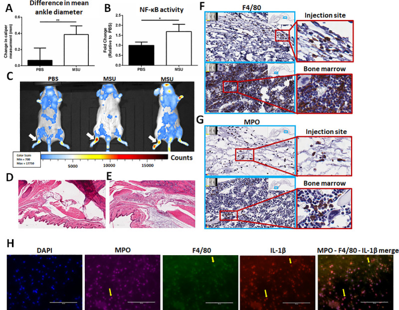
Fig 1

(A) Change in mean ankle diameter 15 hrs post-PBS/MSU injection. Values reported as changes in caliper measurement (final–initial) average ± SD. (n = 3; n = 7, respectively) (B) NF-κB activity was compared in injected feet by in vivo imaging (IVIS) between PBS and MSU treatments. Values reported as fold change (FC ± SD) relative to PBS (n = 3 PBS; n = 5 MSU). Analysis by two-tailed, nonparametric, unpaired Mann-Whitney t-test. *p<0.05, **p<0.01. (C) Whole-mouse comparison of bioluminescent signals between PBS (n = 3) and MSU (n = 5) crystal injected mice (white arrows indicate the site of injection). (D-E) Hematoxylin&Eosin (H&E) stains at 100x magnification in Aperioimage software of (D) PBS-injected and (E) MSU-injected ankle joint spaces. (F) F4/80 immunohistochemistry (IHC) of joint space (top) and bone marrow (bottom). (G) MPO IHC of joint space (top) and bone marrow (bottom). Images on left are 100x magnification and images on right are 400x magnification. Scanned using in Aperio image software. Bone marrow images represent positive controls for IHC staining. (H) Immunofluorescent staining of DAPI, MPO, F4/80, and IL-1β within the synovium of MSU-injected mice. Yellow arrows highlight co-localization of IL-1β expression for macrophages and neutrophils.

Exercise intensity correlates with severity of inflammation

To determine treadmill speeds correlating with different exercise intensities, a metabolic treadmill and respirometer were used to measure maximum oxygen consumption (VO2 max) in male and female BALB/c mice. Since no differences were observed between sexes in analysis, the data were reported collectively. Low, medium, and high-intensity exercise were defined as 35% VO2 max, 55% VO2 max, and 75% VO2 max, respectively [17]. A polynomial curve of VO2 max values at increasing treadmills speeds was used to interpolate exercise intensity levels. Our results showed that the speeds for 35%, 55%, and 75% VO2 max in this mouse strain were 8 m/min, 11 m/min, and 15 m/min, respectively (S2 Fig). Additionally, although murine exercise studies have shown that physical activity can induce a positive effect on emotional behavior by promoting neurotrophic factor expression [18]. no differences in emotional behavior were observed in the mice examined in this study.

Caliper measurements of ankle diameter obtained before and after MSU crystal injection in male mice revealed a significant increase in the non-exercised control mice (0.82 ± 0.27mm). In comparison, mice that exercised before MSU injection exhibited less inflammation compared to the non-exercised control after 15 hrs, but to varying degrees. The low (0.22 ± 0.20mm; p<0.0001) and moderate-intensity (0.13 ± 0.21mm; p<0.0001) exercise groups displayed comparable levels of ankle swelling, which were both significantly less than the non-exercised control and high-intensity groups (Fig 2A).

Fig 2. Mean difference in ankle diameter and NF-κB activity in MSU-injected mice following exercise regimens of varying intensity.
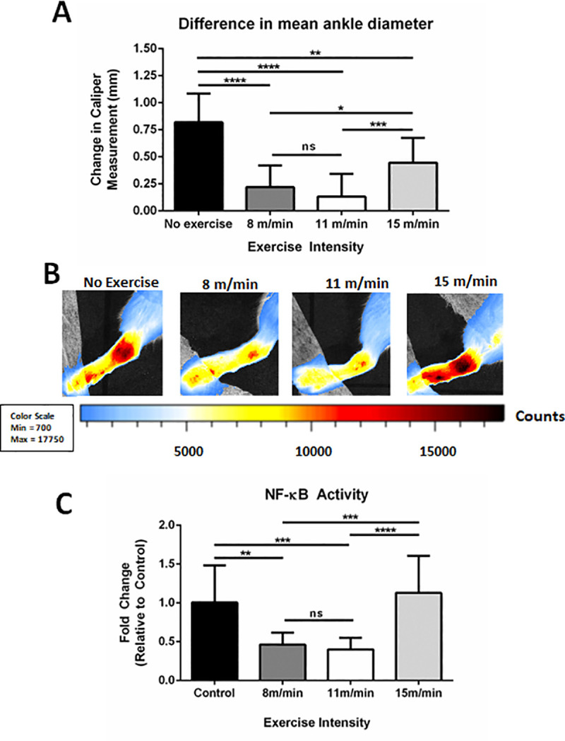
Fig 2

(A) Change in mean ankle diameter (swelling) between non-exercised, 8 m/min, 11 m/min, and 15 m/min groups. Values reported as changes in caliper measurement (final–initial) average ± SD (n = 10, 10, 22, 11, respectively). (B) Representative in vivo imaging system (IVIS) images of MSU injected ankles from non-exercised (n = 10), 8 m/min (n = 10), 11 m/min (n = 22), and 15 m/min (n = 10) groups. Scale of luminescent counts below. (C) Normalized IVIS intensity values as fold changes relative to non-exercised controls (FC ± SD). (n = 10, 10, 22, 11 respectively). Analysis by ANOVA and followed by two-tailed, nonparametric, unpaired Mann-Whitney t-tests. *p<0.05, **p<0.01, ***p<0.001, ****p<0.0001, ns = not significant.

Comparisons of relative NF-κB activity using IVIS revealed a similar trend (Fig 2B). Relative to the non-exercised controls, male NF-κB-luc reporter mice in the low and moderate-intensity groups had 54 ± 15% (p < 0.0001) and 60 ± 15% (p < 0.0001) reduced levels of NF-κB activity, respectively. Furthermore, there was less than a 10% change in NF-κB activity between the non-exercised control and the high-intensity exercise group. Since physical activity as an intervention itself has been shown to correlate with decreased inflammation, male and female mice were exercised at low, moderate, and high intensities; bioluminescent imaging was performed both pre and post-exercise to measure NF-κB activity for comparison to the non-exercised control group. Measurements of systemic NF-κB activity in mice showed no significant differences across groups (S3 Fig), which demonstrates that these exercise regimens do not significantly influence endogenous levels of inflammation. Collectively, these results suggest that exercise intensity can greatly affect the amount of inflammation and NF-κB activity in the synovium following ankle injection with MSU crystals.

Low to moderate-intensity exercise decreases macrophage and neutrophil infiltration in situ

To determine if exercise intensity can affect the degree of macrophage/neutrophil infiltration, we examined the macrophage marker F4/80 and the granulocyte marker MPO on paraffin-embedded sections of the ankle synovial space (Fig 3). F4/80+ cells are decreased in the synovium of low and moderate-intensity exercise groups compared to the non-exercised controls and the high-intensity exercise group (Fig 3A). To quantify these differences, the slides were scanned and digitized for histopathological analysis using Aperio IHC analysis software [10]. F4/80 staining within the intra-articular space shows that there is 68 ± 33% less F4/80 staining in the low-intensity exercise group (p<0.01) and 93 ± 5% less F4/80 staining in the moderate-intensity exercise group (p<0.001) relative to the non-exercised controls and the high-intensity exercise group. (Fig 3B). Further histopathological analysis of the joint space for the granulocyte marker MPO revealed decreased MPO staining in the low-intensity (69 ± 21%, p<0.01) and moderate-intensity (83 ± 30%, p<0.001) groups relative to the high-intensity exercise group and the non-exercised controls (Fig 3C and 3D). In parallel with the F4/80 staining, there was no significant difference in MPO staining between the high-intensity exercise group and the non-exercised controls. These results suggest that exercise intensity plays a significant role in modulating the ability of macrophages and neutrophils to infiltrate the synovium in response to inflammatory stimuli, such as MSU crystals.

Fig 3. Immunohistochemistry for F4/80 and MPO in MSU-injected synovium.
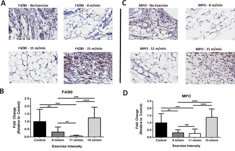
Fig 3

(A) Expression of F4/80 by immunohistochemical (IHC) staining on non-exercised control, 8 m/min, 11 m/min, and 15 m/min exercise conditions. Image magnification at 400x and pictures captured from Aperio Image software. (B) Analysis of F4/80+ pixel intensity of 10 randomized areas in joint space, as expressed by fold change (FC ± SD) relative to non-exercised controls (n = 3 mice/group). (C) Expression of myeloperoxidase (MPO) by IHC on non-exercised control, 8 m/min, 11 m/min, and 15 m/min exercise conditions. Image magnification at 400x with Aperio Image software photomicrographs. (D) Analysis of MPO+ pixel intensity of 10 randomized areas in joint space as expressed by fold change (FC ± SD) relative to non-exercised controls (n = 3 mice/group). Analysis by ANOVA and followed by two-tailed, nonparametric, unpaired Mann-Whitney t-tests. *p<0.05, **p<0.01, ***p<0.001, ****p<0.0001, ns = not significant.

IL-1β expression is decreased locally and inflammasome activation is downregulated systemically following low/moderate-intensity exercise

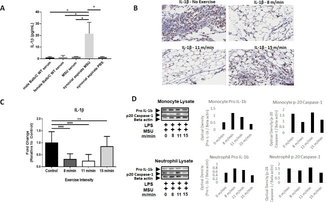
IL-1β is a tightly regulated inflammatory cytokine, which has been previously shown to be a primary mediator of MSU crystal-induced inflammation and inflammasome activation [1921]. To determine the extent exercise intensity can affect IL-1β, systemic and localized IL-1β expression levels were examined. While IL-1β expression levels in serum of MSU-injected NF-κB-luc reporter mice or wild-type control Balb/C mice did not differ significantly from synovial aspirates of PBS-injected mice, localized IL-1β expression in the synovium of NF-κB-luc mice was significantly elevated after MSU injection (Fig 4A). Furthermore, an examination of cleaved/secreted IL-1β by IHC at the site of MSU crystal injection revealed markedly reduced IL-1β staining in the synovial space of low/moderate-intensity exercise (Fig 4B). Quantification using Aperio IHC analysis revealed a 68.5% (p<0.0001) and 86.1% (p<0.0001) reduction in IL-1β staining present locally in the synovium of low or moderate-intensity exercise groups, respectively (Fig 4C). There was no significant difference in IL-1β staining between the high-intensity exercise group and the non-exercised controls. Collectively, these results show that MSU-induced IL-1β expression is suppressed locally with low and moderate exercise.

Fig 4. IL-1β expression in MSU-injected mice and in vitro analysis of inflammasome activation in monocytes and neutrophils.

Fig 4

(A) ELISA analysis of IL-1β expression in serum or synovial aspirates of Balb/c mice. (B) Expression of IL-1β by immunohistochemical (IHC) staining on non-exercised control, 8 m/min, 11 m/min, and 15 m/min exercise conditions. Image magnification at 400x and pictures captured using Aperio Image software. (C) Analysis of F4/80+ pixel intensity of 10 randomized areas in joint space as expressed by fold change (FC ± SD) relative to non-exercised controls (n = 3 mice/group). (D) Following exercise regimens at low, medium, and high intensities, neutrophils and monocytes were isolated from whole blood by immunomagnetic sorting and stimulated in vitro by LPS priming followed by MSU to induce inflammasome activation. Western blots were performed on pooled cell lysates (n = 5 per condition) to measure pro-IL-1β and p20 caspase-1 expression. Beta-actin was used as a loading control. Optical density values expressed relative to beta-actin normalization. Analysis by ANOVA and followed by two-tailed, nonparametric, unpaired Mann-Whitney t-tests. *p<0.05, **p<0.01, ***p<0.001, ****p<0.0001, ns = not significant.

In a previous murine model of gouty inflammation, neutrophil infiltration and IL-1β production at the site of MSU crystal injection were shown to be suppressed in mice lacking various components of the NLRP3 inflammasome [19]. Given the reduced presence of IL-1β, monocytes, and neutrophils at the site of MSU crystal injection, inflammasome activity was examined in peripheral monocytes and neutrophils to determine if exercise intensity can affect inflammasome activation upstream of IL-1β production and cellular infiltration. Mice were exercised at low, medium, and high intensities daily for 2 weeks. Whole blood samples from each condition were isolated by positive selection with immunomagnetic sorting and macrophages or neutrophils were treated with LPS and MSU in vitro to stimulate inflammasome activation. Cell lysates were analyzed by Western blotting for changes in pro-IL-1β production and activation of p20 caspase-1 following isolation with immunomagnetic beads. With LPS and MSU stimulation, both neutrophils and monocytes had elevated levels of pro-IL-1β following low/moderate intensity exercise relative to high intensity and controls without exercise (Fig 4D). Additionally, previous studies have shown that inflammasome activation is directly correlated with the level of p20 active caspase-1 released from cells into the surrounding extracellular space [19]. Thus, higher levels of p20 caspase-1 in the cellular lysate indicates that p20 caspase-1 is not secreted, which suggests decreased inflammasome activation. Mice that exercised at 11 m/min had elevated p20 caspase-1 remaining in the neutrophil and monocyte lysates compared to the other experimental groups (Fig 4D). These data suggest that both low and moderate exercise regimens can result in physiologically dysregulated inflammasome activation when exposed to catalysts to this response in vitro, which may be associated with reduced levels of inflammatory recruitment of neutrophils and monocytes locally in MSU crystal-induced arthritis.

Low and moderate-intensity exercise decreases systemic levels of CXCL1 and TLR2 expression on peripheral neutrophils

Previous studies in humans have shown that moderate physical activity (~65% VO2 max) can decrease the surface expression of TLRs on peripheral monocytes and neutrophils [21]. Furthermore, TLR2 signaling has been highly implicated in mediating gout pathogenesis [2224]. Using flow cytometry, we quantified TLR2 expression on peripheral monocytes and neutrophils following exercise and MSU crystal-induced gout (Fig 5A). A finalized gating strategy for TLR2 detection was determined by using compensation control analysis with both unstained and single stained cells (S4 Fig). A significant decrease in TLR2 expression on neutrophils was observed in both low-intensity (47.81 ± 10.98%; p<0.001) and moderate-intensity (27.59 ± 18.15%; p<0.0001) exercise groups compared to the non-exercised controls (57.91 ± 6.79%) (Fig 5B). Similarly, the median-fluorescent intensity (MFI) of TLR2 on neutrophils was significantly decreased in the low-intensity (13.41 ± 15.56%; p<0.001) and moderate-intensity (38.69 ± 24.73%; p<0.0001) groups compared to the non-exercised controls (Fig 5C). There was less than 1% difference in MFI between the high-intensity exercise mice and the non-exercised controls. These results confirm that TLR2 expression on neutrophils is significantly affected by exercise intensity and provides a plausible mechanism for the observed decreases in inflammation and NF-κB activity in vivo in this model.

Fig 5. Analysis of and CXCL1 serum levels and TLR2 expression on peripheral neutrophils.
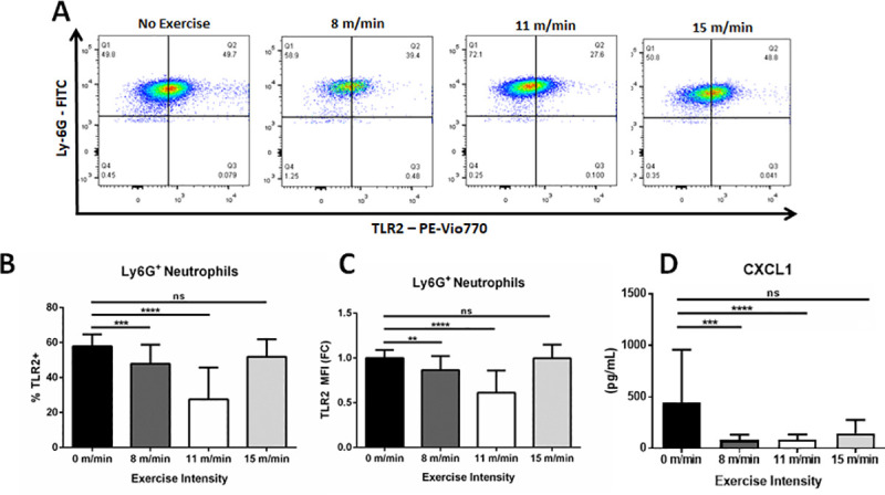
Fig 5

(A) Flow cytometry of TLR2 expression on Ly6G+ / CD115- neutrophils from the periphery of non-exercised control, 8 m/min, 11 m/min, and 15 m/min groups. Images are representative of multiple samples. (B) Analysis of percent TLR2+ neutrophils and (C) TLR2 MFI from non-exercised, 8 m/min, and 11 m/min conditions. (n = 10, 15, 15, and 10 samples run in duplicate, respectively). (D) ELISA analysis measuring CXCL1 expression in serum. Statistical analysis by ANOVA and followed by two-tailed, nonparametric, unpaired Mann-Whitney t-tests. Error bars ±SD. *p<0.05, **p<0.01, ***p<0.001, ****p<0.0001, ns = not significant.

Our histopathological results demonstrate a decrease in neutrophil infiltrates at the site of MSU crystal injection with low/moderate physical activity. CXCL1 is a neutrophil chemoattractant that recruits neutrophils and thereby helps facilitate inflammatory responses [25]. Our results show that whole-blood expression of CXCL1was significantly suppressed in the low-intensity (80.18 ± 13.61 pg/mL; p<0.01) and moderate-intensity (84.65 ± 11.95 pg/mL p<0.01) groups compared to the non-exercised controls (440.9 ± 110.3 pg/mL), respectively (Fig 5D). No significant differences were observed between high-intensity exercise mice and the non-exercised controls. These data demonstrate that low/moderate intensity exercise results in a systemic suppression of CXCL1 expression with the stimulation of MSU crystal-induced arthritis, which can lead to the inhibition of neutrophil recruitment locally to ankle joint synovial space.

Increased physical activity in human gout patients correlates with decreased markers of inflammation and better patient prognosis

While exercise has recently been shown to be efficacious in reducing pathogenic inflammatory burdens in several autoimmune diseases, the impact in patients with gout has not been studied extensively. To examine the effects of physical activity in a gout patient population, physical activity questionnaires and clinical data were analyzed from a cohort of patients with a gout diagnosis (n = 30). The patient population consisted of 25 males and 5 females; however, all females were post-menopausal. While the male predilection of gout is 10:1 prior to menopause, this is reduced significantly in post-menopausal cohorts presumably due to the uricosurics effect of female sex hormones [13]. Furthermore, since individual analysis showed no significant correlation to sex in any metric measured, the data from this cohort was reported collectively. At the scheduled visit, gout patients not experiencing a flare at the time of visit were recruited from our clinics; IPAQ surveys were completed to assess current levels of daily/weekly physical activity and clinical data was obtained, including BMI, age, years since diagnosis, attacks per year, perceived pain at the time of visit and in the past 4 weeks, and CRP levels. The IPAQ survey, which has been previously used in studies of rheumatologic diseases [26], separated the gout patients into physically active (n = 16) and physically inactive cohorts (n = 14) (p<0.001; Fig 6A). While average age, BMI, or years since diagnosis did not significantly differ between cohorts (Fig 6B–6D), physically active gout patients had over 12-fold fewer gout flares per year (p<0.01; Fig 6E), 10-fold less CRP (p<0.01; Fig 6F), a 4.6-fold decrease in perceived pain at the time of visit (p<0.01; Fig 6G), and a 2.8-fold decrease in perceived pain over the past 4-week period (p<0.05; Fig 6H). These data indicate that increased physical activity is indeed beneficial in patients with gout.

Fig 6. Questionnaires and clinical data collected from gout patients.
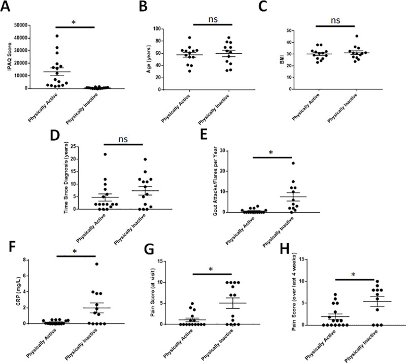
Fig 6

Physically active = 16 (IPAQ>1400); physically inactive = 14 (IPAQ<1400). Data analyzed with individual unpaired t-tests with Welch’s correction and one-way ANOVA without Gaussian distribution and multiple comparisons to analyze the mean rank of each data set with that of every other to obtain p-values. p≤0.05 considered statistically significant. *p<0.05, **p<0.01, ***p<0.001, ****p<0.0001, ns = not significant.

Discussion

Our results show that regimens of low-to-moderate intensity exercise can significantly reduce the inflammation observed in an acute model of murine gout. Mice receiving low-intensity and moderate-intensity exercise had measurably less swelling and NF-κB activity than the non-exercised controls. Furthermore, we showed that low-to-moderate exercise significantly decreases infiltration of F4/80+ macrophages and MPO+ neutrophils into the synovium of MSU crystal-injected mice. As a possible mechanism explaining these results, we show that mice receiving low-intensity and moderate-intensity exercise have significantly fewer TLR2+ neutrophils and reduced systemic expression of the neutrophil chemoattractant CXCL1 relative to non-exercised controls.

Research studies specifically examining the effects of exercise in a canine model of gouty arthritis date back almost half a century, were performed only once, and contained very small sample sizes [27,28]. Agudelo and Dorwart reported increased articular inflammation and tissue infiltration in the exercised condition versus the non-exercised condition [28,29]. This is likely attributable to experimental differences in MSU crystal administration: Agudelo and Dorwart injected MSU crystals prior to initiating joint movement whereas mice in this study were physically conditioned to pre-determined exercise intensities, exercised regularly for two weeks, and then administered MSU crystals. Additionally, these studies used passively controlled movement of the effected joint in an otherwise resting animal while our study evaluated the effects of physical activity with active ambulatory exercise routines.

MSU crystals induce cellular stress resulting in NLRP3 inflammasome activation and IL-1β secretion [20,30]. In addition, in vitro studies with human and murine macrophages have shown that free fatty acids can signal through TLR2 and prime the MSU-induced immune response synergistically leading to ASC-caspase-1 driven IL-1β release [20]. Our results show decreased TLR2 expression on the surface of neutrophils, which suggests that exercise downregulates this priming element of inflammasome activation. Moreover, we demonstrated in vitro that low/moderate exercise leads to an upregulation of p20 caspase-1 and/or pro-IL1β expression intracellularly, suggesting dysregulated inflammasome signaling. Furthermore, previous studies have shown that chronic, low-level exposure to LPS priming of cells in vitro leads to a suppression of inflammasome activation and IL-1β expression [31]. Since physical activity is associated with an acute pro-inflammatory response [32], this can potentially function as a daily chronic exposure to inflammation physiologically and result in immune tolerance to subsequent challenges with MSU crystals. Collectively, these data suggest that exercise-induced downregulation of TLR2 expression and inflammasome priming may result in a tolerogenic effect and reduced inflammatory responses. Thus, we propose that the reduced inflammation observed with MSU crystal injection is not an anti-inflammatory response, but rather an immunomodulated and regulated immune response resulting from immune tolerance derived from chronic exposures to small bouts of inflammation following exercise. Using this hypothesis, it can be speculated that exercise can immuno-optimize physiologically by preventing hyperactive immune responses leading to inflammation and clinical symptoms following antigenic challenge, which would correlate with the wealth of epidemiological data concluding that people with higher levels of physical activity have a significantly decreased frequency of clinical illness.

While the clinical practice guidelines for gout intervention released by The European League Against Rheumatism (EULAR; 2016) suggest that regular physical activity may help combat the mortality associated with chronic hyperuricemia, no insight is provided into intensity, duration, or type of exercise. Current ACR and EULAR recommendations for the treatment of gout have only recently started to recommend exercise for the treatment of gout; however, no recommendations have been made regarding the type or intensity of exercise [6,33]. Considering the improved gout patient clinical course and CRP levels observed in our human cohort and the increasing prevalence and incidence of gout, this further highlights the importance of incorporating low or moderate-intensity exercise as an adjunct treatment option [34]. Whether exercise can affect the frequency of gout flares in a mouse model has yet to be determined and will require the establishment and characterization of a chronic gout model, which more accurately represents disease occurrence in humans.

In summary, this study reveals that low-intensity and moderate-intensity exercise can prevent the inflammatory response in acute models of murine gout. Low and moderate-intensity exercise can reduce tissue swelling, NF-κB activity, tissue infiltration by macrophages and neutrophils, TLR2 expression on peripheral neutrophils, and systemic expression of CXCL1. Although this is not the first study to examine the relationship between exercise and gout, our work helps define the therapeutic potential of low/moderate-intensity aerobic exercise in the management of gout and other inflammatory diseases. In subsequent experiments to the work outlined here, we also examined the effects of exercise during and after the induction of gout in preliminary studies to determine if exercise can therapeutically inhibit the pathology associated with flares. NF-κB-RE-luc mice were exercised at the optimal regimen defined herein (11 m/min) and MSU crystals were injected intra-articularly in several rounds with a rest/recovery period in-between. The results indicated complete resolution of localized NF-κB activity in mice given repeated MSU injections without measures to raise serum uric acid levels (S5 Fig); thus, a chronic model of gout could not be established, which precluded further characterization.

Elucidating the role exercise plays in health and disease is fundamental to our understanding of human physiology and inflammatory disease pathogenesis. As our knowledge of the benefits of exercise increases at the molecular level, so too will our ability to incorporate its effectiveness as a tool for improving health outcomes in the clinical setting. Considered collectively and in a translational context, we feel that the results of our study show that exercise would be helpful to a patient with gout that is in a recovery period between flares to hopefully prevent or limit future ones, as a preventative measure. While rest and decreased movement/weight may still be recommended to a patient with a flare presenting with a red, painful, and swollen foot, we envision a standardized exercise regimen being prescribed during times of clinical inactivity to help the severity and frequency of future occurrences. Future translational studies examining the effects low and moderate-intensity exercise in patients with gout could facilitate new discoveries in our understanding and treatment of this disease.

Supporting information

S1 Fig

Confirmation of MSU crystals in the ankle joints of MSU injected mice (top row). MSU crystals were visualized by polarizing light and phase contrast microscopy following resuspension in PBS, diluted 1:50, or PBS without MSU. Mice were given intra-articular injections of PBS or MSU and assessed 16 hrs later. (middle row) Pictures taken of feet/ankles show increased swelling with MSU injection relative to contralateral feet/ankles or PBS controls (white arrows). (bottom row) Synovial aspirates of PBS-injected mice show no detectable cells or MSU crystals. Conversely, MSU injection produced detectable MSU crystals and cells were observed actively engulfing crystals (white arrow).

(TIF)

S2 Fig. Determination of exercise intensity by respirometry.

Male and female Balb/c mice (n = 14) were exercised in five minutes intervals at increasing speeds with a 5% incline. The VO2 consumption was recorded every 30 seconds. Values were plotted and a polynomial curve was applied to graph and interpolate speeds correlating with 35% (low-intensity), 55% (medium-intensity), and 75% (high-intensity) VO2 max. Speeds were determined to be 8 m/min, 11 m/min, and 15 m/min, respectively.

(TIF)

S3 Fig. Exercise alone does not significantly impact NF-κB activity.

Male and female Balb/c mice (n = 14) were exercised at 8 m/min, 11 m/min, and 15 m/min daily for 2 weeks. Systemic NF-κB activity was measured by IVIS before beginning the exercise regimen and 24 hrs after the completion of the final session. Analysis by ANOVA and followed by two-tailed, nonparametric, unpaired Mann-Whitney t-tests produced no statistically different results.

(TIF)

S4 Fig. Flow cytometry gating strategy for the detection of TLR2.

Gating strategy of successive flow cytometry gates to detect TLR2 positive expression. The total cell population was first gated on forward and side scatter. Subsequently, neutrophils were gated by Ly6G+ / CD11b+ detection. TLR2 expression was then determined by PE-Vio770 fluorochrome detection relative to the signals observed in unstained and single stained cells.

(TIF)

S5 Fig. Repeated MSU injections do not contribute to enhanced NF-κB activation.

Bioluminescent imaging of NF-κB-RE-luc mouse feet measured and quantitated via IVIS. Repeated intra-articular injections of MSU crystals (20 μg) were made where indicated. Analysis of localized NF-κB activity suggests complete resolution prior to subsequent challenges.

(TIF)

S1 Raw data

(ZIP)

S1 File

(DOC)

Acknowledgments

We would like to thank the Comparative Pathology & Mouse Phenotyping Shared Resource staff, Department of Veterinary Biosciences staff, and the Comprehensive Cancer Center staff at The Ohio State University.

Data Availability

All relevant data are within the paper and its Supporting Information files.

Funding Statement

Both Nicholas Young and Naomi Schlesinger had research support initially derived from Ardea Biosciences (this support was subsequently provided via Ironwood Pharmaceuticals) examining the role of exercise in inflammation, which directly supported these research endeavors. The Center for Clinical and Translational Science (CCTS) at OSUWMC is supported by UL1TR001070 from the National Center for Advancing Translational Science. The Comparative Pathology & Mouse Phenotyping Shared Resource at The Ohio State University is supported in part by grant P30 CA016058, National Cancer Institute. The funders had no role in study design, data collection and analysis, decision to publish, or preparation of the manuscript.

References

  • 1.Macera CA, Hootman JM, Sniezek JE. Major public health benefits of physical activity. Arthritis Rheum 2003;49:122–8. 10.1002/art.10907 [DOI] [PubMed] [Google Scholar]
  • 2.Warburton DER, Nicol CW, Bredin SSD. Health benefits of physical activity: the evidence. CMAJ 2006;174:801–9. 10.1503/cmaj.051351 [DOI] [PMC free article] [PubMed] [Google Scholar]
  • 3.Ford ES. Does Exercise Reduce Inflammation? Physical Activity and C-Reactive Protein Among U.S. Adults. Epidemiology 2015;13:561–8. 10.1097/01.EDE.0000023965.92535.C0 [DOI] [PubMed] [Google Scholar]
  • 4.Benatti FB, Pedersen BK. Exercise as an anti-inflammatory therapy for rheumatic diseases-myokine regulation. Nat Rev Rheumatol 2015;11:86–97. 10.1038/nrrheum.2014.193 [DOI] [PubMed] [Google Scholar]
  • 5.Holla J, Fluit M, Van Schaardenburg D, et al. Recreational exercise in rheumatic diseases. Int J Sports Med 2009;30:814–20. 10.1055/s-0029-1233467 [DOI] [PubMed] [Google Scholar]
  • 6.Khanna D, Fitzgerald JD, Khanna PP, et al. 2012 American college of rheumatology guidelines for management of gout. part 1: Systematic nonpharmacologic and pharmacologic therapeutic approaches to hyperuricemia. Arthritis Care Res 2012;64:1431–46. 10.1002/acr.21772 [DOI] [PMC free article] [PubMed] [Google Scholar]
  • 7.Qaseem A, Harris RP, Forciea MA, et al. Management of acute and recurrent gout: A clinical practice guideline from the American College of Physicians. Ann Intern Med 2017;166:58–68. 10.7326/M16-0570 [DOI] [PubMed] [Google Scholar]
  • 8.Torres R, Macdonald L, Croll SD, et al. Hyperalgesia, synovitis and multiple biomarkers of inflammation are suppressed by interleukin 1 inhibition in a novel animal model of gouty arthritis. Ann Rheum Dis 2009;68:1602–8. 10.1136/ard.2009.109355 [DOI] [PubMed] [Google Scholar]
  • 9.Young N a., Bruss MS, Gardner M, et al. Oral Administration of Nano-Emulsion Curcumin in Mice Suppresses Inflammatory-Induced NFκB Signaling and Macrophage Migration. PLoS One 2014;9:e111559 10.1371/journal.pone.0111559 [DOI] [PMC free article] [PubMed] [Google Scholar]
  • 10.Young NA, Wu LC, Bruss M, et al. A chimeric human-mouse model of Sjogren’s syndrome. Clin Immunol 2015;156:1–8. 10.1016/j.clim.2014.10.004 [DOI] [PMC free article] [PubMed] [Google Scholar]
  • 11.Young NA, Sharma R, Friedman AK, et al. Aberrant muscle antigen exposure in mice is sufficient to cause myositis in a treg cell-deficient milieu. Arthritis Rheum 2013;65:3259–70. 10.1002/art.38184 [DOI] [PMC free article] [PubMed] [Google Scholar]
  • 12.Carlsen H, Moskaug JO, Fromm SH, et al. In Vivo Imaging of NF- B Activity. J Immunol 2002;168:1441–6. 10.4049/jimmunol.168.3.1441 [DOI] [PubMed] [Google Scholar]
  • 13.Singh JA. Racial and gender disparities among patients with gout. Curr Rheumatol Rep 2013;15:1–15. 10.1007/s11926-012-0307-x [DOI] [PMC free article] [PubMed] [Google Scholar]
  • 14.Busso N, So A. Mechanisms of inflammation in gout. Int J Biochem Cell Biol 2010;42:479 10.1016/j.biocel.2010.02.001 [DOI] [PubMed] [Google Scholar]
  • 15.Mitroulis I, Kambas K, Ritis K. Neutrophils, IL-1β, and gout: Is there a link? Semin Immunopathol 2013;35:501–12. 10.1007/s00281-013-0361-0 [DOI] [PubMed] [Google Scholar]
  • 16.Martin WJ, Grainger R, Harrison A, et al. Differences in MSU-induced superoxide responses by neutrophils from gout subjects compared to healthy controls and a role for environmental inflammatory cytokines and hyperuricemia in neutrophil function and survival. J Rheumatol 2010;37:1228–35. 10.3899/jrheum.091080 [DOI] [PubMed] [Google Scholar]
  • 17.Schefer V, Talan MI. Oxygen consumption in adult and aged C57BL/6J mice during acute treadmill exercise of different intensity. Exp Gerontol 1996;31:387–92. 10.1016/0531-5565(95)02032-2 [DOI] [PubMed] [Google Scholar]
  • 18.Sleiman SF, Henry J, Al-Haddad R, et al. Exercise promotes the expression of brain derived neurotrophic factor (BDNF) through the action of the ketone body β- hydroxybutyrate. Elife 2016;5:1–21. 10.7554/eLife.15092 [DOI] [PMC free article] [PubMed] [Google Scholar]
  • 19.Martinon F, Pétrilli V, Mayor A, et al. Gout-associated uric acid crystals activate the NALP3 inflammasome. Nature 2006;440:237–41. 10.1038/nature04516 [DOI] [PubMed] [Google Scholar]
  • 20.So AK, Martinon F. Inflammation in gout: Mechanisms and therapeutic targets. Nat Rev Rheumatol 2017;13:639–47. 10.1038/nrrheum.2017.155 [DOI] [PubMed] [Google Scholar]
  • 21.Lancaster GI, Khan Q, Drysdale P, et al. The physiological regulation of toll-like receptor expression and function in humans. J Physiol 2005;563:945–55. 10.1113/jphysiol.2004.081224 [DOI] [PMC free article] [PubMed] [Google Scholar]
  • 22.Joosten LAB, Netea MG, Mylona E, et al. Engagement of fatty acids with toll-like receptor 2 drives interleukin-1β production via the ASC/caspase 1 pathway in monosodium urate monohydrate crystal-induced gouty arthritis. Arthritis Rheum 2010;62:3237–48. 10.1002/art.27667 [DOI] [PMC free article] [PubMed] [Google Scholar]
  • 23.Joosten LAB, Abdollahi-Roodsaz S, Dinarello CA, et al. Toll-like receptors and chronic inflammation in rheumatic diseases: new developments. Nat Rev Rheumatol 2016;12:344–57. 10.1038/nrrheum.2016.61 [DOI] [PubMed] [Google Scholar]
  • 24.Liu-Bryan R, Scott P, Sydlaske A, et al. Innate immunity conferred by Toll-like receptors 2 and 4 and myeloid differentiation factor 88 expression is pivotal to monosodium urate monohydrate crystal-induced inflammation. Arthritis Rheum 2005;52:2936–46. 10.1002/art.21238 [DOI] [PubMed] [Google Scholar]
  • 25.Yuan M, Zhu H, Xu J, et al. Tumor-derived CXCL1 promotes lung cancer growth via recruitment of tumor-associated Neutrophils. J Immunol Res 2016;2016 10.1155/2016/6530410 [DOI] [PMC free article] [PubMed] [Google Scholar]
  • 26.Hernández-hernández V, Ferraz-amaro I, Díaz-gonzález F. Influence of disease activity on the physical activity of rheumatoid arthritis patients. Rheumatol (United Kingdom) 2014;53:722–31. 10.1093/rheumatology/ket422 [DOI] [PubMed] [Google Scholar]
  • 27.Agudelo C a Wise CM. Gout: diagnosis, pathogenesis, and clinical manifestations. Curr Opin Rheumatol 2001;13:234–9. 10.1097/00002281-200105000-00015 [DOI] [PubMed] [Google Scholar]
  • 28.Dorwart BB, Hansell JR, Schumacher HR. Effects of cold and heat on urate crystal-induced synovitis in the dog. Arthritis Rheum 1974;17:563–71. 10.1002/art.1780170510 [DOI] [PubMed] [Google Scholar]
  • 29.Agudelo CA, Schumacher HR, Phelps P. Effect of Exercise on Urate Crystal-Induced Inflammation in Canine Joints. 1972;15:609–16. [DOI] [PubMed] [Google Scholar]
  • 30.Abais JM, Xia M, Zhang Y, et al. Redox Regulation of NLRP3 Inflammasomes: ROS as Trigger or Effector? Antioxidants Redox Signal 2015;22:1111–29. 10.1089/ars.2014.5994 [DOI] [PMC free article] [PubMed] [Google Scholar]
  • 31.Gurung P, Li B, Subbarao Malireddi RK, et al. Chronic TLR Stimulation Controls NLRP3 Inflammasome Activation through IL-10 Mediated Regulation of NLRP3 Expression and Caspase-8 Activation. Sci Rep 2015;5:1–10. 10.1038/srep14488 [DOI] [PMC free article] [PubMed] [Google Scholar]
  • 32.Brown M, McClean CM, Davison GW, et al. The acute effects of walking exercise intensity on systemic cytokines and oxidative stress. Eur J Appl Physiol 2018;118:2111–20. 10.1007/s00421-018-3930-z [DOI] [PMC free article] [PubMed] [Google Scholar]
  • 33.Lunzer R. 2016 updated EULAR evidence-based recommendations for the management of gout. J fur Miner 2016;23:108 10.1136/annrheumdis-2016-209707 [DOI] [PubMed] [Google Scholar]
  • 34.So AK. Why better treatment of gout is needed. Clin Exp Rheumatol 2016;34:63–5. [PubMed] [Google Scholar]

Decision Letter 0

Fulvio D'Acquisto

8 Apr 2020

PONE-D-20-02077

Physical Activity Suppresses Acute Inflammation in a Gout Model by Downregulation of TLR2 on Circulating Neutrophils as well as Inhibition of Serum CXCL1 and is Associated with Decreased Pain and Inflammation in Gout Patients

PLOS ONE

Dear Dr. Young,

Thank you for submitting your manuscript to PLOS ONE. After careful consideration, we feel that it has merit but does not fully meet PLOS ONE’s publication criteria as it currently stands. Therefore, we invite you to submit a revised version of the manuscript that addresses the points raised during the review process.

We would appreciate receiving your revised manuscript by May 23 2020 11:59PM. When you are ready to submit your revision, log on to https://www.editorialmanager.com/pone/ and select the 'Submissions Needing Revision' folder to locate your manuscript file.

If you would like to make changes to your financial disclosure, please include your updated statement in your cover letter.

To enhance the reproducibility of your results, we recommend that if applicable you deposit your laboratory protocols in protocols.io, where a protocol can be assigned its own identifier (DOI) such that it can be cited independently in the future. For instructions see: http://journals.plos.org/plosone/s/submission-guidelines#loc-laboratory-protocols

Please include the following items when submitting your revised manuscript:

  • A rebuttal letter that responds to each point raised by the academic editor and reviewer(s). This letter should be uploaded as separate file and labeled 'Response to Reviewers'.

  • A marked-up copy of your manuscript that highlights changes made to the original version. This file should be uploaded as separate file and labeled 'Revised Manuscript with Track Changes'.

  • An unmarked version of your revised paper without tracked changes. This file should be uploaded as separate file and labeled 'Manuscript'.

Please note while forming your response, if your article is accepted, you may have the opportunity to make the peer review history publicly available. The record will include editor decision letters (with reviews) and your responses to reviewer comments. If eligible, we will contact you to opt in or out.

We look forward to receiving your revised manuscript.

Kind regards,

Fulvio D'Acquisto, PhD

Academic Editor

PLOS ONE

Journal Requirements:

When submitting your revision, we need you to address these additional requirements.

1. Please ensure that your manuscript meets PLOS ONE's style requirements, including those for file naming. The PLOS ONE style templates can be found at

http://www.journals.plos.org/plosone/s/file?id=wjVg/PLOSOne_formatting_sample_main_body.pdf and http://www.journals.plos.org/plosone/s/file?id=ba62/PLOSOne_formatting_sample_title_authors_affiliations.pdf

2.Please provide additional details regarding participant consent. In the ethics statement in the Methods and online submission information, please ensure that you have specified whether consent was written or verbal/oral. If consent was verbal/oral, please specify: 1) whether the ethics committee approved the verbal/oral consent procedure, 2) why written consent could not be obtained, and 3) how verbal/oral consent was recorded. If your study included minors, please state whether you obtained consent from parents or guardians in these cases.

3. Please include in your Methods section the date ranges over which you recruited participants to this study."

4. To comply with PLOS ONE submissions requirements, please provide method(s) of animal sacrifice/euthanasia in the Methods section of your manuscript.'"

5. Please note that all PLOS journals ask authors to adhere to our policies for sharing of data and materials: https://journals.plos.org/plosone/s/data-availability. According to PLOS ONE’s Data Availability policy, we require that the minimal dataset underlying results reported in the submission must be made immediately and freely available at the time of publication. As such, please remove any instances of 'unpublished data' or 'data not shown' in your manuscript and replace these with either the relevant data (in the form of additional figures, tables or descriptive text, as appropriate), a citation to where the data can be found, or remove altogether any statements supported by data not presented in the manuscript.

6. We note that you have indicated that data from this study are available upon request. PLOS only allows data to be available upon request if there are legal or ethical restrictions on sharing data publicly. For more information on unacceptable data access restrictions, please see http://journals.plos.org/plosone/s/data-availability#loc-unacceptable-data-access-restrictions.

In your revised cover letter, please address the following prompts:

a) If there are ethical or legal restrictions on sharing a de-identified data set, please explain them in detail (e.g., data contain potentially sensitive information, data are owned by a third-party organization, etc.) and who has imposed them (e.g., an ethics committee). Please also provide contact information for a data access committee, ethics committee, or other institutional body to which data requests may be sent.

b) If there are no restrictions, please upload the minimal anonymized data set necessary to replicate your study findings as either Supporting Information files or to a stable, public repository and provide us with the relevant URLs, DOIs, or accession numbers. For a list of acceptable repositories, please see http://journals.plos.org/plosone/s/data-availability#loc-recommended-repositories.

We will update your Data Availability statement on your behalf to reflect the information you provide.

7. Thank you for stating the following financial disclosure:

"Both Nicholas Young and Naomi Schlesinger had research support initially derived from Ardea Biosciences (this support was subsequently supplemented via Ironwood Pharmaceuticals) examining the role of exercise in inflammation, which directly supported these research endeavors. We would like to extend our gratitude to the Clinical Research Center and the Research Match Program through the Center for Clinical and Translational Science (CCTS) at OSUWMC. The CCTS is supported by UL1TR001070 from the National Center for Advancing Translational Science. Comparative Pathology & Mouse Phenotyping Shared Resource at The Ohio State University is supported in part by grant P30 CA016058, National Cancer Institute."

Please state what role the funders took in the study.  If the funders had no role, please state: "The funders had no role in study design, data collection and analysis, decision to publish, or preparation of the manuscript."

If this statement is not correct you must amend it as needed.

Please include this amended Role of Funder statement in your cover letter; we will change the online submission form on your behalf.

8. Thank you for stating the following in the Financial Disclosure section:

"Both Nicholas Young and Naomi Schlesinger had research support initially derived from Ardea Biosciences (this support was subsequently supplemented via Ironwood Pharmaceuticals) examining the role of exercise in inflammation, which directly supported these research endeavors. We would like to extend our gratitude to the Clinical Research Center and the Research Match Program through the Center for Clinical and Translational Science (CCTS) at OSUWMC. The CCTS is supported by UL1TR001070 from the National Center for Advancing Translational Science. Comparative Pathology & Mouse Phenotyping Shared Resource at The Ohio State University is supported in part by grant P30 CA016058, National Cancer Institute."

We note that you received funding from a commercial source: 'Ironwood Pharmaceuticals'

Please provide an amended Competing Interests Statement that explicitly states this commercial funder, along with any other relevant declarations relating to employment, consultancy, patents, products in development, marketed products, etc.

Within this Competing Interests Statement, please confirm that this does not alter your adherence to all PLOS ONE policies on sharing data and materials by including the following statement: "This does not alter our adherence to PLOS ONE policies on sharing data and materials.” (as detailed online in our guide for authors http://journals.plos.org/plosone/s/competing-interests).  If there are restrictions on sharing of data and/or materials, please state these. Please note that we cannot proceed with consideration of your article until this information has been declared.

Please include your amended Competing Interests Statement within your cover letter. We will change the online submission form on your behalf.

9. Thank you for stating the following in the Competing Interests section:

"Naomi Schlesinger has grants from Pfizer and AMGEN; she is also on the advisory board and consulting for Novartis, Horizon Pharma, Selecta Biosciences, Olatec, and Mallinckrodt Pharmaceuticals. The remaining authors declare no conflict of interest."

Please confirm that this does not alter your adherence to all PLOS ONE policies on sharing data and materials, by including the following statement: "This does not alter our adherence to  PLOS ONE policies on sharing data and materials.” (as detailed online in our guide for authors http://journals.plos.org/plosone/s/competing-interests).  If there are restrictions on sharing of data and/or materials, please state these. Please note that we cannot proceed with consideration of your article until this information has been declared.

Please include your updated Competing Interests statement in your cover letter; we will change the online submission form on your behalf.

Please know it is PLOS ONE policy for corresponding authors to declare, on behalf of all authors, all potential competing interests for the purposes of transparency. PLOS defines a competing interest as anything that interferes with, or could reasonably be perceived as interfering with, the full and objective presentation, peer review, editorial decision-making, or publication of research or non-research articles submitted to one of the journals. Competing interests can be financial or non-financial, professional, or personal. Competing interests can arise in relationship to an organization or another person. Please follow this link to our website for more details on competing interests: http://journals.plos.org/plosone/s/competing-interests

10. PLOS ONE now requires that authors provide the original uncropped and unadjusted images underlying all blot or gel results reported in a submission’s figures or Supporting Information files. This policy and the journal’s other requirements for blot/gel reporting and figure preparation are described in detail at https://journals.plos.org/plosone/s/figures#loc-blot-and-gel-reporting-requirements and https://journals.plos.org/plosone/s/figures#loc-preparing-figures-from-image-files. When you submit your revised manuscript, please ensure that your figures adhere fully to these guidelines and provide the original underlying images for all blot or gel data reported in your submission. See the following link for instructions on providing the original image data: https://journals.plos.org/plosone/s/figures#loc-original-images-for-blots-and-gels.

In your cover letter, please note whether your blot/gel image data are in Supporting Information or posted at a public data repository, provide the repository URL if relevant, and provide specific details as to which raw blot/gel images, if any, are not available. Email us at plosone@plos.org if you have any questions.

[Note: HTML markup is below. Please do not edit.]

Reviewers' comments:

Reviewer's Responses to Questions

Comments to the Author

1. Is the manuscript technically sound, and do the data support the conclusions?

The manuscript must describe a technically sound piece of scientific research with data that supports the conclusions. Experiments must have been conducted rigorously, with appropriate controls, replication, and sample sizes. The conclusions must be drawn appropriately based on the data presented.

Reviewer #1: Partly

Reviewer #2: Yes

**********

2. Has the statistical analysis been performed appropriately and rigorously?

Reviewer #1: Yes

Reviewer #2: Yes

**********

3. Have the authors made all data underlying the findings in their manuscript fully available?

The PLOS Data policy requires authors to make all data underlying the findings described in their manuscript fully available without restriction, with rare exception (please refer to the Data Availability Statement in the manuscript PDF file). The data should be provided as part of the manuscript or its supporting information, or deposited to a public repository. For example, in addition to summary statistics, the data points behind means, medians and variance measures should be available. If there are restrictions on publicly sharing data—e.g. participant privacy or use of data from a third party—those must be specified.

Reviewer #1: Yes

Reviewer #2: Yes

**********

4. Is the manuscript presented in an intelligible fashion and written in standard English?

PLOS ONE does not copyedit accepted manuscripts, so the language in submitted articles must be clear, correct, and unambiguous. Any typographical or grammatical errors should be corrected at revision, so please note any specific errors here.

Reviewer #1: Yes

Reviewer #2: Yes

**********

5. Review Comments to the Author

Please use the space provided to explain your answers to the questions above. You may also include additional comments for the author, including concerns about dual publication, research ethics, or publication ethics. (Please upload your review as an attachment if it exceeds 20,000 characters)

Reviewer #1: The Authors showed the important role of some inflammatory pathways, in the beneficial effect of exercise in the gout pathology.

The paper is potentially interesting, there are however, some issue that need to be better clarified.

The Authors declare that they have used both male and female mice. However, the results seem not to be divided for the gender. Did the authors obtain the same results for both male and female mice? In the female mice, did the Authors perform the experiments during a specific phase correlated to the menstrual cycle?

This issue should be better discussed since several inflammatory and also pain-associated pathologies, especially those mediated by immune system can be gender dependent.

Interestingly, recent findings showed that voluntary exercise is associated with production of a ketone body beta hydroxybutyrate (BHB) that exerts beneficial effect on emotional behavior through BDNF enhancement (Sleiman et al., ELIFE, 2016), and recently it has been demonstrated that BHB can exert a potent anti-inflammatory and antineuropathic activity (Youm et al., Nat Med, 2015; Qian et al BJP, 2017; Boccella et al., FASEB-J 2019). Moreover, emotional behavior and inflammation have been recently linked (D'Acquisto F, Dialogues Clin Neurosci, 2017 for review). Do the Authors considered those aspect, at least by measuring the glucose and BHB levels in the trained mice? It is clear that this was not the topic of this specific paper, but there could be some cross-talk in these pathways and it would be intriguing to have a "common soil". The question is: did the Authors observed any behavioral changes also in the trained mice?

The quality of the figures could be enhanced

Reviewer #2: Comments to the manuscript ID: PONE-D-20-02077

In this manuscript (Ref. ID: PONE-D-20-02077) entitled “Physical Activity Suppresses Acute Inflammation in a Gout Model by Downregulation of TLR2 on Circulating Neutrophils as well as Inhibition of Serum CXCL1 and is Associated with Decreased Pain and Inflammation in Gout Patients”, Jablonski K. and colleagues show that physical activity at low-moderate extent is able to counteract the pathologic response induced by intra-articular injection of MSU crystals in a mouse model of Gout arthritis. In particular, low-moderate physical activity reduces the degree of ankle swelling and inflammation at site of injection by limiting NF-kB activity and prevents the infiltration of immune cells, mainly macrophages and neutrophils, in the joint. Specifically, the authors report that peripheral neutrophils expressing TLR2 are less abundant in mice subjected to low-moderate exercise compared to control group or high exercise rate group and this effect could be explained by the reduction in serum level of chemokine CXCL1 involved in neutrophils recruitment. Finally, the authors add some data collected from gout patients in which physical activity contributed to improve CRP levels and more in general the pathological score confirming the importance of performing physical activity in both pre-clinical and clinical settings. The manuscript is well organized and the results described are sounded and supported by the experimental data. However, there are some criticisms that need a deep revision. My comments are listed below:

- In their title the authors claim that Physical Activity Suppresses Acute Inflammation. However, animals are subjected to physical activity daily for two weeks before proceeding to MSU injection and, indeed, before inducing an arthritic damage. Therefore, in my opinion the protective effect mediated by physical activity seems to be preventive rather than therapeutic. I would personally suggest to replace the term “suppresses” with “prevents”. In alternative, did the authors consider to test the effect of physical activity on pain and joint inflammation during and/or after arthritis induction?

- For their model characterization, the authors report data showing the degree of ankle swelling, NF-kB activity, immune cells infiltration and pro-inflammatory cytokine production following MSU-injection. However, there are not data showing pain perception in these animals before and after exercise and MSU-injection. Did the author consider to evaluate allodynia and/or hyperalgesia thresholds in these animals in order to check whether physical exercise is able to prevent hyperalgesia typical of arthritis flares?

-In the immunohistochemistry and immunofluorescence paragraphs of methods section, I was wondering why the authors did not perfuse-fixed their mice before collecting tissue for imaging experiments. This could explain the poor quality of the images presented in Figure 1. In this Figure the resolution seems very low and particularly for panels F, G and H the background is too strong making tough the message uptake. This issue should be considered by improving the quality of the image or repeating the staining with a better protocol for tissue fixation.

- In Figure 1 and 3 the authors perform an immunostaining for MPO to evaluate the degree of neutrophil infiltration in the ankle joint, bone marrow and MSU-injected synovium. In my opinion, it should be more appropriate to measure MPO activity rather than its expression meanwhile neutrophil infiltration might be quantified by counting the immunocomplex formation following immunostaining with Ly6G antibody.

-In Figure 4D, it’s not clear how the authors collected peripheral monocytes and neutrophils to stimulate them with LPS and/or MSU. In the text (line 314), they claim that pooled splenocytes were isolated and thereafter monocytes and neutrophils were stimulated meanwhile in the immunoblotting paragraph of method section it is reported that these cells were immunomagnetically separated from whole blood leukocytes. Which was the source of these cells? Spleen or peripheral blood? Moreover, which kind of immunomagnetic beads have been used? Had stimulating growth factors been used to culture these cells prior to stimulate them? Please add some details to clarify this point.

- Are the immunoblots representative of how many samples for each condition? Figure legend reports that n=5 pooled cell lysates were used for condition. This means that only one sample was tested in the immunoblot? Furthermore, a graphical quantification could help readers in the results comprehension.

- Quality of Figure 5A seems very poor and makes not possible to visualize the scale bar used for TLR2-PE-Vio770 quantification. It seems that gate position is cutting in the middle a population of neutrophils not expressing TLR2. Moreover, gating strategy originating neutrophils population should be showed at least as supplemental material. Finally, why did the authors consider to evaluate peripheral cells rather than cells collected from synovial washes?

- For the clinical data, it’s not clear which ones were the inclusion criteria for patient recruitment a part from gout presence and the lack of flares at the moment of visit. Please include a clear statement in the method paragraph to summarize the criteria adopted to include patients in the study (age, BMI, presence of other autoimmune diseases, contemporary assumption of medication at the moment of the study in particular steroids or other anti-inflammatory drugs, etc.,).

-Please add in the method section or in the legends describing imaging experiments how many slides or pictures were taken for each treatment and if quantification was performed in blind.

-In the Discussion section, the authors should include some considerations on the potential mechanism/s of action by which physical activity is producing the effects reported in their results section. In particular, study conducted by F. D'Acquisto and colleagues show that mice housed in an enrich environment and, therefore stimulated to play and indeed perform physical activity, are endowed with an immune system that responds more effectively and faster to infective insults compared to the respective controls. In this manuscript, although the model is reproducing an acute inflammatory situation, physical activity seems to suppress the immune response. Please add some comments on the mechanisms of action supporting the importance of physical exercise in gout patients.

**********

6. PLOS authors have the option to publish the peer review history of their article (what does this mean?). If published, this will include your full peer review and any attached files.

If you choose “no”, your identity will remain anonymous but your review may still be made public.

Do you want your identity to be public for this peer review? For information about this choice, including consent withdrawal, please see our Privacy Policy.

Reviewer #1: No

Reviewer #2: No

[NOTE: If reviewer comments were submitted as an attachment file, they will be attached to this email and accessible via the submission site. Please log into your account, locate the manuscript record, and check for the action link "View Attachments". If this link does not appear, there are no attachment files to be viewed.]

While revising your submission, please upload your figure files to the Preflight Analysis and Conversion Engine (PACE) digital diagnostic tool, https://pacev2.apexcovantage.com/. PACE helps ensure that figures meet PLOS requirements. To use PACE, you must first register as a user. Registration is free. Then, login and navigate to the UPLOAD tab, where you will find detailed instructions on how to use the tool. If you encounter any issues or have any questions when using PACE, please email us at figures@plos.org. Please note that Supporting Information files do not need this step.

PLoS One. 2020 Oct 1;15(10):e0237520. doi: 10.1371/journal.pone.0237520.r002

Author response to Decision Letter 0


26 Jul 2020

(Note: please see "Response to reviewers" document included with the re-submission files for the embedded figures referenced here; figures could not be placed in this submission format)

Response to reviewers

Dear Dr. D'Acquisto,

Thank you for including the reviews of our manuscript entitled "Physical Activity Prevents Acute Inflammation in a Gout Model by Downregulation of TLR2 on Circulating Neutrophils as well as Inhibition of Serum CXCL1 and is Associated with Decreased Pain and Inflammation in Gout Patients" (PONE-D-20-02077R1) to which we respectfully submit our revised manuscript. Below, we have addressed the reviewer comments point-by-point. All reviewer comments are in italics and the author(s) responses are included subsequently. We hope to have addressed the informative reviews in sufficient detail to accept our revised manuscript for publication.

Reviewer #1

Comments to the Authors:

The Authors showed the important role of some inflammatory pathways, in the beneficial effect of exercise in the gout pathology. The paper is potentially interesting, there are however, some issue that need to be better clarified.

Response:

We thank the reviewer for taking the time to critique our submission and are pleased to learn that they feel it is a potentially interesting study. We have clarified the concerns raised specifically below in the point-by-point responses.

Comments to the Authors:

The Authors declare that they have used both male and female mice. However, the results seem not to be divided for the gender. Did the authors obtain the same results for both male and female mice? In the female mice, did the Authors perform the experiments during a specific phase correlated to the menstrual cycle?

This issue should be better discussed since several inflammatory and also pain-associated pathologies, especially those mediated by immune system can be gender dependent.

Response:

The cohorts and sexes examined in this study were more purposefully predetermined than was described in the previous version of the manuscript. Furthermore, in cases where male and female data were available, they were examined individually and only reported collectively if the data did not demonstrate a significant sex-bias in analysis. This is clarified in the manuscript resubmission (see “track changes” version of the resubmission) and will be outlined below. Male mice were selectively used in this study to investigate inflammatory pathogenesis and disease pathology in order to better reflect the human patient population. Epidemiological data shows that the male sex-bias in the disease reaches disparities of 10:1 when considering pre-menopausal cohorts (1). Consequently, only male mice were injected with MSU for this study.

Alternatively, in the experiments determining treadmill speeds that correlate with different exercise intensities, male and female BALB/c mice were used. Since no differences were observed between sexes in analysis, the data was reported collectively.

To examine the effects of physical activity in a human gout patient population, physical activity questionnaires and clinical data were analyzed from a cohort of patients with a gout diagnosis (n=30). The patient population consisted of 25 males and 5 females; however, all females were post-menopausal. While the male predilection of gout is 10:1 prior to menopause, this is reduced significantly in post-menopausal cohorts presumably due to the uricosurics effect of female sex hormones (1). Since individual analysis showed no significant correlation to sex in any metric measured, the data from this cohort was reported collectively as well.

When female mice were used in the murine treadmill studies to determine VO2max and to examine the anti-inflammatory effects of moderate exercise without MSU injection, the stage of the menstrual cycle was not determined. In collective analysis and assuming that different stages were randomly represented in the cohort, there was no significant difference to male counterparts, which indicates that sex-bias is not an influencing factor in that dataset.

References:

1. Singh, J.A., Racial and gender disparities among patients with gout. Curr Rheumatol Rep, 2013. 15(2): p. 307.

Comments to the Authors:

Interestingly, recent findings showed that voluntary exercise is associated with production of a ketone body beta hydroxybutyrate (BHB) that exerts beneficial effect on emotional behavior through BDNF enhancement (Sleiman et al., ELIFE, 2016), and recently it has been demonstrated that BHB can exert a potent anti-inflammatory and antineuropathic activity (Youm et al., Nat Med, 2015; Qian et al BJP, 2017; Boccella et al., FASEB-J 2019). Moreover, emotional behavior and inflammation have been recently linked (D'Acquisto F, Dialogues Clin Neurosci, 2017 for review). Do the Authors considered those aspect, at least by measuring the glucose and BHB levels in the trained mice? It is clear that this was not the topic of this specific paper, but there could be some cross-talk in these pathways and it would be intriguing to have a "common soil". The question is: did the Authors observed any behavioral changes also in the trained mice?

Response:

While we agree that the examination of emotional behavior and the resulting production of biological immunomodulators is beyond the scope of this study, we share the intrigue raised by the reviewer. One important clarification prior to further discussion below is that these mice were not exercising voluntarily. In murine exercise studies, the physical activity regimens can be passively or actively regulated. With passive exercise, an exercise wheel is placed in the cage and the mice are free to use it when they like. This type of experimental set-up requires individualized mouse caging and a system to monitor wheel revolutions to determine distance traveled and speed, which were logistical and financial barriers preventing our adoption of these techniques. On the other hand, in this study, the Exer3/6 treadmill and controller with an electric grid by Columbus Instruments was used. The exercise is not voluntary due to the electrical grid. The comprehensive details of this training are captured in our approved IACUC protocol for this study and will be summarized below for the reviewer’s reference.

Prior to experimental analysis, mice began treadmill training and reached the target speeds for experimental evaluation. Most mice adapt well to walking on the treadmill, and weight loss is not anticipated. However, a baseline weight was obtained and the mice were weighed on a weekly basis. No significant weight loss was observed in any of our mice throughout the study. Before exercising, mice were acclimated to the treadmill for at least 10 minutes with the belt not moving and the electric grid off. Then, the belt was activated at approximately 5 meter/min and exercising began with electric grid still off for an additional 5 minutes, so that the mice could get used to running with minimal distress. After switching the electric grid on, the same speed was maintained for additional 5 minutes and then gradually accelerated to target speeds. The grid provides a stimulus of 163V at 1mA with a shock provided every second, which is a moderate intensity to the animal and is included in our IACUC protocol in adherence to the basic principles of humane endpoints and using removal criteria that meet current standards of animal care (1). A mouse staying on the electric grid for 5 consecutive seconds would be considered exhausted and removed from the treadmill.

Since no differences in emotional behavior were observed in the mice throughout the course of this study and this specific investigation is beyond the scope of the manuscript, levels of glucose and BHB were not measured/reported and the specific effects of emotional behavior over inflammation were not investigated. The Sleiman et al. manuscript highlighted by the reviewer above is referenced in the revised submission and the absence of significant changes in the emotional behavior of the mice throughout this study is duly noted in the text (see “track changes” version of the resubmission).

Reference:

1. Nemzek, Jean A, Xiao, Hong-Yan, Minard, Anne E, Bolgos, Gerald L, Remick, Daniel G. Humane Endpoints in Shock Research. Shock: January 2004, Volume 21 - Issue 1 p 17-25.

Comments to the Authors:

The quality of the figures could be enhanced

Response:

All of the figures created for submission of this manuscript were made using Adobe Photoshop Version: 21.1.2. All final figure resolutions were greater than or equal to 300 pixels/inch (ppi) in RGB color mode with 16 bit graphics, an enhanced color profile (working RGB: sRGB IEC61966-2.1), and a square pixel aspect ratio. For the histology tissue sections of each figure panel, the slides were digitally scanned using Aperio ImageScope digital analysis version 9.1 software. Since there are no issues with resolution and clarity on our end, we believe this is an artifact of the upload through the journal submission site due to the large file size of each figure. For the resubmission, new figure files were generated and uploaded with modified/enlarged aspect ratios. Additionally, each file was compressed to facilitate uploading to the on-line submission site. We can assure the reviewer that we will work in coordination with the journal to ensure the final publication has images/figures matching the quality on our end.

The final image file sizes are: 5,913 KB for Figure 1 (1,499 KB after compression to reduce file size), 2,873 KB for Figure 2 (283 KB after compression to reduce file size), 3,739 KB for Figure 3 (801 KB after compression to reduce file size), 5,636 KB for Figure 4 (646 KB after compression to reduce file size), 1,996 KB for Figure 5 (143 KB after compression to reduce file size), 4,297 KB for Figure 6 (81 KB after compression to reduce file size), 3,197 KB for Supplemental Figure 1 (504 KB after compression to reduce file size), 3,187 KB for Supplemental Figure 2 (102 KB after compression to reduce file size), 2,660 KB for Supplemental Figure 3 (113 KB after compression to reduce file size).

Reviewer #2

Comments to the Authors:

In this manuscript (Ref. ID: PONE-D-20-02077) entitled “Physical Activity Suppresses Acute Inflammation in a Gout Model by Downregulation of TLR2 on Circulating Neutrophils as well as Inhibition of Serum CXCL1 and is Associated with Decreased Pain and Inflammation in Gout Patients”, Jablonski K. and colleagues show that physical activity at low-moderate extent is able to counteract the pathologic response induced by intra-articular injection of MSU crystals in a mouse model of Gout arthritis. In particular, low-moderate physical activity reduces the degree of ankle swelling and inflammation at site of injection by limiting NF-kB activity and prevents the infiltration of immune cells, mainly macrophages and neutrophils, in the joint. Specifically, the authors report that peripheral neutrophils expressing TLR2 are less abundant in mice subjected to low-moderate exercise compared to control group or high exercise rate group and this effect could be explained by the reduction in serum level of chemokine CXCL1 involved in neutrophils recruitment. Finally, the authors add some data collected from gout patients in which physical activity contributed to improve CRP levels and more in general the pathological score confirming the importance of performing physical activity in both pre-clinical and clinical settings. The manuscript is well organized and the results described are sounded and supported by the experimental data. However, there are some criticisms that need a deep revision. My comments are listed below:

Response:

We thank Reviewer #2 for their time in the consideration of our manuscript and for the positive review provided. Based on the reviewer critiques, the manuscript has been edited as explained below.

Comments to the Authors:

In their title the authors claim that Physical Activity Suppresses Acute Inflammation. However, animals are subjected to physical activity daily for two weeks before proceeding to MSU injection and, indeed, before inducing an arthritic damage. Therefore, in my opinion the protective effect mediated by physical activity seems to be preventive rather than therapeutic. I would personally suggest to replace the term “suppresses” with “prevents”. In alternative, did the authors consider to test the effect of physical activity on pain and joint inflammation during and/or after arthritis induction?

Response:

The point raised by the reviewer is correct regarding the exercise functioning as a preventative intervention more than as a therapeutic one. Considering that the exercise regimen was performed entirely prior to MSU challenge in the data included in the manuscript, the assessment that this is a preventative intervention is accurate. Consequently, the title has been changed from “suppresses” to “prevents”.

In our complete study, we actually investigated the effects of exercise both before and during/after the induction of gout. In the set of experiments included in the manuscript, the mice were exercised prior to gout induction to examine the influence of exercise frequency and intensity on the inflammatory response. However, since patients do not walk into the clinic before they have gout, we also examined the effects of exercise during/after the induction of gout to determine whether exercise can minimize the pathology associated with flares. In a second set of experiments, we attempted to design a novel animal model where gout would be induced in several rounds with a rest/recovery period in-between, which should better represent the chronic inflammatory milieu that persists in patients with gout. Using the optimal exercise regimen defined in the first set of experiments (11 m/min), we exercised the mice during a recovery period to determine if this has an effect over a subsequent flare of gout. MSU crystals were injected intra-articularly in NFκB-RE-luc mice every 10 days for four cycles. IVIS measurements of localized NFκB activity were taken 14-16 hrs following MSU injection and 4 days later during a rest/recovery period. Longitudinal analysis of localized NFκB activity showed what is representative of a complete resolution in-between each round, as reflected by similar levels at baseline when compared to the rest/recovery periods (Fig. 1). These results indicate that repeated MSU injections alone were insufficient to induce the chronic levels of inflammation required to establish a chronic gout model.

Since gout patients suffer from elevated uric acid levels, we hypothesized that inducing hyperuricemia in conjunction with repeated MSU challenges would be sufficient to establish a chronic mouse model of gout. In subsequent experiments, subcutaneous injections of uric acid (50 mg/mL uric acid resuspended in 1M sodium hydroxide; 500 µL per injection) were performed daily (approximately 30 mg per mouse) in an attempt to raise systemic levels of uric acid above 6.8 mg/dL, which is the saturation point in serum. Unfortunately, the mice (N = 10) did not tolerate the daily subcutaneous injections due to the lower pH of the solution and all met the early removal criteria of our animal protocol after 7 days due to skin tightening and subdermal inflammation/irritation at the sight of injection (Fig. 2). Two mice faired even more poorly and died of adverse events (exact cause unknown; found dead in the cage) following 2 days of injections. These results indicated that another method of elevating serum uric acid levels would be required to establish a chronic mouse model of gout.

In an additional attempt to induce elevated uric acid levels, we followed a previously established model of hyperuricemia in mice by adding 2% oxonic acid to the drinking water. Oxonic acid is an inhibitor of uricase, which is an enzyme that permits the metabolism and secretion of uric acid in mice. In de-ionized water, oxonic acid (2g) was dissolved in 100 mL diH2O. The solution was heated to 60C for 10 min and stirred gently while keeping covered in order to prevent loss of water via evaporation. The solution was a pH of around 5.5 at this point, but was brought to pH 7.85 ± 0.05 with 8M NaOH, which was the closest to pH 7 with no precipitation observed. The solution was cooled to room temperature and placed in water bottles for the mice. Water was changed weekly for each cage.

Serum was collected after 4 weeks and analyzed. Compared to mice with normal water, 2% oxonic acid only minimally raised serum uric acid levels and the difference was not statistically significant (Fig. 3). Based on previous studies, this should have elevated uric acid levels more significantly by this point; thus, we decided to explore alternative strategies since this was not observed in our cohort.

Following a protocol performed by Johnson et al. (published in P.S.E.B.M., vol. 131, 1968) in rats, we contracted a commercially available manufacturer (TestDiet® http://www.testdiet.com/) to prepare a unique formulation of mouse chow containing 5% oxonic acid and 1% uric acid. According to the Johnson et al. study, serum uric acid levels were raised 5-fold in five days and plateaued at a 7-fold increase after one week. Additionally, at necropsy on day 20, they found MSU crystal deposition in the kidneys of this animal model. Although a significant increase in serum urate was detectable, they found that food intake was decreased 20%, weight gain was decreased by 50%, and urine volumes increased 8-9 times when compared to controls. However, these side-effects did not result in any adverse events or intolerance/death in the cohort compared to the control group. Consequently, we closely monitored our mice on this diet and anticipated the potential possibility of some adverse effects and/or unintended consequences. Our rationale to move forward with this approach was based on 3 factors: i) the diet was sustainable to long-term induction of hyperuricemia, ii) MSU crystal deposition was detected in the kidneys in previous studies, and iii) this method was shown to be superior to the administration of oxonic acid or uric acid alone.

NFκB-RE-luc mice were fed the 5% oxonic acid and 1% uric acid TestDiet® formulation of their standard mouse chow and monitored daily via gross examination. After 3 days, lethargy and weight loss were evident and mice did not seem to be eating the food. Mash was made in Petri dishes with water and the mice improved food intake (mash preps were replaced as needed – once to twice daily), but weight loss and lack of ambulatory function was continually observed. At one week, mice met early removal criteria for our animal protocol and had to be removed from the study. Serum was collected to measure uric acid levels and mice were examined by gross necropsy. Just below the stomach in the small intestine, gas bubbles and an inflated tract were observed in all mice (Fig. 4), which may have contributed to decreased food intake. Measurement of serum uric acid levels after 7 days of this 5% oxonic acid and 1% uric acid diet resulted in a significant induction of uric acid from 125 to 215 µM (p ≤ 0.01; Fig. 5). Since human uric acid levels deviate in healthy males compared to those with hyperuricemic gout by approximately 40% (around 5 mg/dL in healthy adults and 8.5 mg/dL in those with gout by estimation), this 42% increase from 125 to 215 µM is comparable to human disease. In conclusion, we were able to induce the target level of hyperuricemia, but the mice did not tolerate the diet.

Since a chronic model of gout could not be established and complete inflammatory resolution was observed in mice given repeated MSU injections without measures to raise serum uric acid levels, only the acute model data was included in the manuscript. These data are briefly summarized in the discussion section of the revised submission (see track changes version of the revised submission; supplemental figure 5).

Taken together, we feel that the results of our study show that exercise would be helpful to a patient with gout that is in a recovery period between flares to hopefully prevent or limit future ones, as a preventative measure. While rest and decreased movement/weight may still be recommended to a patient with a flare presenting with a red, painful, and swollen foot, we envision a standardized exercise regimen being prescribed during times of clinical inactivity to help the severity and frequency of future occurrences.

Comments to the Authors:

For their model characterization, the authors report data showing the degree of ankle swelling, NF-kB activity, immune cells infiltration and pro-inflammatory cytokine production following MSU-injection. However, there are not data showing pain perception in these animals before and after exercise and MSU-injection. Did the author consider to evaluate allodynia and/or hyperalgesia thresholds in these animals in order to check whether physical exercise is able to prevent hyperalgesia typical of arthritis flares?

Response:

As described above, the effects of exercise were examined both before and during/after the induction of gout. However, only the data in the acute gout model is included in the manuscript. In this model, mice were exercised for 2 weeks to be acclimated to the treadmill. Subsequently, MSU challenge occurred immediately following the final exercise session. Thus, the mice were not on the treadmill with inflamed ankles/feet, so an assessment of pain was not possible under those conditions. Furthermore, the experimental endpoint was within 16 hours of the MSU injection, including a 12 hour period of time that was the dark cycle time of the animal research facility. When the animals were observed during this limited window, there were no signs of distress or pain and ambulatory function was normal. Consequently, we do not feel that the evaluation of allodynia or hyperalgesia would have yielded substantive data. In addition, while pain is associated with gouty arthritis (in both mice and humans), the purpose of this study was to examine immunological factors in the context of the disease pathology rather than the associated pain response.

Comments to the Authors:

In the immunohistochemistry and immunofluorescence paragraphs of methods section, I was wondering why the authors did not perfuse-fixed their mice before collecting tissue for imaging experiments. This could explain the poor quality of the images presented in Figure 1. In this Figure the resolution seems very low and particularly for panels F, G and H the background is too strong making tough the message uptake. This issue should be considered by improving the quality of the image or repeating the staining with a better protocol for tissue fixation.

Response:

The intra-articular joint space injection site of MSU crystals also was the location of the resulting inflammatory infiltrate. While the histopathology showed cells both in the joint space in in surrounding tissue areas, the tissues were not perfused in order to preserve the inflammatory cells in the joint space. Otherwise, cellular architecture would not have been representative of the in vivo pathology. All of the figures created for submission of this manuscript were made using Adobe Photoshop Version: 21.1.2. and the final figure resolutions were greater than or equal to 300 pixels/inch (ppi) in RGB color mode with 16 bit graphics, an enhanced color profile (working RGB: sRGB IEC61966-2.1), and a square pixel aspect ratio. The histology tissue sections of each figure panel were digitally scanned using Aperio ImageScope digital analysis version 9.1 software. Since there are no issues with resolution and clarity on our end, we believe this is an artifact of the upload through the journal submission site due to the large file size of each figure. For the resubmission, new figure files were generated and uploaded as condensed files. We can assure the reviewer that we will work in coordination with the journal to ensure the final publication has images/figures matching the quality on our end.

In this study, all the tissue slides were submitted to the Comparative Pathology & Mouse Phenotyping Shared Resource at The Ohio State University as a contracted service for immunohistochemical (IHC) staining. In this process, antibody optimization testing was first performed to determine a final concentration for staining. With IHC, we typically have higher background with this antibody, as is the case with some IHC antibodies. However, despite some non-specific background staining, the cells positive for staining are clearly distinguishable, which was considered by the staff at the Comparative Pathology & Mouse Phenotyping Shared Resource in the histopathological analysis and recommendation of antibody dilution to use for the final/experimental staining of our slides. Furthermore, since the tissue sections contained bone, decalcification was required in TBD-2 before dissection for paraffin processing and IHC staining. The decalcification process often influences IHC quality and contributes to higher background levels, even with antibody optimization.

Comments to the Authors:

In Figure 1 and 3 the authors perform an immunostaining for MPO to evaluate the degree of neutrophil infiltration in the ankle joint, bone marrow and MSU-injected synovium. In my opinion, it should be more appropriate to measure MPO activity rather than its expression meanwhile neutrophil infiltration might be quantified by counting the immunocomplex formation following immunostaining with Ly6G antibody.

Response:

Provided the decalcification processing for these tissue sections, the Comparative Pathology & Mouse Phenotyping Shared Resource at The Ohio State University recommended to use an MPO antibody for IHC because commercially-available Ly6G IHC antibodies that would work under these conditions were not able to be identified. Myeloperoxidase (MPO) is most abundantly expressed in neutrophils and suitable to identify these cells by IHC. In addition, MPO is associated with NLRP3 inflammasome activity, which is a known inflammatory mechanism upregulated in gout.

Immunocomplexes would be defined by an antigen (presumably MSU) and an antibody. While we agree with the reviewer that an investigation of the potential influence of the adaptive immune system and the role that B cells play in the process would indeed be interesting, we feel that the scope of the study was limited to acute inflammation and the innate response in the context of macrophages and neutrophils specifically, which are the primary cells known to be involved in this acute inflammatory response. Consequently, the evaluation of immunocomplex formation is beyond the scope of our study and was not considered in our experiments or manuscript text.

Comments to the Authors:

In Figure 4D, it’s not clear how the authors collected peripheral monocytes and neutrophils to stimulate them with LPS and/or MSU. In the text (line 314), they claim that pooled splenocytes were isolated and thereafter monocytes and neutrophils were stimulated meanwhile in the immunoblotting paragraph of method section it is reported that these cells were immunomagnetically separated from whole blood leukocytes. Which was the source of these cells? Spleen or peripheral blood? Moreover, which kind of immunomagnetic beads have been used? Had stimulating growth factors been used to culture these cells prior to stimulate them? Please add some details to clarify this point.

Response:

The macrophages and neutrophils used in ex vivo LPS/MSU stimulation were derived from whole blood samples. The splenocytes were used for compensation controls (not for experimental analysis) and the text in the manuscript mistakenly had this labeled incorrectly. In the supplemental figure included with the resubmission (please see supplemental Figure 4), the gating strategy is provided and the results from compensation control staining of splenocytes are included. The figure legend (Figure 4) captured the conditions more accurately, but was also edited for clarity in the manuscript resubmission. The objective of this experiment was to assess the potential influence of exercise conditioning on subsequent inflammasome activation in the cells of the peripheral immune system, which was achieved via ex vivo LPS/MSU stimulation. Consequently, whole blood was used and monocytes or neutrophils were isolated by positive selection via immunomagnetic sorting. Since the yields of monocytes and neutrophils from the peripheral blood were expectedly low from individual mice, 5 mice from each experimental condition were pooled so that a sufficient concentration could be analyzed by Western Blot analysis. In the materials and methods, the STEMCELL Technologies immunomagnetic bead kits are indicated for monocyte or neutrophil isolation following manufacturer’s protocol. In lieu of specific growth factors, LPS and MSU were used to prime cells and stimulate inflammasome activation, which is a standard technique for in vitro inflammasome studies.

Comments to the Authors:

Are the immunoblots representative of how many samples for each condition? Figure legend reports that n=5 pooled cell lysates were used for condition. This means that only one sample was tested in the immunoblot? Furthermore, a graphical quantification could help readers in the results comprehension.

Response:

As detailed above, the protein yields of monocyte or neutrophil isolation by immunomagnetic bead isolation would have been insufficient as individual samples for Western Blot analysis. Therefore, this precluded individual analysis and required a pooled sample to be run. In the revised submission, the Western Blot bands are quantified by densitometry and expressed as an optical density ratio relative to the housekeeping gene (beta-actin). Please refer to the revised submission text and the new version of Figure 4.

Comments to the Authors:

Quality of Figure 5A seems very poor and makes not possible to visualize the scale bar used for TLR2-PE-Vio770 quantification. It seems that gate position is cutting in the middle a population of neutrophils not expressing TLR2. Moreover, gating strategy originating neutrophils population should be showed at least as supplemental material. Finally, why did the authors consider to evaluate peripheral cells rather than cells collected from synovial washes?

Response:

As mentioned above, the images are of high quality and resolution on our end and any issues otherwise are presumably due to a problem with uploading on the journal website. We will work hand-in-hand with the journal editors to ensure that we have significantly improved image quality in the final publication. To aid in uploading in the revised submission, the images were condensed to reduce the file size. Furthermore, we can understand the point raised by the reviewer about the selection of gating position for TLR2-PE-Vio770. To address this concern, we have included a gating strategy figure as a supplement (see supplemental figure 4 in the revised submission), which outlines the compensation control analysis used to make the decisions on the final gating strategy. This confirms that the gates were not arbitrarily selected, despite dissecting the middle of a population of cells. The selection to evaluate peripheral neutrophils rather than those from synovial washes was made because of the question we were answering with this experiment. Specifically, is TLR2 downregulated in neutrophils in the immune system periphery as a result of physical activity, which would potentially provide an explanation why less neutrophils are found in the ankle with exercise pre-conditioning. We already knew by our histopathological observations that less neutrophils were present locally in the ankle; consequently, this experiment was examining what was happening in the immune system periphery that may affect their recruitment to that site of inflammation following MSU crystal injection.

Comments to the Authors:

For the clinical data, it’s not clear which ones were the inclusion criteria for patient recruitment a part from gout presence and the lack of flares at the moment of visit. Please include a clear statement in the method paragraph to summarize the criteria adopted to include patients in the study (age, BMI, presence of other autoimmune diseases, contemporary assumption of medication at the moment of the study in particular steroids or other anti-inflammatory drugs, etc.,).

Response:

Please refer to the revised submission for a clarification of the inclusion/exclusion criteria used for the selection of gout patients for this study.

Comments to the Authors:

Please add in the method section or in the legends describing imaging experiments how many slides or pictures were taken for each treatment and if quantification was performed in blind.

Response:

In the methods section in the revised submission, it is indicated that the images were not performed in blinded analysis. Due to the small study team involved in this work, all study members were actively involved in the care of the mice, exercise conditioning, tissue collection, and imaging of the mice, which precluded the ability to effectively blind the data in acquisition/analysis. To mediate this confounding factor in data analysis, the data was generated collectively with all experimental conditions analyzed simultaneously in real-time throughout the study. In addition, the number of pictures for the imaging analysis is included in the figure legends in the revised submission.

Comments to the Authors:

In the Discussion section, the authors should include some considerations on the potential mechanism/s of action by which physical activity is producing the effects reported in their results section. In particular, study conducted by F. D'Acquisto and colleagues show that mice housed in an enrich environment and, therefore stimulated to play and indeed perform physical activity, are endowed with an immune system that responds more effectively and faster to infective insults compared to the respective controls. In this manuscript, although the model is reproducing an acute inflammatory situation, physical activity seems to suppress the immune response. Please add some comments on the mechanisms of action supporting the importance of physical exercise in gout patients.

Response:

This a very astute and relevant point raised by the reviewer. Consequently, we have added additional text to the discussion in the revised submission that addresses the importance of physical exercise in gout patients. Considered collectively and in a translational context, we feel that the results of our study show that exercise would be helpful to a patient with gout that is in a recovery period between flares to hopefully prevent or limit future ones, as a preventative measure. While rest and decreased movement/weight may still be recommended to a patient with a flare presenting with a red, painful, and swollen foot, we envision a standardized exercise regimen being prescribed during times of clinical inactivity to help the severity and frequency of future occurrences.

Attachment

Submitted filename: 6-18-2020__Response to reviewers.docx

Decision Letter 1

Fulvio D'Acquisto

29 Jul 2020

Physical Activity Prevents Acute Inflammation in a Gout Model by Downregulation of TLR2 on Circulating Neutrophils as well as Inhibition of Serum CXCL1 and is Associated with Decreased Pain and Inflammation in Gout Patients

PONE-D-20-02077R1

Dear Dr. Young,

We’re pleased to inform you that your manuscript has been judged scientifically suitable for publication and will be formally accepted for publication once it meets all outstanding technical requirements.

Within one week, you’ll receive an e-mail detailing the required amendments. When these have been addressed, you’ll receive a formal acceptance letter and your manuscript will be scheduled for publication.

An invoice for payment will follow shortly after the formal acceptance. To ensure an efficient process, please log into Editorial Manager at http://www.editorialmanager.com/pone/, click the 'Update My Information' link at the top of the page, and double check that your user information is up-to-date. If you have any billing related questions, please contact our Author Billing department directly at authorbilling@plos.org.

If your institution or institutions have a press office, please notify them about your upcoming paper to help maximize its impact. If they’ll be preparing press materials, please inform our press team as soon as possible -- no later than 48 hours after receiving the formal acceptance. Your manuscript will remain under strict press embargo until 2 pm Eastern Time on the date of publication. For more information, please contact onepress@plos.org.

Kind regards,

Fulvio D'Acquisto, PhD

Academic Editor

PLOS ONE

Additional Editor Comments (optional):

Reviewers' comments:

Acceptance letter

Fulvio D'Acquisto

10 Sep 2020

PONE-D-20-02077R1

Physical Activity Prevents Acute Inflammation in a Gout Model by Downregulation of TLR2 on Circulating Neutrophils as well as Inhibition of Serum CXCL1 and is Associated with Decreased Pain and Inflammation in Gout Patients

Dear Dr. Young:

I'm pleased to inform you that your manuscript has been deemed suitable for publication in PLOS ONE. Congratulations! Your manuscript is now with our production department.

If your institution or institutions have a press office, please let them know about your upcoming paper now to help maximize its impact. If they'll be preparing press materials, please inform our press team within the next 48 hours. Your manuscript will remain under strict press embargo until 2 pm Eastern Time on the date of publication. For more information please contact onepress@plos.org.

If we can help with anything else, please email us at plosone@plos.org.

Thank you for submitting your work to PLOS ONE and supporting open access.

Kind regards,

PLOS ONE Editorial Office Staff

on behalf of

Professor Fulvio D'Acquisto

Academic Editor

PLOS ONE

Associated Data

    This section collects any data citations, data availability statements, or supplementary materials included in this article.

    Supplementary Materials

    S1 Fig

    Confirmation of MSU crystals in the ankle joints of MSU injected mice (top row). MSU crystals were visualized by polarizing light and phase contrast microscopy following resuspension in PBS, diluted 1:50, or PBS without MSU. Mice were given intra-articular injections of PBS or MSU and assessed 16 hrs later. (middle row) Pictures taken of feet/ankles show increased swelling with MSU injection relative to contralateral feet/ankles or PBS controls (white arrows). (bottom row) Synovial aspirates of PBS-injected mice show no detectable cells or MSU crystals. Conversely, MSU injection produced detectable MSU crystals and cells were observed actively engulfing crystals (white arrow).

    (TIF)

    S2 Fig. Determination of exercise intensity by respirometry.

    Male and female Balb/c mice (n = 14) were exercised in five minutes intervals at increasing speeds with a 5% incline. The VO2 consumption was recorded every 30 seconds. Values were plotted and a polynomial curve was applied to graph and interpolate speeds correlating with 35% (low-intensity), 55% (medium-intensity), and 75% (high-intensity) VO2 max. Speeds were determined to be 8 m/min, 11 m/min, and 15 m/min, respectively.

    (TIF)

    S3 Fig. Exercise alone does not significantly impact NF-κB activity.

    Male and female Balb/c mice (n = 14) were exercised at 8 m/min, 11 m/min, and 15 m/min daily for 2 weeks. Systemic NF-κB activity was measured by IVIS before beginning the exercise regimen and 24 hrs after the completion of the final session. Analysis by ANOVA and followed by two-tailed, nonparametric, unpaired Mann-Whitney t-tests produced no statistically different results.

    (TIF)

    S4 Fig. Flow cytometry gating strategy for the detection of TLR2.

    Gating strategy of successive flow cytometry gates to detect TLR2 positive expression. The total cell population was first gated on forward and side scatter. Subsequently, neutrophils were gated by Ly6G+ / CD11b+ detection. TLR2 expression was then determined by PE-Vio770 fluorochrome detection relative to the signals observed in unstained and single stained cells.

    (TIF)

    S5 Fig. Repeated MSU injections do not contribute to enhanced NF-κB activation.

    Bioluminescent imaging of NF-κB-RE-luc mouse feet measured and quantitated via IVIS. Repeated intra-articular injections of MSU crystals (20 μg) were made where indicated. Analysis of localized NF-κB activity suggests complete resolution prior to subsequent challenges.

    (TIF)

    S1 Raw data

    (ZIP)

    S1 File

    (DOC)

    Attachment

    Submitted filename: 6-18-2020__Response to reviewers.docx

    Data Availability Statement

    All relevant data are within the paper and its Supporting Information files.


    Articles from PLoS ONE are provided here courtesy of PLOS

    RESOURCES