Abstract
Rising antibiotic resistance urgently begs for novel targets and strategies for antibiotic discovery. Here, we report that over-activation of the periplasmic DegP protease, a member of the highly conserved HtrA family, can be a viable strategy for antibiotic development. We demonstrate that tripodal peptidyl compounds that mimic DegP-activating lipoprotein variants allosterically activate DegP and inhibit the growth of an Escherichia coli strain with a permeable outer membrane in a DegP-dependent fashion. Interestingly, these compounds inhibit bacterial growth at a temperature at which DegP is not essential for cell viability, mainly by over-proteolysis of newly synthesized proteins. Co-crystal structures show that the peptidyl arms of the compounds bind to the substrate-binding sites of DegP. Overall, our results represent an intriguing example of killing bacteria by activating a non-essential enzyme, and thus expand the scope of antibiotic targets beyond the traditional essential proteins or pathways.
Subject terms: Proteases, Target validation
Hyunjin Cho et al. show that peptidyl compounds activating the periplasmic DegP protease inhibit the growth of Escherichia coli by promoting the proteolysis of newly synthesized proteins. This study presents an intriguing strategy to combat antibiotic resistance by activating a non-essential bacterial enzyme, thus expanding the scope of traditional antibiotic targets.
Introduction
Antibiotic resistance is one of the most urgent and growing public health threat. The level of resistance against effective antibiotics has been rising over the last several decades, whereas the number of new antibiotic drugs has dramatically decreased1. Furthermore, there has been limited innovation in antibiotic development: most antibiotics target only four major cellular pathways—maintenance of membrane, and biosynthesis of nucleic acids, of proteins, and of cell wall peptidoglycan—and newly developed antibiotics are mostly derivatives of old scaffolds, which are more susceptible to existing resistance mechanisms2–4. To overcome the current antibiotic crisis, it is of utmost importance to promptly develop new antibiotics with novel targets and mechanisms of action.
Targeting bacterial proteases provides unique opportunities in antibiotic discovery5. Protease inhibitors have been successfully applied to treat many human diseases, but antibiotics targeting proteases have not yet been used in clinic6,7. Typically, a protease targeting occurs through inhibition by an active-site inhibitor that mimicks native substrates. Distinctively, a class of antibiotics, acyldepsipeptides (ADEPs), kills bacteria by over-activating the cytoplasmic ClpP protease8,9. Particularly, in combination with other antibiotics, ADEPs kill persistent bacteria that are extremely difficult to eradicate with conventional antibiotics10. As such, although ADEPs have the potential to be used as alternatives to orthodox antibiotics, this novel approach of activating proteases has not been further explored, largely due to the lack of information on proper target proteases and activating molecules.
Here, we report data showing that the chemical over-activation of DegP, a highly conserved periplasmic heat-shock protease11, can be an unprecedented strategy for development of new antibiotics. We reasoned DegP may be a good target for this approach for several reasons. First, DegP is an allosteric protease, whose activity is carefully regulated12–15, and a hyperactive DegP variant kills bacteria16. Therefore, molecules that allosterically activate DegP may be exploited to trigger toxicity. Second, because DegP is an inherently promiscuous protease that degrades misfolded proteins, over-activated DegP may destroy various proteins that mediate essential processes in the bacterial envelope, such as cell wall maintenance, membrane construction, or cell division17. Third, although loss-of-function mutations in the degP gene can suppress the toxicity of activators, they may also reduce bacterial fitness against stress and pathogenesis. Indeed, DegP proteolysis is essential for cell viability at high temperatures that generate misfolded proteins18,19, and the deletion of the homologous gene often reduces the virulence of pathogenic bacteria in animal models20,21.
Results and discussion
Design of the DegP-activating molecules
We previously reported that a variant of an outer membrane lipoprotein, Lpp+Leu, which contains an additional leucine at the C-terminus, can function as an allosteric activator of wild-type DegP22. Because Lpp is a trimeric protein23 and only the C-terminal region of Lpp+Leu is critical for activity modulation16,22, we reasoned that tripodal compounds that mimic this C-terminal region may function as DegP activators. Crystal structure of the Lpp trimer shows that all Tyr56 and Arg57 residues reside in the same plane (Fig. 1a)23. To mimic this planar structure, we synthesized tripodal peptidyl compounds, TMB_(Cys_peptide), in which three peptidyl arms are attached to benzylic positions of a scaffold, 1,3,5-trimethylbenzene (TMB), via the N-terminal cysteine (Fig. 1b and Supplementary Fig. 1 and Supplementary Table 1)24,25.
Fig. 1. Design of the DegP-activating compounds mimicking Lpp+Leu.
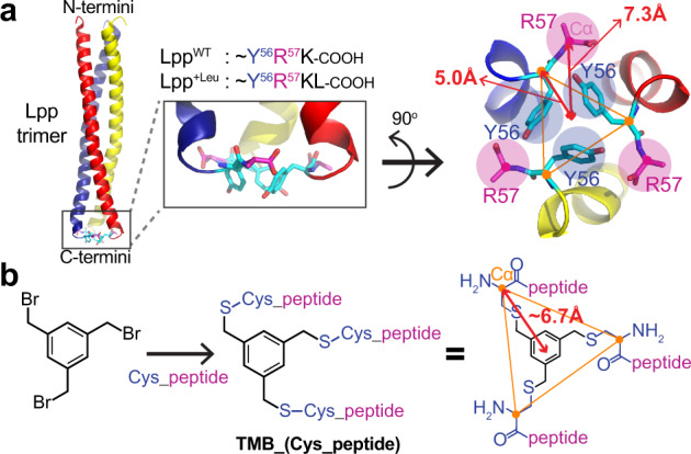
a Crystal structure of the Lpp trimer (PDB code, 1EQ7; cartoon presentation) with a close-up view of the C-terminal region. All Tyr56 (cyan sticks) and Arg57 (magenta sticks) residues roughly sit in the same plane. Distances from the center to the Tyr56-Cα and Arg57-Cα are 5.0 Å and 7.3 Å, respectively. b Synthesis of the tripodal peptidyl compounds, TMB_(Cys_peptide). Peptides are attached to the benzyl positions of 1,3,5-trimethylbenzene (TMB) via the N-terminal cysteine. Distance from the center to the cysteine-Cα in the extended tripodal compound is about 6.7 Å.
Compounds with three peptidyl arms are good DegP activators
We initially tested compounds that contain various sizes of peptides from the Lpp+Leu C-terminus, ~KYRKL, using an activation assay, in which we measured the cleavage rate of a poor model substrate, the reporter14,22. The reporter peptide is made of a short sequence from RseA and efficiently cleaved only in the presence of a good activator. Interestingly, activation was observed with TMB_CYRKL and TMB_CKYRKL, but not with TMB_CRKL or TMB_CKL (Figs. 2a, b). In particular, TMB_CYRKL increased the cleavage rate by at least ten times more than Lpp+Leu and TMB_CKYRKL. Activation was followed by inhibition at higher concentrations of compounds and maximal activation occurred at around 100 µM. A previous report has shown that a good model substrate, 18–58, which is derived from the lysozyme, binds to both the active site and the PDZ1 domain of DegP, and allosterically activates DegP14. In an activation assay with the reporter peptide, 18–58 displayed two phases, an initial activation followed by inhibition, indicating competition for binding to the active site of DegP. The concentration-dependent activation and inhibition suggest that the tripodal compounds also bind to the active site of DegP. Alanine substitution of each residue of the pentapeptide (CYRKL) revealed that tyrosine is most important for activation (Fig. 2c).
Fig. 2. Tripodal peptidyl compounds activate the DegP protease.
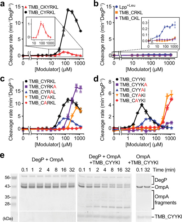
Data are presented as dot plots with mean ±1 SD (n = 3 independent experiments). a, b Activation effects of compounds were monitored by measuring cleavage rates of the reporter peptide (100 μM) by DegP (1 μM for TMB_CKYRKL and TMB_CYRKL (a); 10 μM for Lpp+Leu, TMB_CRKL, and TMB_CKL (b)) with increasing concentrations of Lpp+Leu or tripodal compounds. The insets show close-up views. c Effect of alanine substitution on each position of the pentapeptide (CYRKL) for DegP activation. d DegP activation effects of TMB_CYYKI or its variants with an alanine substitution were monitored as described in Fig. 2a. e Degradation of denatured OmpA (2 μM) by DegP (5 or 0 μM) was monitored by SDS-PAGE in the absence or presence of TMB_CYYKI (10 μM).
Previously, we reported that an Lpp variant carrying YKI at the C-terminus (LppYKI) instead of RKL in Lpp+Leu has a better activation profile with its maximal activation at around 20 µM and a stronger growth inhibition at a heat-shock temperature22. We also tested the tripodal compound with the CYYKI arms (TMB_CYYKI). As expected, we obtained a similar biphasic curve with the maximal activation at around 10 µM, suggesting that this compound interacts with DegP more tightly than TMB_CYRKL (Fig. 2d, black circle). Alanine scanning of the peptide showed that the C-terminal isoleucine and two tyrosines are critical for the activation and tight binding of DegP (Fig. 2d). The importance of the residues was further confirmed by the comparison with TMB_CYYKL and TMB_CYRKI (Supplementary Fig. 2). Previous report showed that substrate binding accompanies several allosteric behaviors of DegP: proteolytic activation, assembly of cage-like proteolytic chamber and positively cooperative binding of a substrate14. Aside from the allosteric activation, we also analyzed the effects of the two compounds on assembly of cages and cooperative binding of a substrate, and found that both TMB_CYRKL and TMB_CYYKI assemble DegP cages, but that they differently affect the binding of model substrates (Supplementary Fig. 3).
Next, we tested whether the tripodal compound promoted degradation of a protein substrate, OmpA, which is an abundant outer membrane protein. Denatured OmpA was degraded not by DegP alone, but by TMB_CYYKI-activated DegP, indicating that the compound induces degradation of a protein that is not efficiently recognized and degraded by DegP (Fig. 2e and Supplementary Fig. 4). Good substrates can activate DegP, but their activation effects disappear when they are completely cleaved14. By contrast, TMB_CYYKI functions as a permanent activator; it remained almost intact and still activated DegP after longer incubation times (1–4 h) with DegP (Supplementary Fig. 5). Taken together, we demonstrated that the tripodal compounds with pentapeptide arms that mimick the Lpp variants are permanent activators of DegP.
Tripodal compounds promote DegP-dependent growth inhibition
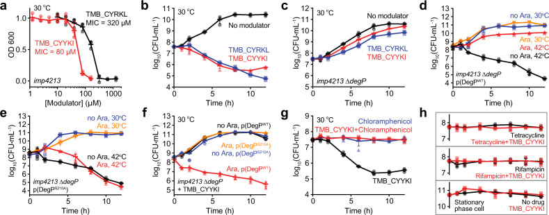
Next, we tested the cellular effects of these tripodal compounds, TMB_CYRKL and TMB_CYYKI. Outer membrane (OM) of gram-negative bacteria often functions as a permeability barrier to molecules that are bigger than 600 Da26. As expected, TMB_CYRKL and TMB_CYYKI, whose molecular weights are ~2 kDa, did not inhibit growth of wild-type Escherichia coli (W3110; Supplementary Fig. 6a). However, they inhibited growth of an OM-permeable mutant strain, the imp4213 strain that is frequently used to study OM-impermeable antibiotics27, with minimum inhibitory concentrations (MICs) of 320–640 µM and 80 µM, respectively (Fig. 3a and Supplementary Fig. 6b). Surprisingly, growth inhibition was observed at 30 °C, at which DegP is not essential for bacterial survival and LppYKI does not trigger growth defects22. These results suggest that the tripodal compounds turn a temperature-dependent essential protease into a temperature-independent toxic enzyme and that they have stronger cellular activity than the Lpp variants.
Fig. 3. Tripodal compounds inhibit bacterial growth in a DegP-dependent fashion.
Data are presented as dot plots with mean ±1 SD (n = 3 independent experiments). a Growth of the imp4213 cells in the presence of TMB_CYYKI or TMB_CYRKL. Overnight cultures in LB broth were diluted 100-fold in the presence of different concentrations of TMB_CYYKI or TMB_CYRKL. Growth was monitored by OD600 after 12 h at 30 °C. b Time-kill curves of the imp4213 strain with TMB_CYYKI, TMB_CYRKL or no compound. Cells at log phase (OD600 = 0.01) were incubated at 30 °C in the absence or presence of TMB_CYYKI (160 µM) or TMB_CYRKL (640 µM). Cells at different times were taken out to determine colony forming units (CFUs). c Time-kill curves of the imp4213 ∆degP strain with TMB_CYYKI, TMB_CYRKL, or no compound at 30 °C. d, e Time-kill curves of the imp4213 ∆degP strain that has a plasmid expressing either DegPWT (d) or DegPS210A (e) in the absence or presence of arabinose (Ara) at 30 °C or 42 °C. f Time-kill curves of the imp4213 ∆degP strain containing a DegPWT- or DegPS210A-plasmid in the presence of TMB_CYYKI (2x MIC) at 30 °C. g, h Time-kill curves of the imp4213 strain at 30 °C in the presence of TMB_CYYKI (160 µM), chloramphenicol (20 µg/ml), rifampicin (0.016 μg/ml), tetracycline (2 μg/ml), or both an antibiotic and TMB_CYYKI. Those of the cells at stationary phase in the absence or presence of TMB_CYYKI are also shown.
To obtain time-kill curves, we incubated cells at log-phase with compounds at 2x MICs and counted viable cells at several time points. Notably, TMB_CYRKL and TMB_CYYKI reduced the number of viable cells by two orders of magnitude at both 30 °C and 42 °C (Fig. 3b and Supplementary Fig. 6c). The degP-deletion strain (imp4213 ∆degP), which does not grow at a high temperature (42 °C; Supplementary Fig. 6d), grew normally at 30 °C when treated with the compounds, indicating that their cellular toxicity requires the DegP function (Fig. 3c). To further confirm that the toxicity is dependent of DegP, we inserted into the imp4213 ∆degP strain the plasmid that expresses either wild-type DegP (DegPWT) or a catalytically inactive variant (DegPS210A) in the presence of arabinose. The ectopic expression of DegPWT, not DegPS210A, suppressed the heat shock stress at a high temperature (42 °C; Figs. 3d, e). When we treated these two strains with TMB_CYYKI at 30 °C in the absence or presence of arabinose, we observed that TMB_CYYKI inhibited growth of the strain expressing DegPWT, but not DegPS210A (Fig. 3f), suggesting that the toxicity of TMB_CYYKI requires catalytically active DegP.
It is unknown what proteins the over-activated DegP mainly degrades. Because DegP does not normally degrade folded proteins and most proteins in the periplasm and OM are exported as unfolded proteins across the cytoplasmic membrane after ribosomal synthesis28, the newly synthesized and translocated proteins may become more accessible targets for the DegP over-proteolysis than the folded periplasmic proteins. If the over-activated DegP becomes toxic by degradation of folded proteins, the activator compounds would reduce cell viability regardless of whether cells are actively growing or not. We tested this idea by combining TMB_CYYKI with a ribosome inhibitor, chloramphenicol. Chloramphenicol alone (20 µg/ml) maintained the number of viable cells roughly at the same level for 12 h, and interestingly, TMB_CYYKI in a combination of chloramphenicol also showed the similar static pattern without considerable reduction of cell numbers at both 30 °C and 42 °C (Fig. 3g, and Supplementary Fig. 7). Consistent with this result, no reduction of viable cells was observed in combination with other antibiotics, tetracycline and rifampicin that inhibit protein synthesis and RNA synthesis, respectively, as well as in a single treatment of TMB_CYYKI to cells at stationary phase at which protein synthesis is substantially reduced (Fig. 3h). These results suggest that the toxicity of TMB_CYYKI is mainly the result of excessive degradation of newly synthesized proteins.
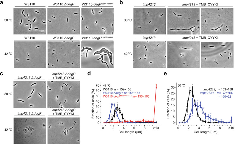
Previously we reported that cells overexpressing a hyperactive DegP variant, DegPR207P/Y444A, died uniformly with an elongated morphology at both higher and lower temperatures, whereas the ∆degP cells were mainly shorter and smaller at lethal high temperatures (Fig. 4a)16. Cells treated with TMB_CYYKI showed both elongated and normal morphology at both 30 °C and 42 °C (Fig. 4b), and, as expected, TMB_CYYKI did not induce any morphological difference in the ∆degP cells (Fig. 4c). We also quantified cell length and confirmed that, at 42 °C, the ∆degP cells and the DegPR207P/Y444A-overexpressing cells are smaller and much longer, respectively, than the wild-type cells (Fig. 4d). The imp4213 cells treated with TMB_CYYKI were slightly longer than those without TMB_CYYKI, while they were not as much longer as the DegPR207P/Y444A-overexpressing cells (Fig. 4e). Although DegPR207P/Y444A and TMB_CYYKI-activated DegP appear to differently affect cells, these results suggest that TMB_CYYKI-driven growth inhibition is not the result of misfolded protein stress.
Fig. 4. TMB_CYYKI induces an elongated morphology.
a–c Representative micrographs of E. coli W3110 (wild-type), ∆degP, or degPR207P/Y444A strains (a), the imp4213 strain in the absence or presence of TMB_CYYKI (160 µM; b), or the imp4213 ∆degP strain in the absence or presence of TMB_CYYKI (160 µM; c), at 30 °C or 42 °C after 4 h of incubation from OD600 = 0.1. d, e Quantification of cell length (n = 152–221) of the wild-type, ∆degP, or degPR207P/Y444A strains at 42 °C (d), or the imp4213 strain at 30 °C in the absence or presence of TMB_CYYKI (160 µM; e). Data are presented as dot plots with mean ±1 SD (n = 3 independent experiments).
Activators bind to the substrate-binding sites of DegP
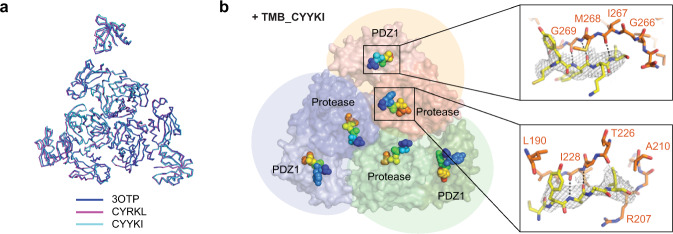
Two DegP-binding motifs of 18–58, a cleavage-site motif and a PDZ1-binding motif, bind to the active site region and the PDZ1 pocket of DegP, respectively (Supplementary Fig. 8a)14. To determine if the tripodal compounds bind to these substrate-binding sites or new allosteric sites, we solved the crystal structures of a DegP active-site mutant, DegPS210A, with TMB_CYYKI or TMB_CYRKL at 3.6 Å and 4.2 Å resolution (PDB 6JJK and 6JJO), respectively, by molecular replacement using the peptide-deleted DegP dodecamer model (3OTP14; Table 1). Overall DegP structures were almost identical to that of the dodecameric DegP with an active conformation (rmsd 0.697 Å and 0.986 Å, respectively, for 2309 Cα atoms in six DegP subunits; Fig. 5a and Supplementary Fig. 8b). Additional electron density maps were found at positions where two degrons of 18–58 are present in the dodecamer model (Fig. 5b and Supplementary Fig. 8c). Because CYRKL and CYYKI have hydrophobic C-terminal residues that can occupy both the S1 pocket of the active site and the hydrophobic pocket of PDZ1, we believe that these peptides are located in the two substrate-binding sites in the same fashion as two degrons of 18–58. Although the lack of electron density for the central benzene ring hampers determination of the binding mode, we suggest that only one arm of the tripodal compounds in the co-crystal structures binds to either substrate-binding sites of DegP, because the distances between two cysteine-Cα atoms in the CYYKI peptides of the crystal structure are too long to justify any of the three plausible modes for the bi- or tri-partite interactions (Supplementary Fig. 8d, 8e). Nevertheless, the simultaneous binding of multiple arms in solution cannot be ruled out, because a slight change of peptide conformation may allow the connection of two pentapeptides to the benzylic scaffold without changing the position of the two C-terminal hydrophobic residues (Supplementary Fig. 8f).
Table 1.
Data collection and refinement statistics (molecular replacement).
| DegPS210A TMB_CYYKI (6JJK) | DegPS210A TMB_CYRKL (6JJO) | |
|---|---|---|
| Data collection | ||
| Space group | C2 | C2 |
| Cell dimensions | ||
| a, b, c (Å) | 214.9, 122.4, 140.2 | 217.2, 123.5, 140.6 |
| α, β, γ (°) | 90, 117.7, 90 | 90, 118, 90 |
| Resolution (Å) | 50–3.60 (3.66–3.60)a | 50–4.20 (4.27–4.20)a |
| Rmerge (%) | 10.2 (52.7)a | 11.2 (66.5)a |
| I/σI | 36.0 (5.9)a | 33.2 (5.9)a |
| Completeness (%) | 99.9 (100.0)a | 99.8 (100.0)a |
| Redundancy | 7.3 (7.5)a | 7.2 (7.1)a |
| Refinement | ||
| Resolution (Å) | 35–3.60 | 35–4.20 |
| No. reflections | 37,191 | 24,436 |
| Rwork/Rfree | 19.9 / 26.5 | 21.6 / 27.8 |
| No. atoms | 16,997 | 16,907 |
| Protein | 16,997 | 16,907 |
| Ligand/ion | 0 | 0 |
| Water | 0 | 0 |
| B-factors | 126.0 | 151.78 |
| Protein | 126.0 | 151.78 |
| Ligand/ion | 0 | 0 |
| Water | 0 | 0 |
| R.m.s. deviations | ||
| Bond lengths (Å) | 0.003 | 0.003 |
| Bond angles (°) | 0.61 | 0.58 |
aValues in parentheses are for the highest-resolution shell.
Fig. 5. Co-crystal structures reveal that the tripodal compounds mainly bind to substrate-binding sites of DegP.
a Superposition of three trimers from the previously reported dodecameric structure (PDB code, 3OTP) and our structures with TMB_CYRKL or TMB_CYYKI. b Pentapeptides in DegPS210A•TMB_CYYKI are shown in spheres (rainbow colors from the blue N-terminus to the red C-terminus) on the two substrate-binding sites of DegP. Three DegP monomers are colored differently in surface presentation. Two insets show close-ups of peptapeptides (yellow sticks with electron density maps) and nearby DegP residues (orange sticks) at the PDZ1 pocket (above) and the active-site region (below) of DegP.
The three-arm compound shows the highest activity
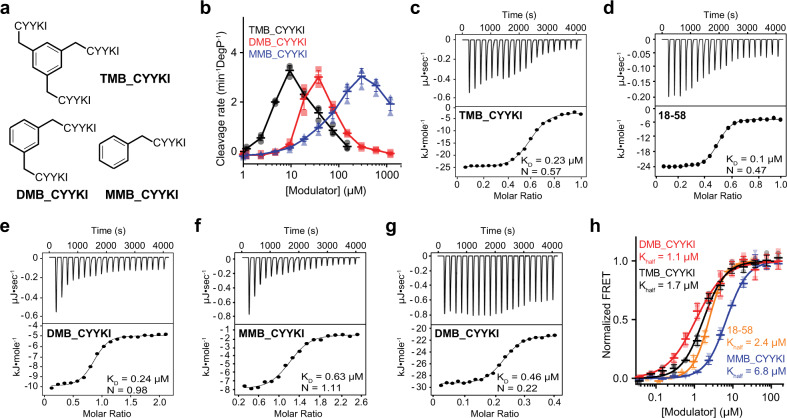
To test whether less than three arms are sufficient for their function, we synthesized compounds with two CYYKI arms (DMB_CYYKI; DMB, 1,3-dimethylbenzene) or one arm (MMB_CYYKI; MMB, methylbenzene) (Fig. 6a). Several results suggested that the three-arm compound has the highest activity and mediates interactions different from one- or two-arm compounds. First, DMB_CYYKI and MMB_CYYKI had MIC values that are at least 10-fold higher than that of TMB_CYYKI (Supplementary Fig. 9). Second, they required higher concentrations for maximal activation, about 40 µM for DMB_CYYKI and 300 µM for MMB_CYYKI, indicating that their interactions for activation are 4-fold and 30-fold weaker, respectively, than that of TMB_CYYKI (Fig. 6b). Finally, isothermal titration calorimetry (ITC) experiments demonstrated a different binding stoichiometry of TMB_CYYKI from DMB_CYYKI and MMB_CYYKI. TMB_CYYKI showed similar stoichiometry (N = 0.57; Fig. 6c) with 18–58 (N = 0.47; Fig. 6d), whereas DMB_CYYKI and MMB_CYYKI showed the N values near 1 (0.98 for DMB_CYYKI and 1.11 for MMB_CYYKI; Figs. 6e, f). We also observed large peaks at low molar ratio of DMB_CYYKI and MMB_CYYKI. To further analyze this region, we titrated DMB_CYYKI in 0–0.4 molar ratio in a separate ITC experiment and found another N-value (0.22; Fig. 6g). Although it is unclear what this unusual N-value conveys, we did not see similar large peaks with TMB_CYYKI or 18–58. Because 18–58 binds to DegP at a 1:1 ratio in crystal structure, we believe that the ITC result reflects not the enthalpy change of the 18–58 binding itself, but that of the overall conformational changes that are triggered by the 18–58 binding. Despite these differences, all the peptidyl compounds showed very similar effects on DegP cage assembly: Half-maximal assembly required 1–2 µM of 18–58, TMB_CYYKI, or DMB_CYYKI, or only 7 µM MMB_CYYKI (Fig. 6h), indicating that a single peptidyl arm is sufficient for cage assembly. Taken together, the crystal structures suggest that the tripodal compounds mainly bind to the substrate-binding sites of DegP. Although only one peptidyl arm appears to interact with DegP in the crystal structure, three peptidyl arms are required for optimal activity with a binding mode different from those of one- or two-arm compounds.
Fig. 6. The compound with three arms has the best activity and properties different from those with one or two arms.
a Structures of compounds with three, two, or one peptidyl arm(s) (TMB_CYYKI, DMB_CYYKI, or MMB_CYYKI, respectively). b Activation effects of TMB_CYYKI, DMB_CYYKI, and MMB_CYYKI were monitored by measuring cleavage rates of the reporter peptide (100 µM) by DegP (1 µM) with increasing concentrations of compounds. Data are presented as dot plots with mean ± 1 SD (n = 3 independent experiments). c–g Isothermal titration calorimetry (ITC) experiments of TMB_CYYKI (c), 18–58 (d), DMB_CYYKI (e, g for 0–2 and 0–0.4 molar ratio, respectively), and MMB_CYYKI (f) with DegPS210A. Data were fitted to the single binding site model. h DegP assembly was monitored by measuring fluorescence resonance energy transfer (FRET) of the donor- and acceptor-labeled DegP variants (0.1 M total) with increasing concentrations of 18–58, TMB_CYYKI, DMB_CYYKI, or MMB_CYYKI. Data were fitted to the Hill equation. Data are presented as dot plots with mean ± 1 SD (n = 3 independent experiments).
In conclusion, our results demonstrate that over-activation of the DegP protease can be a new approach to find antibacterial drugs (Fig. 7). Unlike the previously reported activators14,29, the tripodal compounds can inhibit bacterial growth and activate DegP without their own degradation. Although allosteric inhibitors of enzymes have been suggested to be of high priority for drug discovery against proteases6, allosteric activators of proteases with antibiotic effects are shown only with our compounds and ADEPs. Our results also suggest that, while the conventional approach in antibiotic discovery is to inhibit essential proteins or pathways, targeting a non-essential enzyme by over-activation can also be an effective strategy. Although OM impermeability, high MICs, and the peptidyl nature of the tripodal compounds may limit the clinical development of our compounds, our established assays can serve as a tool to help screen chemical libraries to identify activator compounds that are more suitable for clinical development. The information we obtained concerning the binding mode of the compounds suggests that the activator molecules should target the substrate-binding sites of DegP. Moreover, because a mammalian homolog, HtrA2/Omi, functions in mitochondrial protein quality control and is associated with neurodegeneration and aging30,31, it is also of interest to find HtrA2 activators that may help treat associated human diseases either by restoring mitochondrial homeostasis or by disrupting mitochondrial integrity, as shown by activators of mitochondrial ClpP for killing cancer cells32.
Fig. 7. A model for bacterial killing by DegP over-activation.
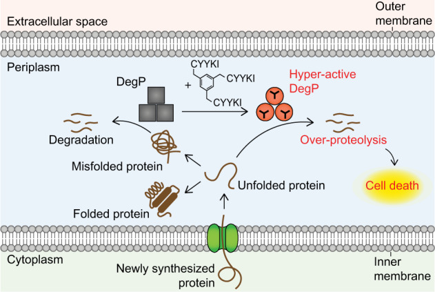
DegP mainly degrades misfolded proteins, but its over-activation by the tripodal compounds triggers excessive degradation of newly synthesized proteins and leads to bacterial death.
Methods
Bacterial strains and growth conditions
Bacterial strains and plasmids are listed in Supplementary Tables 2 and 3, respectively. E. coli imp4213 (RFM795; CGSC#: 14179), which is streptomycin-resistant, was obtained from E. coli genetic stock center at Yale University. The imp4213 ∆degP strain was constructed by P1 transduction33, in which the ΔdegP::pheS-kanR allele from the E. coli SK322 donor strain was transferred to E. coli imp4213 recipient strain. E. coli was grown in Luria-Bertani (LB) medium with aeration (200 rpm) at 37 °C overnight and diluted 1:100 in fresh LB. DegPWT and its variants were prepared as previously described14.
Peptide synthesis
Peptides were synthesized by solid phase peptide synthesis (SPPS) as previously described34. Wang resin and Fmoc protected amino acids (GL Biochem) were used in SPPS. First C-terminal amino acid (5 equiv.) and 4-Dimethylaminopyridine (DMAP, 0.1 equiv.; Sigma-Aldrich) dissolved in DMF were mixed with Wang resin for 20 min, and then N,N′-Diisopropylcarbodiimide (DIC, 4 equiv.; TCI) was added. The reaction mixture was incubated at room temperature for 40 h. Beads were washed three times with DMF and DCM. Acetic anhydride (10 equiv.; Sigma-Aldrich) and DMAP (0.1 equiv.) dissolved in DCM were added to beads for capping for 1 hour, and then the Fmoc group was removed using the deprotection solution (20% (v/v) piperidine (Alfa Aesar) in DMF) for 20 min after washing. The remaining amino acids were attached by coupling (5 equiv. HATU (GL Biochem) and 10 equiv. N,N-Diisopropylethylamine (Samchun) in DMF for 30 min), capping (Acetic anhydride: N,N-Diisopropylethylamine: DMF in a 5: 2.5: 92.5 ratio for 7 min), and deprotecting reactions. Coupling and deprotection were monitored by TNBS test. The global deprotection of all the protecting groups and the cleavage from the beads were performed with a cleavage cocktail (Trifluoroacetic acid (Samchun): water: 1,2-Ethanedithiol (Sigma-Aldrich): Triisopropylsilane (Sigma-Aldrich) in a 940:25: 25:10 ratio for 2 h). The peptides were concentrated by solvent evaporation using nitrogen or air blowing and then precipitated by cold ether/hexane (1:1). Precipitates were dried and dissolved in water containing 0.1% TFA. Peptides were purified by reverse-phase high-performance liquid chromatography (RP-HPLC, Agilent 1260) and analyzed by matrix-assisted laser desorption/ionization (MALDI, Bruker) or LC-MS (Agilent 1260 infinity).
Activator synthesis
The 1,3,5-Tris(bromomethyl)benzene (10 μmol; Sigma-Aldrich) in acetonitrile (470 μL; Fisher) was added dropwise to a solution of the peptide (35 μmol) in NaHCO3 (100 mM pH 8, 700 μL; Duksan). The reaction mixture was stirred for 1 h at room temperature. Product (TMB_(Cys_peptide)) was purified by HPLC and analyzed by MALDI. Conjugation of thiol, not amine, was confirmed by the absence of alkylation of the compounds with iodoacetamide. DMB_CYYKI and MMB_CYYKI were synthesized by using 1,3-bis(bromomethyl)benzene and benzyl bromide (Sigma-Aldrich), respectively.
DegP activation assay with the reporter peptide
DegP activation experiments were performed as previously described14,22. All the in vitro assays were performed in a buffer containing 50 mM sodium phosphate (pH 8) and 100 mM NaCl. Two-fold serial dilutions of the activator were incubated with DegPWT (1, 3, or 10 μM) for 5 min. After incubation, the reporter peptide (Abz-KASPVSLGYNO2D; Abz, 2-aminobenzoic acid; YNO2, 3-nitrotyrosine; 100 μM final) was added. DegP activation was monitored by increased fluorescence by using a micro-plate reader (excitation 320 nm, emission 430 nm; Infinite F200pro, Tecan). Assays were performed in triplicates.
Activator degradation assay
An activator (10 μM) was mixed with DegP (10 μM). Aliquots were taken after 0.17, 1, 2, or 4 h. Each aliquot of the reaction solutions was quenched by the addition of TFA (1% final). The amount of activator for each sample was quantified using HPLC. Water (0.05% TFA) and methanol were used as the HPLC buffer.
OmpA degradation assay
OmpA (2 μM final; 100-fold dilution from the 8 M urea solution) was added to the reaction solutions of DegP (5 μM) and activator (0 or 10 μM). Each aliquot of the reaction solutions taken after 0.1, 1, 2, 4, 8, 16, or 32 min at 37 °C was quenched by the addition of SDS (2% final). OmpA protein degradation was monitored by the SDS-PAGE.
Fluorescence resonance energy transfer
Fluorescence resonance energy transfer (FRET) assay was performed as previously described14,22. Equimolar amounts of fluorescence donor- and acceptor-labeled DegPS210A (total 0.1, 1 or 3 μM) was mixed in a buffer containing 50 mM sodium phosphate (pH 8) and 100 mM NaCl. Two-fold serial dilutions of activators were added to the DegP mixtures. Alexa Fluor 555 C2-maleinide and 647 C2-maleinide (life technologies) were used as fluorescence dyes. Cage assembly of DegP was monitored by increased FRET ratio using the micro-plate reader (excitation 520 nm, emission 570 or 670 nm, Infinite M200pro, Tecan). Assays were performed in triplicates.
Fluorescence anisotropy
Fluorescence anisotropy assay was performed as previously described14,22. Two-fold serial dilutions of activators were mixed with a fluorescence-labeled peptide (flC18–58 or fl45–58, 50 nM; Oregon Green 488 maleimide, Invitrogen) and DegPS210A (10 μM for flC18–58; 80 μM for fl45–58). Anisotropy was monitored by using a micro-plate reader (excitation 485 nm, emission 535 nm, Infinite F200pro, Tecan). The assay was performed in triplicates.
Minimum inhibitory concentration
The susceptibility of E. coli to activators was analyzed by determining the MICs. Two-fold serial dilutions of activators in LB broth were added to bacteria (OD600 = 0.1) in 96-well plates. Plates were incubated with shaking at 30 or 42 °C. After 12 h of incubation, the MIC was defined as the lowest concentration of the activator to inhibit the growth of bacteria by using a micro-plate reader (Absorbance 600 nm, Infinite M200pro, Tecan). The assay was performed in triplicates.
Time-kill experiment
Time-kill experiment was performed by counting the viable bacteria at each time point. The E.coli imp4213 strain was cultured with streptomycin overnight and diluted 1:100 in fresh LB broth. When the OD600 value became 0.01, the activator was added at 2x MIC. The sample was incubated with shaking (180 rpm) at 30 °C or 42 °C, and aliquots were taken after 0, 1, 2, 4, 6, 8, 10, or 12 h. These aliquots were serially diluted (ten-fold) and plated on LB agar plates for colony counts. Experiments were performed in triplicates. The kill curves were plotted with the CFU/mL against time.
Experiments with chloramphenicol (20 μg/ml, 10x MIC; Bio basic), tetracycline (2 μg/ml, 2x MIC; Bio basic), rifampicin (0.016 μg/ml, 2x MIC; Sigma-Aldrich) were performed in the same way. Experiments with cells in the stationary phase, however, were performed without dilution of the overnight culture.
Imaging
The W3110 (WT) and imp4213 cells were grown overnight in LB broth at 37 °C and W3110 ∆degP, imp4213 ∆degP and W3110 degPR207P/Y444A cells were grown at 30 °C. Cells were diluted to OD600 = 0.1. The activator was added in the final concentration of 2x MIC. The sample was incubated with shaking (180 rpm) at 30 °C or 42 °C. After 4 h, 20 μL of the cell culture was placed between a coverslip and a 1.5% low-melting-temperature agarose gel pad (Agarose LE). Samples were placed on an inverted microscope (Olympus, IX-71) with a ×100 oil-immersed objective lens (Olympus). Phase contrast images were acquired using a cooled EMCCD camera (Andor iXon DU897). Metamorph software (Molecular Devices) was used to find the focus and control the measurements. For length quantification, a few hundred cells were taken from three independent replicate experiments (W3110, n = 153, 152, and 156; W3110 ∆degP, n = 155, 155, and 158; W3110 degPR207P/Y444, n = 158, 156, and 165; imp4213, n = 156, 153, and 156; imp4213 + TMB_CYYKI, n = 173, 221, and 160).
Crystallization and data collection
DegPS210A (198 µM) and an activator (TMB_CYRKL or TMB_CYYKI; 297 µM) were incubated in a buffer containing 5 mM phosphate (pH 8.0) for 1 h at 295 K. Crystals of DegPS210A with TMB_CYRKL were grown using the hanging-drop vapor diffusion method by mixing equal volume (1 μL) of the protein-activator solutions and the reservoir solutions. A reservoir solution consisting of PEG3350 (11%) and tacsimate (0.1 M, pH 3.5) was used to grow crystals of DegPS210A with TMB_CYRKL. Crystals reached maximum size within 7 days at 295 K. Crystals of DegPS210A with TMB_CYYKI were grown, using a reservoir solution with PEG3350 (10%) and tacsimate (0.1 M, pH 3.4). The crystals were soaked in Paratone-N (Hampton Research) and incubated for 15 min before being flash-frozen in a nitrogen stream at 100 K. Native data for the DegPS210A with TMB_CYRKL or TMB_CYYKI were collected at the 5 C beamline and 7 A beamline of Pohang Accelerator Laboratory (Pohang, South Korea), respectively. The detector was an ADSC QUANTUM 270 and ADSC QUANTUM 315r (ADSC, USA), respectively, using a single wavelength of 0.9790 Å. The raw data were processed and scaled using the program suite HKL200035.
Structure determination and refinement
The structures of the DegPS210A with each activators were solved by the molecular replacement method using the hexamer model (chain A-F) of DegPS210A from E. coli (PDB ID: 3OTP)14. The original 3OTP structure contained a peptide substrate, and the peptide was removed for the calculation of molecular replacement. A cross-rotational search followed by a translational search was performed using the Phaser program36. Subsequent manual model building was carried out using the COOT program and restrained refinement was performed using the PHENIX program37. Several rounds of model building, simulated annealing, positional refinement with NCS restraints, and individual B-factor refinement were performed. Two structures determined in this study display Ramachandran statistics with 95.1% and 96.8% of residues in the most favored regions and 4.9% and 3.1% of residues in the allowed regions of the Ramachandran diagram, respectively. Table 1 lists the refinement statistics. The crystallographic asymmetric unit contained two trimers of DegPS210A with each modulator. Structural figures were drawn with PyMOL.
The atomic coordinates and structure factors for the DegPS210A•TMB_CYYKI and DegPS210A•TMB_CYRKL complexes were deposited in Protein Data Bank (PDB ID code 6JJK and 6JJO respectively).
Isothermal titration calorimetry
ITC experiments were performed using Affinity ITC instruments (TA Instruments, New Castle, DE, USA) at 298 K. DegPS210A mutant (18.10 μM for 0–2 molar ratio and 109.33 μM for 0–0.4 molar ratio), which were prepared in a buffer containing 5 mM phosphate (pH 8.0) were degassed at 295 K prior to measurements. Using a micro-syringe, 2.5 μL of DMB_CYYKI (300 μM) was added at intervals of 200 s to the DegP solution in the cell with gentle stirring. As for other ligands, 2.5 μL of MMB_CYYKI (453 μM), TMB_CYYKI (218.46 μM) or 18–58 substrate peptide (80 μM) was added at intervals of 200 s to the DegP solution (27.33 μM, 37.45 μM, and 20 μM, respectively). Data were fitted to the single binding site model. In experiments with 0–2 and 0–2.6 molar ratio of DMB_CYYKI and MMB_CYYKI, respectively, the data with 0–0.3 molar ratio were excluded for the fitting.
Statistics and reproducibility
Data in graphs with error bars are presented as means ± standard deviation (S.D.) from three independent experiments (n = 3). All Specific information on the number of replicates and statistical analyses are included in the figure legends. Data were processed with GraphPad Prism version 6.07.
Reporting summary
Further information on research design is available in the Nature Research Reporting Summary linked to this article.
Supplementary information
Description of Additional Supplementary Files
Acknowledgements
The imp4213 strain (RFM795) was obtained from Coli Genetic Stock Center from Yale University. We thank Ga-eul Eom, Hyunbin Lee, Inseok Song, Daeje Seo, Heejin Roh for helpful discussions and technical assistance, and Yan Lee, Youngjun Song, and Taeyang An for assistance in synthesizing compounds. We also thank the staffs of the 5C and 7A beamlines at the Pohang Light Source. This research was supported by Basic Science Research Program through the National Research Foundation of Korea (NRF) funded by the Ministry of Education (NRF-2014R1A1A2056722).
Author contributions
S.K. and H.C. conceived and designed the work, analyzed and interpreted the data, and wrote the paper. H.C. performed the majority of experiments with helps from H.P. except for crystallographic analysis, ITC, and cell imaging. Y.C. and H.H.L. performed the crystallographic work. K.M. performed the ITC experiments. J.B.S. obtained the imaging data. All authors approved the paper.
Data availability
The coordinates and structure factors of DegPS210A•TMB_CYYKI and DegPS210A•TMB_CYRKL have been deposited in the Protein Data Bank under the accession codes 6JJK and 6JJO, respectively. Source data for graphs in this study are available in Supplementary Data 1 (in excel format).
Competing interests
The authors declare no competing interests.
Footnotes
Publisher’s note Springer Nature remains neutral with regard to jurisdictional claims in published maps and institutional affiliations.
Supplementary information
Supplementary information is available for this paper at 10.1038/s42003-020-01266-9.
References
- 1.Levy SB, Marshall B. Antibacterial resistance worldwide: causes, challenges and responses. Nat. Med. 2004;10:S122–S129. doi: 10.1038/nm1145. [DOI] [PubMed] [Google Scholar]
- 2.Fischbach MA, Walsh CT. Antibiotics for emerging pathogens. Science. 2009;325:1089–1093. doi: 10.1126/science.1176667. [DOI] [PMC free article] [PubMed] [Google Scholar]
- 3.Lewis K. Platforms for antibiotic discovery. Nat. Rev. Drug Discov. 2013;12:371–387. doi: 10.1038/nrd3975. [DOI] [PubMed] [Google Scholar]
- 4.Brown ED, Wright GD. Antibacterial drug discovery in the resistance era. Nature. 2016;529:336–343. doi: 10.1038/nature17042. [DOI] [PubMed] [Google Scholar]
- 5.Culp E, Wright GD. Bacterial proteases, untapped antimicrobial drug targets. J. Antibiot. (Tokyo) 2017;70:366–377. doi: 10.1038/ja.2016.138. [DOI] [PubMed] [Google Scholar]
- 6.Drag M, Salvesen GS. Emerging principles in protease-based drug discovery. Nat. Rev. Drug Discov. 2010;9:690–701. doi: 10.1038/nrd3053. [DOI] [PMC free article] [PubMed] [Google Scholar]
- 7.Deu E, Verdoes M, Bogyo M. New approaches for dissecting protease functions to improve probe development and drug discovery. Nat. Struct. Mol. Biol. 2012;19:9–16. doi: 10.1038/nsmb.2203. [DOI] [PMC free article] [PubMed] [Google Scholar]
- 8.Brotz-Oesterhelt H, et al. Dysregulation of bacterial proteolytic machinery by a new class of antibiotics. Nat. Med. 2005;11:1082–1087. doi: 10.1038/nm1306. [DOI] [PubMed] [Google Scholar]
- 9.Lee BG, et al. Structures of ClpP in complex with acyldepsipeptide antibiotics reveal its activation mechanism. Nat. Struct. Mol. Biol. 2010;17:471–478. doi: 10.1038/nsmb.1787. [DOI] [PubMed] [Google Scholar]
- 10.Conlon BP, et al. Activated ClpP kills persisters and eradicates a chronic biofilm infection. Nature. 2013;503:365–370. doi: 10.1038/nature12790. [DOI] [PMC free article] [PubMed] [Google Scholar]
- 11.Clausen T, Kaiser M, Huber R, Ehrmann M. HTRA proteases: regulated proteolysis in protein quality control. Nat. Rev. Mol. Cell Biol. 2011;12:152–162. doi: 10.1038/nrm3065. [DOI] [PubMed] [Google Scholar]
- 12.Krojer T, et al. Structural basis for the regulated protease and chaperone function of DegP. Nature. 2008;453:885–890. doi: 10.1038/nature07004. [DOI] [PubMed] [Google Scholar]
- 13.Jiang J, et al. Activation of DegP chaperone-protease via formation of large cage-like oligomers upon binding to substrate proteins. Proc. Nati. Acad. Sci. USA. 2008;105:11939–11944. doi: 10.1073/pnas.0805464105. [DOI] [PMC free article] [PubMed] [Google Scholar]
- 14.Kim S, Grant, Robert A, Sauer RobertT. Covalent linkage of distinct substrate degrons controls assembly and disassembly of DegP proteolytic cages. Cell. 2011;145:67–78. doi: 10.1016/j.cell.2011.02.024. [DOI] [PMC free article] [PubMed] [Google Scholar]
- 15.Kim S, Sauer RT. Cage assembly of DegP protease is not required for substrate-dependent regulation of proteolytic activity or high-temperature cell survival. Proc. Nati. Acad. Sci. USA. 2012;109:7263. doi: 10.1073/pnas.1204791109. [DOI] [PMC free article] [PubMed] [Google Scholar]
- 16.Kim S, Sauer RT. Distinct regulatory mechanisms balance DegP proteolysis to maintain cellular fitness during heat stress. Genes Dev. 2014;28:902–911. doi: 10.1101/gad.238394.114. [DOI] [PMC free article] [PubMed] [Google Scholar]
- 17.Silhavy, T. J., Kahne, D. & Walker, S. The bacterial cell envelope. Cold Spring Harb. Perspect. Biol. 2, a000414 (2010). [DOI] [PMC free article] [PubMed]
- 18.Lipinska B, Fayet O, Baird L, Georgopoulos C. Identification, characterization, and mapping of the Escherichia coli htrA gene, whose product is essential for bacterial growth only at elevated temperatures. J. Bacteriol. 1989;171:1574–1584. doi: 10.1128/JB.171.3.1574-1584.1989. [DOI] [PMC free article] [PubMed] [Google Scholar]
- 19.Strauch KL, Johnson K, Beckwith J. Characterization of degP, a gene required for proteolysis in the cell envelope and essential for growth of Escherichia coli at high temperature. J. Bacteriol. 1989;171:2689–2696. doi: 10.1128/JB.171.5.2689-2696.1989. [DOI] [PMC free article] [PubMed] [Google Scholar]
- 20.Skorko-Glonek J, et al. HtrA protease family as therapeutic targets. Curr. Pharm. Des. 2013;19:977–1009. doi: 10.2174/1381612811319060003. [DOI] [PubMed] [Google Scholar]
- 21.Skorko-Glonek J, et al. The extracellular bacterial HtrA proteins as potential therapeutic targets and vaccine candidates. Curr. Med. Chem. 2017;24:2174–2204. doi: 10.2174/0929867323666161223145825. [DOI] [PubMed] [Google Scholar]
- 22.Park H, Kim YT, Choi C, Kim S. Tripodal lipoprotein variants with C-terminal hydrophobic residues allosterically modulate activity of the degp protease. J. Mol. Biol. 2017;429:3090–3101. doi: 10.1016/j.jmb.2017.09.011. [DOI] [PubMed] [Google Scholar]
- 23.Shu W, Liu J, Ji H, Lu M. Core structure of the outer membrane lipoprotein from Escherichia coli at 1.9 A resolution. J. Mol. Biol. 2000;299:1101–1112. doi: 10.1006/jmbi.2000.3776. [DOI] [PubMed] [Google Scholar]
- 24.Timmerman P, Beld J, Puijk WC, Meloen RH. Rapid and quantitative cyclization of multiple peptide loops onto synthetic scaffolds for structural mimicry of protein surfaces. Chembiochem. 2005;6:821–824. doi: 10.1002/cbic.200400374. [DOI] [PubMed] [Google Scholar]
- 25.Heinis C, Rutherford T, Freund S, Winter G. Phage-encoded combinatorial chemical libraries based on bicyclic peptides. Nat. Chem. Biol. 2009;5:502–507. doi: 10.1038/nchembio.184. [DOI] [PubMed] [Google Scholar]
- 26.Nikaido H, Vaara M. Molecular basis of bacterial outer membrane permeability. Microbiol. Rev. 1985;49:1–32. doi: 10.1128/MMBR.49.1.1-32.1985. [DOI] [PMC free article] [PubMed] [Google Scholar]
- 27.Sampson BA, Misra R, Benson SA. Identification and characterization of a new gene of Escherichia coli K-12 involved in outer membrane permeability. Genetics. 1989;122:491–501. doi: 10.1093/genetics/122.3.491. [DOI] [PMC free article] [PubMed] [Google Scholar]
- 28.Driessen AJ, Nouwen N. Protein translocation across the bacterial cytoplasmic membrane. Annu. Rev. Biochem. 2008;77:643–667. doi: 10.1146/annurev.biochem.77.061606.160747. [DOI] [PubMed] [Google Scholar]
- 29.Meltzer M, et al. Allosteric activation of HtrA protease DegP by stress signals during bacterial protein quality control. Angew. Chem. Int. Ed. Engl. 2008;47:1332–1334. doi: 10.1002/anie.200703273. [DOI] [PubMed] [Google Scholar]
- 30.Goo HG, Rhim H, Kang S. Pathogenic role of serine protease HtrA2/Omi in neurodegenerative diseases. Curr. Protein Pept. Sci. 2017;18:746–757. doi: 10.2174/1389203717666160311115750. [DOI] [PubMed] [Google Scholar]
- 31.Jones JM, et al. Loss of Omi mitochondrial protease activity causes the neuromuscular disorder of mnd2 mutant mice. Nature. 2003;425:721–727. doi: 10.1038/nature02052. [DOI] [PubMed] [Google Scholar]
- 32.Ishizawa J, et al. Mitochondrial ClpP-mediated proteolysis induces selective cancer cell lethality. Cancer Cell. 2019;35:721–737. doi: 10.1016/j.ccell.2019.03.014. [DOI] [PMC free article] [PubMed] [Google Scholar]
- 33.Thomason LC, Costantino N, Court DL. E. coli genome manipulation by P1 transduction. Curr. Protoc. Mol. Biol. 2007;Chapter 1:Unit 1 17. doi: 10.1002/0471142727.mb0117s79. [DOI] [PubMed] [Google Scholar]
- 34.Eom G, Kim S. Identification of nucleophilic probes for protease-mediated transpeptidation. Molecules. 2018;23:2109. doi: 10.3390/molecules23092109. [DOI] [PMC free article] [PubMed] [Google Scholar]
- 35.Otwinowski, Z. & Minor, W. Processing of X-ray diffraction data collected in oscillation mode, in Methods Enzymol. Vol. 276, 307–326 (Academic Press, 1997). [DOI] [PubMed]
- 36.McCoy AJ, Grosse-Kunstleve RW, Storoni LC, Read RJ. Likelihood-enhanced fast translation functions. Acta Crystallogr. Sect. D. 2005;61:458–464. doi: 10.1107/S0907444905001617. [DOI] [PubMed] [Google Scholar]
- 37.Adams PD, et al. PHENIX: building new software for automated crystallographic structure determination. Acta Crystallogr. Sect. D. 2002;58:1948–1954. doi: 10.1107/S0907444902016657. [DOI] [PubMed] [Google Scholar]
Associated Data
This section collects any data citations, data availability statements, or supplementary materials included in this article.
Supplementary Materials
Description of Additional Supplementary Files
Data Availability Statement
The coordinates and structure factors of DegPS210A•TMB_CYYKI and DegPS210A•TMB_CYRKL have been deposited in the Protein Data Bank under the accession codes 6JJK and 6JJO, respectively. Source data for graphs in this study are available in Supplementary Data 1 (in excel format).


