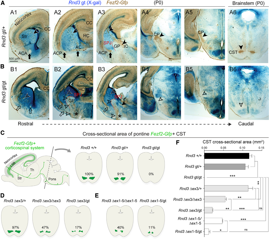
Figure 2. CST Axons Misroute at the SPJ in the Absence of RND3.

(A and B) X-gal staining (blue) and GFP (DAB; brown) on serial coronal sections of P0 Rnd3 gt/+; Fezf2-Gfp (A1–A6) and Rnd3 gt/gt; Fezf2-Gfp (B1–B6) brains depict defects in the Rnd3 gt/gt brain, including reduced cortical thickness (B1–B3); a thin ACA (B1; open arrow); the misrouted ACP to the ventral surface (B2 and B3; open arrows); and misrouted Fezf2-Gfp+ subcerebral projections within the striatum and at the SPJ through the GP (B2 and B3; asterisks and open arrowheads), at the cerebral peduncle (CP) and ventral pons (open arrowheads, B4–B6). Scale bar: 500 μm. CC, corpus callosum; HC, hippocampal commissure; AC, anterior commissure; IC, internal capsule; ACA, anterior branch of AC; ACP, posterior branch of AC.
(C–E) Schematic representations of CST area labeled by GFP (Fezf2-Gfp) in the coronal cross-sections of pontine nuclei from Rnd3 +/+, Rnd3 gt/+, and Rnd3 gt/gt mice (C); Rnd3 Δex3/+; Fezf2-Gfp, Rnd3 Δex3/Δex3; Fezf2-Gfp, and Rnd3 Δex3/gt; Fezf2-Gfp mice (D); and Rnd3 Dex1–5/Dex1–5; Fezf2-Gfp, and Rnd3 Dex1–5/gt; Fezf2-Gfp mice (E) at P0.
(F) Quantitative analysis of CST between mice bearing various Rnd3 alleles. y axis: groups. x axis: CST area in mm2. ANOVA Tukey’s test was applied (means ± SEs,*p < 0.01, **p < 0.001, and ***p < 0.0001. n = 2 for each group).
See also Figures S3–S5.
