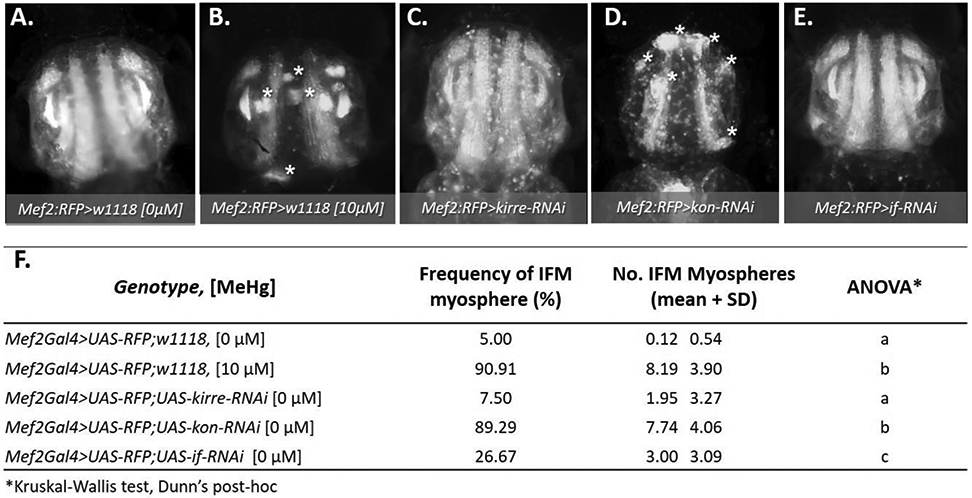
Fig. 3. Morphological effects on IFMs following larval MeHg exposure or muscle-restricted knockdown of myotendinous junction (MTJ) gene candidates.

(A - F) Representative epifluorescence images of 49 - 57 h APF pupal thoraces (n ≥ 20 images per genotype), where asterisks (*) indicate myospheres. (A, B) Mef2:RFP>w1118 (genotype control) with or without prior larval exposure to 10 μM MeHg; (C - E) Mef2:RFP>UAS-RNAi lines against key myogenic genes: (C) myoblast fusion gene, kirre-RNAi, or the MTJ genes, (D) kon-RNAi, or (E) if-RNAi. The number of asterisks in each image correspond to the average number of myospheres observed for each genotype. (F) Quantification of the frequency and average number ± SD of myospheres observed in the IFM for each genotype and treatment. Frequency of myospheres are represented as a percentage of the indicated number of pupae scored per group. Letters indicate pairwise significant differences in average number of myospheres between each group, where p < 0.05, n = 20 – 39, Kruskal-Wallis test with Dunn’s post-hoc for multiple comparisons.
