Abstract
Extrapulmonary tuberculosis (TB) is difficult to diagnose. Here, we report a case of extrapulmonary TB in a 68-year-old woman presented with mental fatigue, poor appetite, and weight loss. 18F-FDG PET/CT revealed elevated 18F-FDG uptake in the left inferior cervical, left supraclavicular, mediastinal, and splenic hilum lymph nodes and spleen, which were suspected of malignant tumor. To further differentiate benign and malignant diseases, 68Ga-FAPI PET/CT was performed. 68Ga-FAPI PET/CT also showed intense 68Ga-FAPI uptake in the previously mentioned FDG-avid lesions. However, biopsy of the left supraclavicular lymph node demonstrated the presence of TB.
Key Words: tuberculosis, 18F-FDG, 68Ga-FAPI, PET/CT
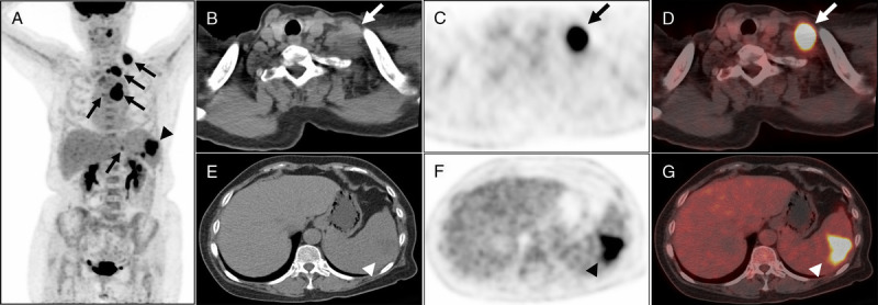
FIGURE 1.

A 68-year-old woman presented with mental fatigue, poor appetite, and weight loss for 1 month. In addition, physical examination revealed enlarged left supraclavicular lymph node. She had no fever, cough, expectoration, or nighttime sweating. Her leukocyte count was 18.1 × 10⁹/L (reference, 3.5–9.5 × 10⁹/L), neutrophil count was 14.5 × 10⁹/L (reference, 1.8–6.3 × 10⁹/L), platelet count was 672 × 109/L (reference, 125–350 × 109/L), and serum creatinine level was within the reference range. 18F-FDG PET/CT was performed to aid in diagnosis. As showed in MIP of 18F-FDG PET, elevated 18F-FDG uptake in left inferior cervical, left supraclavicular, mediastinal, and splenic hilum lymph nodes (A, arrows), and spleen (A, arrowhead) were revealed. Representative lymph node (B–D, arrows; SUVmax, 16.9) and spleen lesions (E–G, arrowhead; SUVmax, 12.0) in the axial CT, PET, and fusion PET/CT images were exhibited. However, it is not easy to make a diagnosis with 18F-FDG PET/CT only.
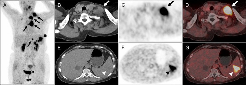
FIGURE 2.

68Ga-FAPI PET/CT was performed to further differentiate benign and malignant diseases. The patient was enrolled in the prospective study evaluating the role of 68Ga-FAPI PET/CT in the management of malignant tumors, which was approved by Shanghai Cancer Center Institutional Review Board (ID 2004216-25), and written informed consent was obtained from the patient. The MIP, axial CT, PET, and fusion PET/CT images of 68Ga-FAPI PET/CT showed intense 68Ga-FAPI uptake in the aforementioned FDG-avid lymph nodes (A–D; arrows, SUVmax, 20.1) and spleen lesions (A, E–G, arrowhead; SUVmax, 11.7). According to the results of 18F-FDG and 68Ga-FAPI PET/CT, the most likely diagnosis was malignant tumor.
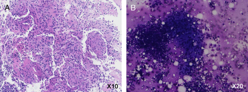
FIGURE 3.

However, biopsy of the left supraclavicular lymph node demonstrated the presence of tuberculosis (TB). Granulomatous nodules composed of epithelioid cells, caseous necrosis, and inflammatory cells or lymphocytes were observed in microscopic section of hematoxylin-eosin stain (A, original magnification ×10) and Liu’s stain (B, original magnification ×20). The 68Ga-FAPI is developed to detect the expression of fibroblast activation protein (FAP).1–3 FAP is an overexpression in more than 90% of epithelial carcinomas and some mesenchymal tumors, and recent studies showed that 68Ga-FAPI might be a broad-spectrum tumor PET agent.4–6 However, high uptake of 68Ga-FAPI was also found in nontumorous lesions, including wound healing, inflammation, fibrosis, and so on.7,8 This case again highlighted that 68Ga-FAPI could gather in nontumorous lesions. Even so, the positive founding of 68Ga-FAPI in extrapulmonary TB lesions indicates that 68Ga-FAPI could serve as a probe in diagnosis and response evaluation of TB.
Footnotes
Conflicts of interest and sources of funding: none declared.
REFERENCES
- 1.Loktev A Lindner T Mier W, et al. . A tumor-imaging method targeting cancer-associated fibroblasts. J Nucl Med. 2018;59:1423–1429. [DOI] [PMC free article] [PubMed] [Google Scholar]
- 2.Lindner T Loktev A Altmann A, et al. . Development of quinoline-based theranostic ligands for the targeting of fibroblast activation protein. J Nucl Med. 2018;59:1415–1422. [DOI] [PubMed] [Google Scholar]
- 3.Loktev A Lindner T Burger EM, et al. . Development of fibroblast activation protein-targeted radiotracers with improved tumor retention. J Nucl Med. 2019;60:1421–1429. [DOI] [PMC free article] [PubMed] [Google Scholar]
- 4.Kratochwil C Flechsig P Lindner T, et al. . (68)Ga-FAPI PET/CT: tracer uptake in 28 different kinds of cancer. J Nucl Med. 2019;60:801–805. [DOI] [PMC free article] [PubMed] [Google Scholar]
- 5.Giesel FL Kratochwil C Lindner T, et al. . (68)Ga-FAPI PET/CT: biodistribution and preliminary dosimetry estimate of 2 DOTA-containing FAP-targeting agents in patients with various cancers. J Nucl Med. 2019;60:386–392. [DOI] [PMC free article] [PubMed] [Google Scholar]
- 6.Meyer C Dahlbom M Lindner T, et al. . Radiation dosimetry and biodistribution of 68Ga-FAPI-46 PET imaging in cancer patients. J Nucl Med. 2020;61:1171–1177. [DOI] [PMC free article] [PubMed] [Google Scholar]
- 7.Luo Y Pan Q Zhang W, et al. . Intense FAPI uptake in inflammation may mask the tumor activity of pancreatic cancer in (68)Ga-FAPI PET/CT. Clin Nucl Med. 2020;45:310–311. [DOI] [PubMed] [Google Scholar]
- 8.Pan Q, Luo Y, Zhang W. Recurrent immunoglobulin G4-related disease shown on (18)F-FDG and (68)Ga-FAPI PET/CT. Clin Nucl Med. 2020;45:312–313. [DOI] [PubMed] [Google Scholar]


