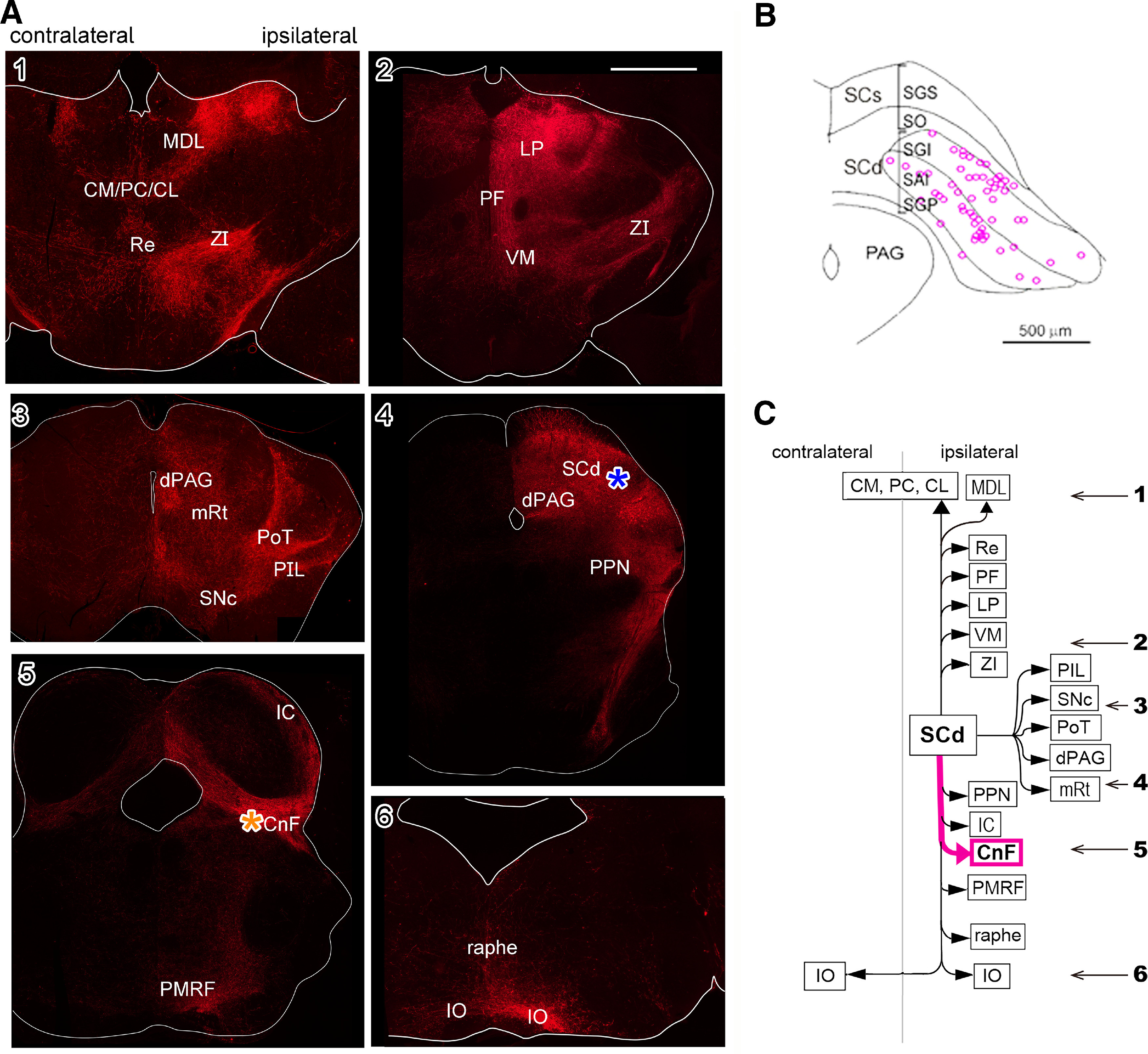
Figure 4.

Axonal trajectories of the uncrossed (defense) pathway neurons visualized by anti-GFP immunohistochemistry with Alexa Fluor 594. A, Uncrossed (defense) pathway. 1–6, Photomicrographs of the frontal sections of the diencephalon and brainstem. Scale bars = 1 mm. B, A diagram showing location of the somata of uncrossed (defense) pathway neurons. C, Targets of the uncrossed (defense) pathway neurons. The numerals with arrows indicate the rostrocaudal levels of photomicrographs in A1–A6. An asterisk indicates the injection site of viral vector in the right MRF around CnF. The same abbreviations as Figure 3 and additional abbreviations: dPAG: dorsal periaqueductal gray matter, IC: inferior colliculus, LP: lateral posterior thalamic nucleus, PIL: posterior intralaminar nucleus, PoT: posterior thalamic nucleus, triangular, Re: reuniens thalamic nucleus. Figure Contributions: Thongchai Sooksawate, Kenta Kobayashi, and Kaoru Isa performed the experiments. Kaoru Isa, Peter Redgrave, and Tadashi Isa analyzed the data.
