Abstract
Aim:
The study aimed to evaluate and compare the push-out bond strength of gutta-percha using AH plus, Endosequence BC, and Roeko seal sealer with lateral condensation and thermoplasticized obturation technique.
Materials and Methods:
Sixty single-rooted premolars were instrumented and samples were randomly assigned into three groups based on the sealer used (Group A-AH Plus, Group B-Endosequence BC, Group C-Roeko Seal) which were further divided into two subgroups-A1, B1, and C1 were obturated by the lateral condensation technique and A2, B2, and C2 using the thermoplasticized technique. Each sample was sectioned horizontally using a diamond disc, representing apical, middle, and coronal thirds, respectively. Root segments were then mounted on an acrylic block, and push-out bond strength of each sample was tested using the universal testing machine.
Statistical Analysis:
One-way ANOVA, Tukey's test, and unpaired t-test.
Results:
For mandibular premolar teeth with a single canal using lateral condensation technique, the highest push-out bond strength was found in the A1 group (7.30 ± 0.61 MPa) at the apical level. While using the thermoplasticized technique, the highest push-out bond strength was found in the B2 group (3.71 ± 0.81 MPa) at the apical level. Overall results showed that the lateral condensation technique showed significantly higher push-out bond strength than thermoplasticized technique (P < 0.028).
Conclusions:
The push-out bond strength of AH Plus sealer was higher than the Endosequence BC sealer and Roeko seal sealer. Lateral condensation technique has shown higher push-out bond strength than the thermoplasticized technique.
Keywords: AH Plus, endosequence BC, lateral condensation technique, push-out test, roeko seal sealer, thermoplasticized technique
INTRODUCTION
Endodontically treated teeth are susceptible to fracture due to dehydration of the dentin during chemomechanical preparation and exertion of excessive pressure during obturation. Therefore, in addition to providing a three-dimensional fluid-tight seal, the obturation process should be aimed at reinforcement of the radicular structure to prevent fracture of the tooth.[1]
A combination of a gutta-percha (GP) core with an endodontic sealer is the most widely accepted method of obturation. The GP, however, undergoes shrinkage on the setting, possesses low modulus of elasticity and does not bond to the dentin, thereby demonstrating no capacity to improve the fracture resistance of teeth.[2]
In addition to their antibacterial effects, sealer cement have the ability to bond to the radicular dentin, thereby reinforcing the tooth structure. However, most of the sealers demonstrate shrinkage on setting as well as dissolution properties, thus allowing microleakage over a period of time.[3]
AH Plus sealer (Dentsply, Maillefer, Ballaigues, Switzerland), though considered to be the gold standard in obturation, demonstrate mild irritation to periradicular tissues and present insufficient antimicrobial properties.[4] These inadequacies have to lead to the introduction of more biocompatible and dimensionally stable sealers such as Endosequence BC sealer and Roeko seal sealer.
Endosequence BC sealer (Brasseler, Savannah, GA, the USA) is a premixed injectable calcium silicate-based sealer which is hydrophilic, radio-opaque, and dimensionally stable in the presence of moisture. In addition, it has the ability to facilitate periapical healing.[5]
Roeko seal sealer (Coltene, GmbH and Co. KG, Germany), on the other hand, is a polydimethylsiloxane-based root canal sealer with excellent flow properties, biocompatibility, radio-opacity, and with the ability to expand slightly on the setting.[6]
In addition to the obturating material used, the technique used for obturation also influences the fracture resistance of radicular dentin. Lateral compaction technique has been popularly used for root canal obturation and serves as a reference for the evaluation of other techniques.[7] Thermoplasticized obturation, however, promises a more dense filling of the root canals and enhances the penetration of sealers and GP in relatively inaccessible areas.[8]
The aim of the present study, therefore, was to evaluate and compare the push-out bond strength of GP using different sealers, i.e., AH plus, Endosequence BC, Roeko seal sealer with lateral condensation, and thermoplasticized obturation technique.
MATERIALS AND METHODS
Sixty freshly extracted single-rooted premolar teeth with a similar apical diameter (#20) were collected and stored in 10% thymol solution. The samples were decoronated to obtain a standardized length of 16 mm. Apical patency was established, and the working length was determined using the radiographic method. The root canals were prepared using ProTaper universal file system up to F3 master apical size to provide a constant apical diameter of a 0.3 mm and taper of 9% in the apical 3 mm.[9]
Intermittent irrigation before and after using each file was performed with 2 ml of 3% NaOCl. After completion of shaping with F3 in each canal the smear layer was removed using 17% ethylenediaminetetraacetic acid for 1 min followed by final flush using 5 ml of normal saline.[9] The canals were then dried with absorbent paper points.
Specimens were randomly assigned into three different groups based on the sealer used (Group A-AH Plus, Group B-Endosequence BC, and Group C-Roeko Seal) and each group was further divided into two subgroups wherein A1, B1, and C1 were obturated using lateral condensation technique and A2, B2 and C2 were obturated using the thermoplasticized technique.
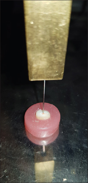
Immediately after obturation, all the teeth were placed in a humidifier at 37°C and 100% humidity for 48 h to allow the sealer to set.[10] Each sample was then sectioned horizontally using a circular diamond disc at low speed with water coolant. Three sections of 2-mm thickness were obtained at 4, 8, and 12 mm from the apex to represent the apical, middle, and coronal third, respectively. The sections below apical 3 mm were discarded due to the presence of aberrant anatomical features.[11] The root segments were then mounted on the acrylic block in an apico – coronal direction. The push-out bond strength of each sample was tested using the universal testing machine (UTM) [Figure 1]. A load was applied at a crosshead speed of 0.5 mm/min using three plungers of different sizes (1 mm, 0.7 mm, and 0.5 mm) for the coronal, middle, and apical sections, respectively in an apico-coronal direction. The push-out bond strength values were measured using the formula.
Figure 1.

Each sample was subjected to compressive loading through universal testing machine until bond failure occurs

The area of a bonded surface of the root canal material was calculated applying the following formula as per Prado et al.[12] [Figure 2].
Figure 2.

Diagrammatic representation of the area of the bonded surface

Where,
π-3.14,
r1-Coronal radius
r2-Apical radius
h-Thickness of the specimen
RESULTS
Data obtained were statistically analyzed using SPSS version 21 (SPSS Inc., Chicago, IL, USA). Means and standard deviation were calculated as shown in Table 1. A P ≤ 0.05 was accepted as statistically significant
Inter-group and intra-group comparison of mean values was done using one-way ANOVA and Tukey's test, respectively. The difference in the mean values between two independent samples was assessed using unpaired t-test
With lateral condensation technique, the highest push-out bond strength was found in the A1 group followed by B1 and C1 and the difference was found to be statistically significant (P < 0.00) [Table 2]. With thermoplasticized technique, the highest push-out bond strength was found in the B2 group followed by A2 and C2 and the difference was found to be statistically significant (P < 0.00) [Table 2]
Overall results showed that the lateral condensation technique showed significantly higher push-out bond strength than thermoplasticized technique (P < 0.028) [Table 2]
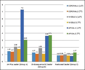
Irrespective of the sealer group, a statistically significant difference in bond strength was observed between coronal and apical levels (P < 0.00) with apical level showing the highest bond strength values. However, there was no statistically significant difference between the results obtained in the coronal and middle levels of roots (P > 0.251) [Graph 1]
AH plus sealer presented the highest push-out bond strength irrespective of the root levels and obturation techniques used, which was followed by Endosequence BC sealer and Roeko seal sealer (P < 0.05) [Graph 1].
Table 1.
Descriptive Statistics for bond strength of all sealer groups using two obturation techniques at coronal, middle and apical level
| Group name | Root level | n | Mean (MPa) | Standard deviation |
|---|---|---|---|---|
| Group A1 | Coronal | 10 | 1.49 | ±0.20 |
| AH plus sealer | Middle | 10 | 2.09 | ±0.19 |
| (LCT) | Apical | 10 | 7.30 | ±0.61 |
| Group A2 | Coronal | 10 | 1.40 | ±0.11 |
| AH plus sealer | Middle | 10 | 1.80 | ±0.22 |
| (TT) | Apical | 10 | 3.05 | ±0.74 |
| Group B1 | Coronal | 10 | 1.27 | ±0.17 |
| Endosequence BC sealer | Middle | 10 | 1.89 | ±0.19 |
| (LCT) | Apical | 10 | 3.63 | ±0.40 |
| Group B2 | Coronal | 10 | 1.10 | ±0.18 |
| Endosequence BC sealer | Middle | 10 | 1.98 | ±0.26 |
| (TT) | Apical | 10 | 3.71 | ±0.81 |
| Group C1 | Coronal | 10 | 0.87 | ±0.19 |
| Roeko seal sealer | Middle | 10 | 1.00 | ±0.11 |
| (LCT) | Apical | 10 | 0.79 | ±0.20 |
| Group C2 | Coronal | 10 | 0.89 | ±0.24 |
| Roeko seal sealer | Middle | 10 | 0.85 | ±0.08 |
| (TT) | Apical | 10 | 1.27 | ±0.46 |
Table 2.
One way ANOVA test for comparison of push out bond strength between the groups.
| Obturation Technique | Sealer Groups | Sum of squares | df | Mean square | F | Sig. |
|---|---|---|---|---|---|---|
| Lateral Condensation Technique (Group 1) | Between Groups (A1,B1,C1) | 152.722 | 3 | 50.907 | 23.982 | 0.000 |
| Thermoplasticized technique (Group 2) | Between Groups (A2,B2,C2) | 56.113 | 3 | 18.704 | 32.220 | 0.000 |
Graph 1.

Comparison of push-out bond strength of all the sealers using lateral condensation technique and thermoplasticized technique at different root levels
DISCUSSION
The success of root canal treatment is dependent on the quality of instrumentation, irrigation, disinfection, and finally, a three-dimensional obturation of the root canal system to create a monoblock effect.[11] In addition to the core material, the root canal sealer plays an important role during obturation, as it obliterates addition to all the imperfections and aberrant anatomical areas, and enhances the adaptation of the obturating material to the canal walls.[13]
Adequate bond strength of root canal sealers to dentin is important for maintaining the integrity of the seal. Along with the core material, it should have the ability to improve the fracture resistance of the tooth.[14]
Bond strength testing is a widely used method for determining the effectiveness of adhesion between obturating materials and the tooth structure.[14] In the present study, the push-out testing method was employed. This method is preferred, as it is sensitive to small variations in stress distribution during load application. Moreover, the results of this test have been shown to be effective and reproducible, which allows root canal sealers to be evaluated even when the bond strength values are low.[14]
In the current study, three plungers, with sizes within a range of 70%–90% of canal diameter, were used to match the diameter of each of the root thirds to avoid excessive pressure on the surrounding root canal walls and also to prevent any notching effect of the plunger into the GP surface.[15]
In the present study, AH plus showed the highest push-out bond strength followed by Endosequence BC and Roeko seal, and the difference was found to be statistically significant.
The probable reason for the highest bond strength value obtained with AH plus sealer could be the formation of a covalent bond by an open epoxide ring of the sealer to any exposed amino groups in the collagen of the dentin. In addition, this sealer shows very low shrinkage on the setting, has long-term dimensional stability and inherent volumetric expansion. It penetrates better into the micro-irregularities of the root canal wall because of its creep capacity and long polymerization period. The bond strength is further improved by mechanical locking between the canal dentine wall and the sealer.[14]
In the present study, Endosequence BC Sealer presented relatively less push out bond strength than AH plus sealer. Endosequence BC sealer is a hydrophilic material, and utilizes the moisture in the smear layer – thus creating hydroxyapatite-like precipitation while setting, and adheres to dentin chemically. Removal of the smear layer, as done in this study, could be a less favorable process and could have created a negative effect on the adaptation of this sealer to the canal walls. The moisture in the dentinal tubules may not be sufficient to help set the material causing lower bond strength of the sealer in root canals dried before obturation.[16] A number of related literatures support these findings by showing that the removal of the smear layer caused significantly less adhesion and more microleakage.[17,18,19] Moreover, the opened dentinal tubules may act as stress raisers, causing failure in the adhesive joint. Moreover, the bond strength values could have been increased by the use of bioceramic GP cones, which would have helped in the formation of a monoblock.
Significantly better bonding was observed with the AH Plus sealer when compared to the Roeko seal, which is in accordance with the study conducted by Tummala et al.[20] This may be attributed to the better wettability and penetration of the AH Plus sealer into the micro-irregularities. Roeko seal, on the other, hand showed poor wetting of the root dentin surface due to the presence of silicone, which possibly produces high surface tension forces, making the spreading of these materials difficult.[21]
In addition to the obturation material used, the obturation technique was also found to influence the bond strength to dentin. Although AH Plus sealer showed significantly higher bond strength values than the other two sealers with cold lateral compaction technique, the values dropped when thermoplasticized technique was used. This could be ascribed to the accelerated polymerization of resin-based sealers with increased temperature, where the fast setting causes a decrease in flow, increase in stiffness hence allowing limited time for relief of shrinkage stresses via resin flow, thus resulting in low bond strength values.[22]
In the present study, the apical third showed significantly higher push-out bond strength values followed by the middle and the coronal third. Although the density of dentinal tubules decreases from the coronal to the apical region, the circular cross-section of the root canal in the apical third may have provided higher resistance to dislodgment forces during the testing. On the other hand, coronal and middle portions, which have an oval or even flattened root canal section, showed no significant difference in the bond strength values. The variations in the root canal anatomy may lead to the misfit of the main GP cone, which may have been the cause for impaired bond strength.[11,23] The results of push-out bond strength studies may differ due to variation in material properties, obturation technique employed, methodology, type of UTM, size, and type of plunger and observer variation. The results of the present study are in collaboration with the studies conducted by Lee et al.,[24] Patil et al.,[25] Gade et al.[10] and Tummala et al.[20] that support the superior bond strength of AH Plus sealer than others still further studies are needed to corroborate the findings of the present study.
CONCLUSIONS
Within the limitation of the study, it may be stated that for mandibular premolar teeth with a single canal-
Using LCT AH plus sealer showed the highest push-out bond strength while using TT Endosequence BC sealer showed the highest push-out bond strength
LCT showed higher push-out bond strength values than TT irrespective of the type of sealer and root level
Push out bond strength was found to be more at the apical level, followed by the middle and coronal level irrespective of the technique used and type of sealer selected
AH plus sealer showed the highest push-out bond strength followed by Endosequence BC sealer and Roeko seal sealer irrespective of technique and root level.
In this study, efforts have been made to reduce the error due to multiple ecological variations and technique, however further studies are needed to corroborate the findings of the current study.
Financial support and sponsorship
Nil.
Conflicts of interest
There are no conflicts of interest.
REFERENCES
- 1.Rashmi NC, Basavanna RS, Gupta D. Influence of a resin based root canal filling material on resistance to fracture of endodontically treated teeth: An in vitro study. OHDM. 2014;13:1013–7. [Google Scholar]
- 2.Mounce ER. “Current Philosophies in Root Canal Obturation”. 2008. [Last accessed on 2020 May 02]. pp. 1–11. Available from: wwwineedcecom .
- 3.Caicedo R, von Fraunhofer JA. The properties of endodontic sealer cements. J Endod. 1988;14:527–34. doi: 10.1016/S0099-2399(88)80084-0. [DOI] [PubMed] [Google Scholar]
- 4.Roggendorf M. Zahnärzteblatt B Sept München Germany. Bavarian Dent J. 2004:32–4. [Google Scholar]
- 5.Takagi S, Chow LC, Hirayama S, Eichmiller FC. Properties of elastomeric calcium phosphate cement-chitosan composites. Dent Mater. 2003;19:797–804. doi: 10.1016/s0109-5641(03)00028-9. [DOI] [PubMed] [Google Scholar]
- 6.De-Deus G, Brandão MC, Fidel RA, Fidel SR. The sealing ability of GuttaFlow in oval-shaped canals: An ex vivo study using a polymicrobial leakage model. Int Endod J. 2007;40:794–9. doi: 10.1111/j.1365-2591.2007.01295.x. [DOI] [PubMed] [Google Scholar]
- 7.Chu CH, Lo EC, Cheung GS. Outcome of root canal treatment using thermafil and cold lateral condensation filling techniques. Int Endod J. 2005;38:179–85. doi: 10.1111/j.1365-2591.2004.00929.x. [DOI] [PubMed] [Google Scholar]
- 8.Guigand M, Glez D, Sibayan E, Cathelineau G, Vulcain JM. Comparative study of two canal obturation techniques by image analysis and EDS microanalysis. Br Dent J. 2005;198:707–11. doi: 10.1038/sj.bdj.4812389. [DOI] [PubMed] [Google Scholar]
- 9.Madhuri GV, Varri S, Bolla N, Mandava P, Akkala LS, Shaik J. Comparison of bond strength of different endodontic sealers to root dentin: An in vitro push-out test. J Conserv Dent. 2016;19:461–4. doi: 10.4103/0972-0707.190012. [DOI] [PMC free article] [PubMed] [Google Scholar]
- 10.Gade VJ, Belsare LD, Patil S, Bhede R, Gade JR. Evaluation of push-out bond strength of endosequence BC sealer with lateral condensation and thermoplasticized technique: An in vitro study. J Conserv Dent. 2015;18:124–7. doi: 10.4103/0972-0707.153075. [DOI] [PMC free article] [PubMed] [Google Scholar]
- 11.Abada H, Farag A, Alhadainy H, Darrag A. Push-out bond strength of different root canal obturation systems to root canal dentin. Tanta Dent J. 2015;12:185–91. [Google Scholar]
- 12.Prado M, Simão RA, Gomes BP. Effect of different irrigation protocols on resin sealer bond strength to dentin. J Endod. 2013;39:689–92. doi: 10.1016/j.joen.2012.12.009. [DOI] [PubMed] [Google Scholar]
- 13.Jainaen A, Palamara JE, Messer HH. Push-out bond strengths of the dentine-sealer interface with and without a main cone. Int Endod J. 2007;40:882–90. doi: 10.1111/j.1365-2591.2007.01308.x. [DOI] [PubMed] [Google Scholar]
- 14.Vilanova WV, Carvalho JR, Jr, Alfredo E, Neto MD, Sousa YT. Effect of intracanal irrigants on the bond strength of epoxy resin-based and methacrylate resin-based sealers to root canal walls. Int Endod J. 2012;45:42–8. doi: 10.1111/j.1365-2591.2011.01945.x. [DOI] [PubMed] [Google Scholar]
- 15.Zmener O, Spielberg C, Lamberghini F, Rucci M. Sealing properties of a new epoxy resin-based root-canal sealer. Int Endod J. 1997;30:332–4. doi: 10.1046/j.1365-2591.1997.00086.x. [DOI] [PubMed] [Google Scholar]
- 16.Shokouhinejad N, Gorjestani H, Nasseh AA, Hoseini A, Mohammadi M, Shamshiri AR. Push-out bond strength of gutta-percha with a new bioceramic sealer in the presence or absence of smear layer. Aust Endod J. 2013;39:102–6. doi: 10.1111/j.1747-4477.2011.00310.x. [DOI] [PubMed] [Google Scholar]
- 17.Yildirim T, Orucoglu H, Cobankara FK. Long-term evaluation of the influence of smear layer on the apical sealing ability of MTA. J Endod. 2008;34:1537–40. doi: 10.1016/j.joen.2008.08.022. [DOI] [PubMed] [Google Scholar]
- 18.Yildirim T, Er K, Sdemir TT, Tahan E, Buruk K, Serper A. Effect of smear layer and root-end cavity thickness on apical sealing ability of MTA as a root-end filling material: A bacterial leakage study. Oral Surg Oral Med Oral Pathol Oral Radiol Endod. 2010;109:67–72. doi: 10.1016/j.tripleo.2009.08.030. [DOI] [PubMed] [Google Scholar]
- 19.Razmi H, Bolhari B, Dashti NK, Fazlyab M. The effect of canal dryness on bond strength of bioceramic and epoxy-resin sealers after irrigation with sodium hypochlorite or chlorhexidine. Iran Endod J. 2016;11:129–33. doi: 10.7508/iej.2016.02.011. [DOI] [PMC free article] [PubMed] [Google Scholar]
- 20.Tummala M, Chandrasekhar V, Rashmi AS, Kundabala M, Ballal V. Assessment of the wetting behavior of three different root canal sealers on root canal dentin. J Conserv Dent. 2012;15:109–12. doi: 10.4103/0972-0707.94573. [DOI] [PMC free article] [PubMed] [Google Scholar]
- 21.Aa A, Hashimi RA. In vitro comparative assessment of the effect of gutta fusion obturation on the push out bond strength of three types of sealers. J Bagh Coll Dent. 2017;29:13–9. [Google Scholar]
- 22.De-Deus G, Di Giorgi K, Fidel S, Fidel RA, Paciornik S. Push-out bond strength of Resilon/Epiphany and Resilon/Epiphany self-etch to root dentin. J Endod. 2009;35:1048–50. doi: 10.1016/j.joen.2009.04.024. [DOI] [PubMed] [Google Scholar]
- 23.Cakici F, Cakici EB, Ceyhanli KT, Celik E, Kucukekenci FF, Gunseren AO. Evaluation of bond strength of various epoxy resin based sealers in oval shaped root canals. BMC Oral Health. 2016;16:106. doi: 10.1186/s12903-016-0301-1. [DOI] [PMC free article] [PubMed] [Google Scholar]
- 24.Lee KW, Williams MC, Camps JJ, Pashley DH. Adhesion of endodontic sealers to dentin and gutta-percha. J Endod. 2002;28:684–8. doi: 10.1097/00004770-200210000-00002. [DOI] [PubMed] [Google Scholar]
- 25.Patil P, Banga KS, Pawar AM, Pimple S, Ganeshan R. Influence of root canal obturation using gutta-percha with three different sealers on root reinforcement of endodontically treated teeth. An in vitro comparative study of mandibular incisors. J Conserv Dent. 2017;20:241–4. doi: 10.4103/JCD.JCD_233_16. [DOI] [PMC free article] [PubMed] [Google Scholar]


