Abstract
Background
To better understand and evaluate clinical usefulness of magnetic resonance imaging (MRI) in diagnosis and treatment of temporomandibular disorders (TMD), parameters for the evaluation are useful.
Purpose
To assess a clinically suitable staging system for evaluation of MRI of the temporomandibular joint (TMJ) and correlate the findings with age and some clinical symptoms of the TMJ.
Material and Methods
Retrospective analysis of 79 consecutive patients with clinical temporomandibular disorder or diagnosed inflammatory arthritis. Twenty-six healthy volunteers were included as controls. Existing data included TMJ pain, limited mouth opening (<30 mm) and corresponding MRI evaluations of the TMJs.
Results
The patients with clinical TMD complaints had statistically significantly more anterior disc displacement (ADD), disc deformation, caput flattening, surface destructions, osteophytes, and caput edema diagnosed by MRI compared to the controls. Among the arthritis patients, ADD, effusion, caput flattening, surface destructions, osteophytes, and caput edema were significantly more prevalent compared to the healthy volunteers. In the control group, disc deformation and presence of osteophytes significantly increased with age, and a borderline significance was found for ADD and surface destructions on the condylar head. No statistically significant associations were found between investigated clinical and MRI parameters.
Conclusion
This study presents a clinically suitable staging system for comparable MRI findings in the TMJs. Our results indicate that some findings are due to age-related degenerative changes rather than pathological changes. Results also show that clinical findings such as pain and limited mouth opening may not be related to changes diagnosed by MRI.
Keywords: Head and neck, temporomandibular joint, magnetic resonance imaging, jaw pain, adults
Introduction
Temporomandibular disorders (TMD) comprise a constellation of signs and symptoms including masticatory dysfunction, disc displacements and inflammatory reactions, and may need a multidisciplinary approach (1). Approximately 5%–12% of the population has TMD symptoms, and about half to two-thirds of these will seek treatment (1). Pain in the temporomandibular joint (TMJ) and the masticatory muscles (MM) provide the main complaints of patients with TMD referred for treatment.
Patients with longstanding TMD complaints are challenging and have often been through several general and specialized dentists and physicians seeking for help. Magnetic resonance imaging (MRI) is not the first diagnostic approach for TMJ pain. However, for oral and maxillofacial specialists, MRI is included in the diagnosis and evaluation for treatment and follow-up of patients with TMD when non-invasive treatment fails to relieve the symptoms. Imaging is essential for exact diagnosis of disc displacement, degenerative disc and bone deformations, inflammatory reactions, and other pathological conditions. MRI visualizes not only bone and soft tissue, but also fluid content within these tissues. Hence, inflammatory reactions in bone and discs as well as disc displacements can be diagnosed more accurately. Several studies have investigated the relationship between clinical and MRI findings regarding TMJ disorders (2–15). However, comparison of different publications based on the relationship between clinical and imaging findings is often difficult or not possible due to lack of comparable clinical and MRI diagnostic parameters.
The aim of the present study was to systematize MRI descriptions compared to clinical symptoms. Impact of aging on imaging diagnostics was considered of further importance.
Material and Methods
The material consisted of 79 patients with TMJ disorders and 26 healthy volunteers representing the control group. The consecutive patients were referred for MRI of the TMJs during 2002–2008 from general dental or medical practitioners to a university department with regional functions in oral and maxillofacial surgery for evaluation of TMJ problems. The patients not responding to 6–12 months of non-invasive treatment including masticatory muscle exercise and splint therapy and their clinical examination revealed the need for more diagnostic information. MRIs of all patients were performed at one department of radiology. Medically compromised patients and patients with chronic headache, migraine, fibromyalgia, and pain associated with dental problems or other inflammatory conditions were excluded from the study. Furthermore, patients whose symptoms originated from masticatory musculature were excluded. An experienced oral and maxillofacial surgeon examined the patients. Main complaints included: (i) current localized pre-auricular pain; (ii) TMJ sounds such as clicking or cracking; (iii) articular dysfunction such as locking or uncoordinated movements; (iv) pre-auricular swelling; and (v) sudden or gradual onset of malocclusion. Clinical variables further investigated in this study were maximum vertical mouth opening and pain on palpation of the TMJs.
The following groups were included:
TMD group: 55 patients (46 women, 9 men; age range = 16–68 years; mean age = 42.5 years) with general TMD complaints, including pain and functional dysfunction;
Arthritis group: 24 patients (18 women, 6 men; age range = 25–67 years; mean age = 45.1 years) with inflammatory arthritis diagnosed according to current criteria. Seventeen patients had rheumatoid arthritis (RA), four patients had ankylosing spondylitis (AS), and three patients had psoriatic arthritis (PsA);
Control group: 26 healthy volunteers (18 women, 8 men; age range = 20–56 years, mean age = 33.2 years) without any present or former symptoms from the TMJs were included after informed written consent. Volunteers with any history of rheumatic disease or other muscle or joint-related disorders were excluded from the study.
The study was conducted in line with the Declaration of Helsinki. Ethical approval from the Institutional Review Board was given by the Regional Committee for Medical and Health Research Ethics, western Norway. The study was acknowledged to be a quality control study (028.09).
Clinical examination
Maximum unassisted mouth opening was registered as inter-incisor distance in millimeters at full mouth opening. Inter-incisor distance on maximum mouth opening < 30 mm was categorized as pathological. TMJ pain was registered through bilateral manual index palpation of the lateral aspect of the condylar head and subsequently registered as pain originating from the TMJs.
The clinical data for the patients in the TMD group and arthritis group were retrospectively collected from the patients’ journal. In the TMD group, information about pain on palpation of the TMJs was missing for six patients, and two patients lacked information about maximum mouth opening in the patient journal.
MRI evaluation
The MRI examination was performed on a GE Signa® 1.5-T, 33 mT raise gradients machine (General Electric, Milwaukee, WI, USA) with a dedicated TMJ coil. Images with closed mouth were obtained for all patients. In order to obtain open mouth images, the patients were asked to bite on a 20-cc syringe (diameter approximately 20 mm). Not all patients (almost half of them) managed to perform the open mouth MRI examination. Hence, only the images with closed mouth were included in the study. No intravenous contrast media were administered. The MRI examinations were retrospectively examined and described by an oral and maxillofacial radiologist blinded for all clinical data. The criteria used are presented in Table 1.
Table 1.
Staging criteria for the MRI parameters according to Moen et al. (15).
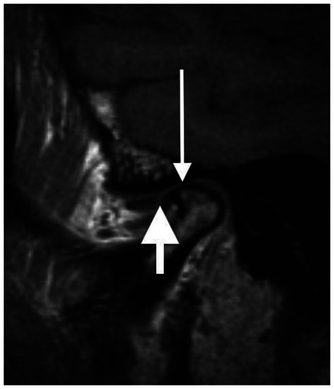
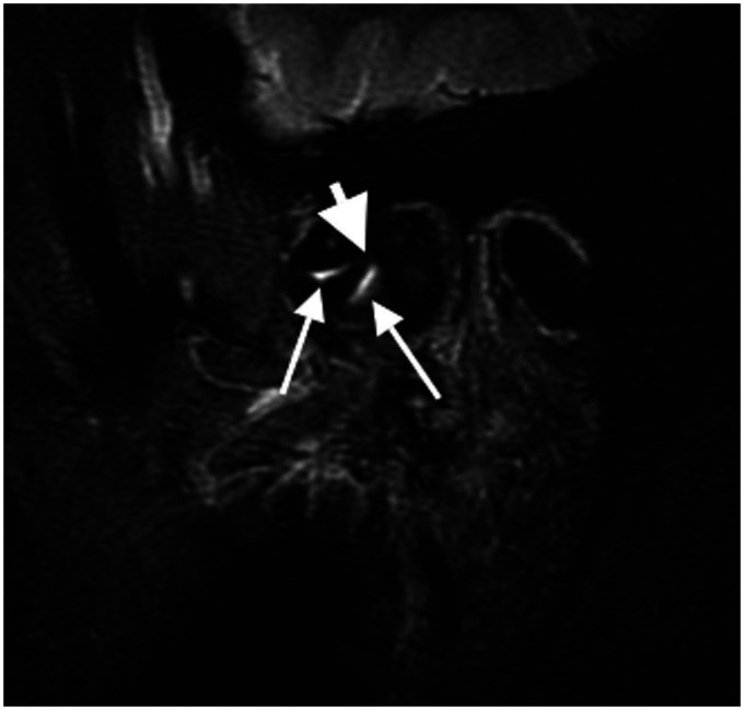
| Disc position | Anterior disc position was defined as the end of the posterior band located anterior to the 10 o’clock position relative to the condyle (16). Anterior dislocation (Figs. 1 and 2) was registered according to Drace and Enzmann (17). Only disc displacement in sagittal direction was described. |
| Disc deformation | Registered as pathologic when the biconcave morphology of the disc was clearly changed. |
| Effusion | Fluid in the synovial compartments (Fig. 2) was, when clearly apparent, registered in three different grades: moderate, marked, or extensive according to Larheim and Westesson (18). |
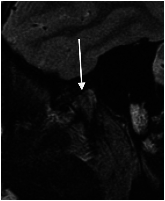
| Caput flattening | Obvious flattening only was registered as pathological (Fig. 3). |
| Surface destructions | Obvious or extensive destructions on the surface of the condylar head were registered as pathological (Fig. 3). |
| Osteophytes | Registered as present or not (Fig. 1). |
| Caput edema | Bone marrow edema in the condylar head was registered by hypo intensive signal on T1 and hyper intensive on T2-weighted images, according to Larheim et al. (19). |
MRI, magnetic resonance imaging.
Findings were recorded as positive if present either unilaterally or bilaterally. In some joints, it was not possible to determine the presence or absence of all the MRI variables from the existing MR images. If an MRI variable was absent in one joint, and not possible to diagnose in the joint on the other side, the patient was excluded in the analysis of that variable.
The patient groups and controls were further classified into two subgroups—age > 40 years and age < 40 years—to test the influence of aging on MRI findings.
The clinical parameters limited mouth opening and pain on palpation of the TMJs were separately tested for association with the MRI variables. When correlating TMJ pain with MRI findings, right and left sides were evaluated separately. Concerning associations with limited mouth opening, pathological MRI findings in at least one of the two TMJs were required.
Fig. 1.
Osteoarthritic joint. Degenerative osteophyte anteriorly (long arrow), anterior displacement of the disc (short arrow), and subchondral sclerosis in the caput.
Fig. 2.
Osteoarthritic joint. Anterior displacement of the disc (9 o’clock) (short arrow showing posterior part of the disc). Effusion in the joint (long arrows).
Fig. 3.
Severe osteoarthritis. Surface destruction and flattening of the caput (white arrow). It is difficult to see the joint due to the extensive degenerative changes.
Statistical methods
In the statistical evaluation of the data, frequencies of the clinical and radiological parameters in each of the patient groups and the control group were calculated. Fisher’s exact test was used to compare the relative frequencies between the groups and to test for association between clinical and MRI parameters. The level of significance was set to 0.05. The statistical software STATA/IC 14.1 (Stata Corp LP, College Station, TX, USA) was used for the analyses.
Results
Clinical findings
Pain on palpation of one or both TMJs were found in 88% of the patients in the TMD group and in 57% of the arthritis group. Furthermore, 42% of the patients in the TMD group had reduced mouth opening (<30 mm) compared to 21% in the arthritis group. None of the included healthy volunteers had reduced mouth opening (<30 mm) or pain on palpation of the TMJs (Tables 2 and 3).
Table 2.
A comparison of clinical and MRI findings in the TMD group and control group (Fisher’s exact test).
| TMD (n=55) | Control (n=26) | P value | |
|---|---|---|---|
| TMJ pain* | 43 (88) | 0 (0) | <0.001 |
| Trismus* | 22 (42) | 0 (0) | <0.001 |
| ADD | 41 (75) | 7 (27) | <0.001 |
| Disc deformation | 37 (67) | 6 (23) | <0.001 |
| Effusion† | 27 (50) | 7 (27) | 0.058 |
| Caput flattening† | 30 (56) | 4 (15) | 0.001 |
| Surface destructions | 29 (53) | 7 (27) | 0.034 |
| Osteophytes | 32 (58) | 6 (23) | 0.004 |
| Caput edema | 26 (47) | 1 (4) | <0.001 |
Values are given as n (%).
*In the TMD group, six patients had no information about pain on palpation of the TMJ registered in the journal (n = 49 for TMJ pain) and two patients lacked information about maximum mouth opening (n = 53 for trismus).
†n = 54 in the TMD group due to difficulties in determining presence or absence of effusion in one patient and caput flattening in another patient from the existing MR images.
ADD, anterior disc displacement; MRI, magnetic resonance imaging; TMD, temporomandibular disorder; TMJ, temporomandibular joint.
Table 3.
A comparison of clinical and MRI findings in the arthritis group and control group (Fisher’s exact test).
| Arthritis (n = 24) | Control (n = 26) | P value | |
|---|---|---|---|
| TMJ pain* | 13 (57) | 0 (0) | <0.001 |
| Trismus | 5 (21) | 0 (0) | 0.020 |
| ADD | 18 (75) | 7 (27) | 0.002 |
| Disc deformation | 12 (50) | 6 (23) | 0.077 |
| Effusion | 14 (58) | 7 (27) | 0.044 |
| Caput flattening | 20 (83) | 4 (15) | <0.001 |
| Surface destructions | 20 (83) | 7 (27) | <0.001 |
| Osteophytes | 17 (71) | 6 (23) | 0.002 |
| Caput edema | 9 (38) | 1 (4) | 0.004 |
Values are given as n (%).
*One patient in the arthritis group lacked information about pain on palpation of the TMJ in the patient journal.
ADD, anterior disc displacement; MRI, magnetic resonance imaging; TMJ, temporomandibular joint.
MRI findings
TMD group
Of the patients in the TMD group, 75% had moderate or extensive anterior disc displacement (ADD), without reduction when diagnosed in occlusion (closed mouth), either unilaterally or bilaterally, and 67% had disc deformations. We found moderate, marked, or extensive amounts of joint fluid or effusion in 50% of the patients. Osteophytes were present in 58% of the patients, 56% of the patients had flattening of the condylar head, and 53% had destructions on the surface of the condyle. Bone marrow edema in the caput was seen in 47% of the patients in the TMD group. Except for effusion, all the MRI parameters were significantly more prevalent among the TMD patients compared to the control group (Table 2).
Arthritis group
Of the patients in the arthritis group, 75% had moderate or extensive ADD without reduction, either unilaterally or bilaterally, and 50% had severe changes in the biconcave morphology of the disc. Of these patients, 58% had moderate, marked or extensive amounts of effusion in the synovial compartments. Superior flattening of the condylar head was found in 83% of the patients, 83% had destructions on the condylar surface, and osteophytes on the condylar head were present in 71% of the patients. Bone marrow edema in the caput was seen in 38% of the patients in the arthritis group. Except for disc deformation, all the MRI variables were significantly more prevalent among the patients in the arthritis group compared to the control group (Table 3).
Influence of age on MRI findings
Of the patients with TMD aged ≥40 years, 80% had disc deformations. This was significantly more than in the younger population (P = 0.043). No other significant differences in MRI findings were found between the patients aged < 40 years and those aged > 40 years in the two patient groups.
In the control group, more individuals aged > 40 years had osteophytes on the condylar head (P = 0.002), disc deformations (P = 0.028), ADD (P = 0.057), and surface destructions on the condylar head (P = 0.057) compared to the younger participants (Table 4). Effusion was more frequent in the younger age group, but the difference was not statistically significant (Table 4).
Table 4.
Differences in MRI findings before and after the age of 40 years in 26 healthy volunteers (Fisher’s exact test).
|
Age (years) |
|||
|---|---|---|---|
| ≤40 (n = 19) | >40 (n = 7) | P value | |
| ADD | 3 (16) | 4 (57) | 0.057 |
| Disc deformation | 2 (11) | 4 (57) | 0.028 |
| Effusion | 6 (32) | 1 (14) | 0.629 |
| Caput flattening | 3 (16) | 1 (14) | >0.900 |
| Surface destructions | 3 (16) | 4 (57) | 0.057 |
| Osteophytes | 1 (5) | 5 (71) | 0.002 |
| Caput edema | 1 (5) | 0 (0) | >0.900 |
Values are given as n (%).
ADD, anterior disc displacement; MRI, magnetic resonance imaging.
Relationship between clinical and MRI findings
Both pain on palpation of the TMJs and limited mouth opening were statistically significantly more prevalent in the patient groups compared to the controls. However, no statistically significant associations were found between investigated clinical and MRI parameters in the two patient groups (Table 5).
Table 5.
Association (P value) between the clinical variables pain on palpation of the TMJ and trismus (mouth opening < 30 mm), and the MRI findings in the two patient groups (Fisher’s exact test).
|
TMD group |
Arthritis group |
|||||
|---|---|---|---|---|---|---|
|
TMJ pain |
TMJ pain |
|||||
| Right | Left | Trismus | Right | Left | Trismus | |
| ADD | 0.366 | >0.900 | 0.755 | > 0.900 | 0.221 | 0.568 |
| Disc deformation | >0.900 | >0.900 | 0.777 | >0.900 | 0.417 | >0.900 |
| Effusion | >0.900 | 0.566 | >0.900 | 0.363 | 0.680 | 0.122 |
| Caput flattening | 0.245 | 0.244 | >0.900 | 0.400 | >0.900 | 0.179 |
| Surface destructions | 0.221 | 0.396 | 0.782 | 0.400 | > 0.900 | 0.179 |
| Osteophytes | 0.384 | 0.773 | >0.900 | 0.680 | >0.900 | 0.608 |
| Caput edema | 0.758 | 0.561 | 0.578 | 0.643 | 0.660 | 0.071 |
ADD, anterior disc displacement; MRI, magnetic resonance imaging; TMD, temporomandibular disorder; TMJ, temporomandibular joint.
Discussion
The results of the present study indicate that MRI findings of osteoarthritis in the TMJ is not necessarily linked to progressive functional disturbances such as limited mouth opening or pain on palpation of the TMJs. Furthermore, most of the MRI variables were significantly more prevalent in the patient groups compared to the controls. However, increased prevalence of some MRI findings with increasing age among the healthy volunteers indicate that some findings are due to age-related degenerative changes rather than pathological changes.
Various severities of ADD are common in patients with general TMD complaints, and prevalence rates around 70%–90%, as in the present study, are normal findings (4,16–19). Almost one-third of the asymptomatic volunteers in the present study had ADD on MRI, which may indicate that ADD not directly or always induces pain in the TMJ. All normal patients did both open and closed position MRI. These normal patients with ADD had discs that more or less followed the movement of the condylar head (being placed anteriorly without any symptoms). The finding that ADD was significantly more common in symptomatic individuals than in controls agrees with previous reports (20–22). To distinguish between ADD with or without reduction on MRI, recordings at both open and closed mouth must be compared according to former literature. It was voluntarily whether or not the patient would bite on a 20-cc syringe, and a large number of patients were not able to bite on the 20-cc syringe due to pain or reduced mouth opening. In the present study, 75% of the patients in the TMD group as well as in the arthritis group were diagnosed with an ADD without reduction. The diagnostic value of “open mouth” MRI may then be questionable. The patient was also asked to open and close the mouth in order to perform dynamic gradient echo sequence. Too few patients complied with the dynamic part of the examination to perform statistical analyses. Clinical experience tells us that for these patients, jaw movements are difficult whatever type of examination. In our department, the use of contrast media was not a diagnostic routine procedure for these patients. It was not used mostly because the longer lasting and more difficult administrating contrast media would make examination of the patients longer and more difficult, as well as risking adverse reactions. It is known that the use of contrast media has been reported and we decided that a possible gain was too low to defend its use. In the present study, we would have to follow the department’s routine.
Of the patients in the arthritis group, 57% had pain on palpation of the TMJs. This was fewer than expected in this patient group and is probably explained by the frequent use of anti-inflammatory drugs on a daily basis for their general disease. However, disc displacements and mandibular condyle deformities were seen in 75% and 83% of the patients in this group, respectively, typical for long-term manifestations of RA in the TMJs (23). Reduced mouth opening was not a general complaint among the arthritis patients.
In the present study, 50% of the patients in the general TMD group had pathological amounts of joint fluid. This is slightly more than reported in previous studies (6,19). An increased amount of effusion was even found in 27% of the controls. The use of the STIR sequence in the present study could explain this discordance, or maybe effusion indicates the beginning of joint pathology.
Takatsuka et al. (21) reported that for mild osteoarthritis confined to flattening, erosion, and osteophytes, there was no significant statistical difference between presence of osteoarthritis and visual analogue scale (VAS) scores. Among the healthy volunteers in the present study, 23% had osteophytes and 27% had obvious destructions on the surface of the condylar head without having any clinical symptoms. An assumption is that natural degeneration and remodeling of the joint due to normal aging could present with the same MRI findings as osteoarthritis. Hence, this could confound the results when MRI findings were tested for association with the clinical variables reduced mouth opening and pain on palpation of the TMJ.
No significant associations could be found in this study between clinical symptoms and MRI findings of the TMJ. This discordance has also been reported in previous studies (2,3,5,9,10). To explain this lack of association between MRI and clinical symptoms, some particular reasons may be postulated. First, statistical power in this study was tested and found adequate, but the number of patients may still be low for establishing certainty of all the results. Second, significant correlations may exist between MRI variables and other clinical parameters such as joint sounds or horizontal range of movement, which were not investigated in the present study. Third, there are no associations between clinical symptoms and MRI findings to be found. However, other studies have reported such associations (11–14).
In the control group, age clearly made a difference in the prevalence of disc deformation and osteophytes on the condylar head. These findings significantly increased with age. There was also a tendency that age had the same impact on ADD and surface destructions of the condylar head (Table 4). However, the results regarding ADD should be read carefully since the MRI evaluations in the present study could not distinguish between ADD with or without reduction. For effusion it was the opposite, with a higher prevalence among the younger volunteers. In the two patient groups, however, it was only for disc deformation in the TMD group that the prevalence significantly increased with age. For the other variables, age did not seem to play any role. These findings imply that TMD and inflammatory arthritis may overshadow normal degenerative changes in the TMJ due to age.
MRI is an excellent tool for visualizing inflammatory reactions in bone and soft tissues. To better understand and evaluate both clinical usefulness of MRI and find how to properly use MRI in this setting, parameters for the evaluation would be useful. A previously published staging system was used in the present study in order to better compare patient groups (15). This allows for future comparable evaluations. Some findings on MRI are due to age-related degenerative changes rather than pathological changes. Results also show that clinical findings such as pain on palpation of the TMJ and reduced mouth opening may not be related to changes diagnosed by MRI. For the surgical specialists, in particular, MRI should be included in the evaluation and follow-up of patients with clinical signs of TMJ problems if non-invasive treatment fails to relieve the symptoms.
In conclusion, the present study shows data demonstrating that MRI findings in the TMJs may enhance clinical diagnoses. The results indicate that some findings are due to age-related degenerative changes rather than pathological changes. Furthermore, ADD without reduction was diagnosed with “closed mouth” MRI in 75%. Results also show that clinical findings such as pain and limited mouth opening may not be related to changes diagnosed by MRI.
Declaration of conflicting interests
The author(s) declared no potential conflicts of interest with respect to the research, authorship, and/or publication of this article.
Funding
The author(s) received no financial support for the research, authorship, and/or publication of this article.
ORCID iD
Jonn Terje Geitung https://orcid.org/0000-0001-9259-1060
References
- 1.Stanisewski K, Lygre H, Bifulco E, et al. Temporomandibular disorders related to stress and HPA-axis regulation. Pain Res Manag 2018; 2018:7020751. [DOI] [PMC free article] [PubMed] [Google Scholar]
- 2.National Institute of Dental and Craniofacial Research. Available at: https://www.nidcr.nih.gov/DataStatistics/FindDataByTopic/FacialPain/ (last accessed 3 July 2014).
- 3.Koh KJ, List T, Petersson A, et al. Relationship between clinical and magnetic resonance imaging diagnoses and findings in degenerative and inflammatory temporomandibular joint diseases: a systematic literature review. J Orofac Pain 2009; 23:123–139. [PubMed] [Google Scholar]
- 4.Limchaichana N, Nilsson H, Ekberg EC, et al. Clinical diagnoses and MRI findings in patients with TMD pain. J Oral Rehabil 2007; 34:237–245. [DOI] [PubMed] [Google Scholar]
- 5.Ohlmann B, Rammelsberg P, Henschel V, et al. Prediction of TMJ arthralgia according to clinical diagnosis and MRI findings. Int J Prosthodont 2006; 19:333–338. [PubMed] [Google Scholar]
- 6.Rudisch A, Innerhofer K, Bertram S, et al. Magnetic resonance imaging findings of internal derangement and effusion in patients with unilateral temporomandibular joint pain. Oral Surg Oral Med Oral Pathol Oral Radiol Endod 2001; 92:566–571. [DOI] [PubMed] [Google Scholar]
- 7.Westesson PL, Brooks SL. Temporomandibular joint: relationship between MR evidence of effusion and the presence of pain and disk displacement. AJR Am J Roentgenol 1992; 159:559–563. [DOI] [PubMed] [Google Scholar]
- 8.Park JW, Song HH, Roh HS, et al. Correlation between clinical diagnosis based on RDC/TMD and MRI findings of TMJ internal derangement. Int J Oral Maxillofac Surg 2012; 41:103–108. [DOI] [PubMed] [Google Scholar]
- 9.Robinson de Senna B, Marques LS, Franca JP, et al. Condyle-disk-fossa position and relationship to clinical signs and symptoms of temporomandibular disorders in women. Oral Surg Oral Med Oral Pathol Oral Radiol Endod 2009; 108:e117–124. [DOI] [PubMed] [Google Scholar]
- 10.Schmitter M, Essig M, Seneadza V, et al. Prevalence of clinical and radiographic signs of osteoarthrosis of the temporomandibular joint in an older persons community. Dentomaxillofac Radiol 2010; 39:231–234. [DOI] [PMC free article] [PubMed] [Google Scholar]
- 11.Emshoff R, Brandlmaier I, Bertram S, et al. Relative odds of temporomandibular joint pain as a function of magnetic resonance imaging findings of internal derangement, osteoarthrosis, effusion, and bone marrow edema. Oral Surg Oral Med Oral Pathol Oral Radiol Endod 2003; 95:437–445. [DOI] [PubMed] [Google Scholar]
- 12.Adame CG, Monje F, Offnoz M, et al. Effusion in magnetic resonance imaging of the temporomandibular joint: a study of 123 joints. J Oral Maxillofac Surg 1998; 56:314–318. [DOI] [PubMed] [Google Scholar]
- 13.Maizlin ZV, Nutiu N, Dent PB, et al. Displacement of the temporomandibular joint disk: correlation between clinical findings and MRI characteristics. J Can Dent Assoc 2010; 76:a3. [PubMed] [Google Scholar]
- 14.Vogl TJ, Lauer HC, Lehnert T, et al. The value of MRI in patients with temporomandibular joint dysfunction: Correlation of MRI and clinical findings. Eur J Radiol 2016; 85:714–719. [DOI] [PubMed] [Google Scholar]
- 15.Moen K, Hellem S, Geitung JT et al. A practical approach to interpretation of MRI of the temporomandibular joint. Acta Radiol 2010;51:1021--1027. [DOI] [PubMed]
- 16.Tasaki MM, Westesson PL, Isberg AM, et al. Classification and prevalence of temporomandibular joint disk displacement in patients and symptom-free volunteers. Am J Orthod Dentofacial Orthop 1996; 109:249–262. [DOI] [PubMed] [Google Scholar]
- 17.Drace JE, Enzmann DR. Defining the normal temporomandibular joint: closed-, partially open-, and open-mouth MR imaging of asymptomatic subjects. Radiology 1990; 177:67–71. [DOI] [PubMed] [Google Scholar]
- 18.Larheim TA, Westesson PL, Sano T. MR grading of temporomandibular joint fluid: association with disk displacement categories, condyle marrow abnormalities and pain. Int J Oral Maxillofac Surg 2001; 30:104–112. [DOI] [PubMed] [Google Scholar]
- 19.Larheim TA, Katzberg RW, Westesson PL, et al. MR evidence of temporomandibular joint fluid and condyle marrow alterations: occurrence in asymptomatic volunteers and symptomatic patients. Int J Oral Maxillofac Surg 2001; 30:113–117. [DOI] [PubMed] [Google Scholar]
- 20.Katzberg RW, Westesson PL, Tallents RH, et al. Anatomic disorders of the temporomandibular joint disc in asymptomatic subjects. J Oral Maxillofac Surg 1996; 54:147–53, discussion 53–55. [DOI] [PubMed] [Google Scholar]
- 21.Takatsuka S, Yoshida K, Ueki K, et al. Disc and condyle translation in patients with temporomandibular disorder. Oral Surg Oral Med Oral Pathol Oral Radiol Endod 2005; 99:614–621. [DOI] [PubMed] [Google Scholar]
- 22.Kumar R, Pallagatti S, Sheikh S, et al. Correlation between clinical findings of temporomandibular disorders and MRI characteristics of disc displacement. Open Dent J 2015; 9:273–281. [DOI] [PMC free article] [PubMed] [Google Scholar]
- 23.Arvidsson LZ, Smith HJ, Flato B, et al. Temporomandibular joint findings in adults with long-standing juvenile idiopathic arthritis: CT and MR imaging assessment. Radiology 2010; 256:191–200. [DOI] [PubMed] [Google Scholar]


