Abstract
Case presentation
We report the first confirmed case of the novel coronavirus disease (COVID-19) in a lactating patient in Chizhou, Anhui Province, China. The lactating patient presented with intermittent fever for 16 days and cough for 10 days. Given her travel history to the epidemic area and the chest CT scan results, the patient was immediately admitted to the isolation ward of the Infectious Disease Department and breastfeeding was discontinued. Pharyngeal swab specimens tested positive for severe acute respiratory syndrome coronavirus 2 (SARS-CoV-2, previously known as 2019-nCoV) in nucleic acid testing. During hospitalization, she also experienced bilateral breast tenderness. After active treatment, the patient ultimately achieved remission and was discharged from the hospital.
Discussion and conclusions
SARS-CoV-2 is transmitted mainly through respiratory droplets and patient contact, rendering the general population to a high risk of infection. The management of mother–child interactions and breastfeeding in women with COVID-19 is a difficult problem. The purpose of this case report is to help clinicians by improving the understanding of COVID-19, particularly in lactating patients.
Keywords: Lactating patient, COVID-19, Infection
Background
The novel coronavirus disease (COVID-19) caused by the severe acute respiratory syndrome coronavirus-2 (SARS-CoV-2) has emerged as a critical public health challenges of the modern age [1, 2]. Until now, reports on lactating patients with COVID-19 are limited. Here, we report the first confirmed case of COVID-19 in a lactating patient in Chizhou, Anhui Province, China; the patient, who was admitted to our hospital, ultimately achieved remission and was discharged. The purpose of this case report is to improve the understanding of the disease for clinicians, with particular emphasis on lactating patients.
Case presentation
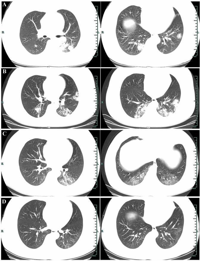
The patient was a 37-year-old woman who presented with intermittent fever for 16 days and cough for 10 days. On January 6, 2020, the patient developed fever (body temperature > 39 °C), sore throat, and fatigue while visiting Wuhan. The patient did not take any medication because she was breastfeeding her 6-month-old infant and she felt that her fever improved after resting. On January 12, 2020, the patient started coughing (mainly dry cough). The patient returned to Chizhou from Wuhan on January 19, 2020, and her cough symptoms gradually worsened. On January 22, 2020, she visited the Respiratory Medicine Department at our hospital. As the patient had returned from Wuhan during the COVID-19 epidemic, she was provided with a medical surgical mask for protection. The complete blood count results of the patient were as follows: white blood cells (WBC): 7.36 × 10⁹/L, neutrophils (N): 72.91%, lymphocytes (L): 19.22% (reference range 20–40%), hemoglobin (HGB): 135.00 g/L, and platelets (PLT): 176.00 × 10⁹/L, and C-reactive protein (CRP) was 40.53 mg/L (reference range 0–10 mg/L). Chest computed tomography (CT) scans showed multiple flaky, consolidated lesions scattered throughout the right lower lobe, the left upper lobe apicoposterior segment, the lingula of the left lung, and the left lower lobe. Some lesions exhibited ground-glass opacity, and internal air bronchograms were also observed (Fig. 1a). Given the patient’s travel history in the epidemic area and the chest CT scan results, the Department of Medical Administration was notified. The patient was then immediately admitted to the isolation ward of the Infectious Disease Department, and breastfeeding was discontinued. For reasons of caution, the 6-month-old infant was taken home, cared for by the father, and given artificial nutrition. A pharyngeal swab specimen was obtained for SARS-CoV-2 nucleic acid testing. The patient denied a history of other special diseases.
Fig. 1.
a Chest CT scans showed multiple flaky, consolidated lesions scattered throughout the right lower lobe, the left upper lobe apicoposterior segment, the lingula of the left lung, and the left lower lobe. b Chest CT showed some lesions had progressed. c Chest CT showed the bilateral lesions were significantly resolved when compared with the previous scans. d Chest CT showed the original focus was obviously absorbed
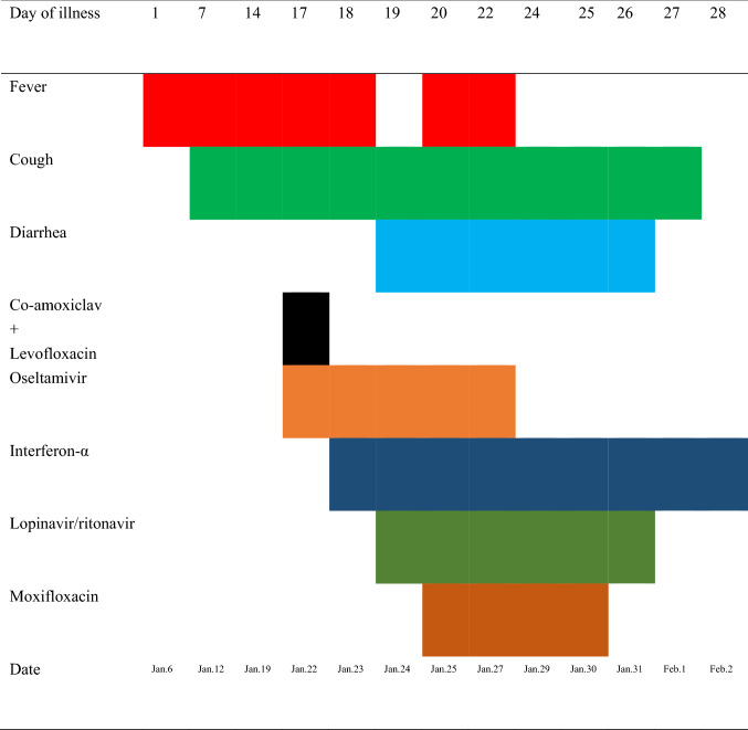
Upon admission, the patient was administered co-amoxiclav at 1.2 g once every 8 h with concurrent levofloxacin (0.5 g IV qd, anti-inflammatory therapy) and oseltamivir (75 mg PO bid, antiviral therapy). Auxiliary examinations were performed. Arterial blood gas analysis on January 23, 2020 showed that the pH was 7.397, partial pressure of carbon dioxide was 20.6 mmHg, partial pressure of oxygen was 157 mmHg, actual base excess was − 10.9 mmol/L, and oxygen saturation was 99.6%. The 2020-01-23 complete blood count plus CRP results were as follows: WBC, 4.38 × 10⁹/L; N, 70.00%; L, 23.20%; monocytes, 6.50%; HGB, 133.00 g/L; PLT, 185.00 × 10⁹/L; CRP 48.40 mg/L. The 2020-01-23 renal and hepatic function test results were normal, and the electrolyte panel results were as follows: potassium 3.41 mmol/L (reference range 3.5–5.5 mmol/L), sodium 141.2 mmol/L, chlorine 101.1 mmol/L, and calcium 2.13 mmol/L (reference range 2.2–2.7 mmol/L). The 2020-01-23 coagulation profile was normal. The 2020-01-23 procalcitonin level was 0.0340 ng/mL. On January 23, 2020, the initial result of SARS-CoV-2 nucleic acid testing of the pharyngeal swab specimen sent to the Chizhou Center for Disease Control and Prevention was positive. A new pharyngeal swab specimen was collected and sent to the Anhui Provincial Center for Disease Control and Prevention for retesting. On January 23, 2020, antibiotics were discontinued. The patient continued to receive oral oseltamivir and was given symptomatic treatment with interferon-α 5 million U twice a day as an inhaled aerosol, oxygen therapy, and fluid replacement. Respiratory virus testing on January 24, 2020 showed that the patient was positive for influenza B virus IgM. On the same day, the SARS-CoV-2 nucleic acid retesting result from the Anhui Provincial Center for Disease Control and Prevention was positive. All family members of the patient were isolated and subjected to SARS-COV-2 pharyngeal swab nucleic acid screening. Fortunately, all results were negative. Patient diagnosis: (1) COVID-19; (2) influenza B infection. Lopinavir/ritonavir (LPV/r) (two tablets PO bid) was added to the treatment regimen. The patient had three episodes of loose stools per day. The patient developed fever again after she had a normal body temperature for 30 h. She also experienced bilateral breast tenderness. The 2020-01-25 complete blood count plus CRP results were as follows: WBC, 6.05 × 10⁹/L; N, 81.20%; L, 13.60%; HBG, 126.00 g/L; PLT 208.00 × 10⁹/L, CRP, 58.20 mg/L. The results of stool analysis and fecal occult blood testing were normal. Considering that the patient was breastfeeding, failure to prompt milk expression may have caused the acute mastitis. The patient’s body temperature gradually returned to normal after anti-inflammatory treatment with moxifloxacin. Oral vitamin B6 was given with consent of the patient to block lactation. From January 27, 2020, two follow-up pharyngeal swabs tested negative for SARS-CoV-2 nucleic acid. On January 29, 2020, follow-up chest CT showed no significant resolution of the lesions, and some lesions had progressed (Fig. 1b). On January 31, 2020, follow-up CRP testing showed a decline, and moxifloxacin was discontinued. On February 1, 2020, LPV/r was discontinued, but the patient continued to receive interferon-α as an inhaled aerosol. On February 2, 2020, follow-up chest CT showed that the bilateral lesions were significantly resolved when compared with the previous scans (Fig. 1c). The patient met the criteria for discharge from the isolation ward. The next day, the patient was discharged from the hospital (Fig. 2). On February 18, the patient came to reexamine the chest CT, and the original focus was obviously absorbed (Fig. 1d).
Fig. 2.
Flowchart of the patient’s symptoms, diagnosis and treatment from January 6 to February 2, 2020
Discussion and conclusions
COVID-19 caused by SARS-COV-2 is a major public health concern worldwide. As the coronavirus pandemic has evolved, pregnant women infected with SARS-CoV-2 are experiencing fear amid uncertainties regarding the care of their fetus or newborn [3]. To solve many problems caused by this epidemic, interdisciplinary research from different departments is needed. Many maternity units around the world are currently considering management protocols for the pregnant or lactating COVID-19 patients [3].
It is well known that breastfeeding benefits both infants and mothers, and is strongly supported by public policy. Human milk provides optimal nutrition and helps to strengthen the immune system of the infants, thereby allowing them to better resist (or even prevent) infections. In addition, breastfeeding can reduce the risk in mothers to develop diabetes, hypertension, and breast cancer [4]. However, for many infected pregnant women with severe acute and highly pathogenic infectious diseases, such as SARS, MERS, and COVID-19, the safety of breastfeeding is still debatable.
Because there is no evidence of vertical transmission of coronavirus between mothers and infants, and the transmission of SARS-CoV-2 from person to person is mainly via the respiratory route [5], SARS-CoV-2 may not be transmitted through breast milk. A recently published report showed that SARS-CoV-2 RNA does not represent replication-competent virus [6]. However, SARS-CoV-2 can be detected in different body fluids. During breastfeeding, the virus may be transmitted to the newborn via respiratory droplets expelled by the infected mother [7]. Although respiratory droplets are a known mechanism of transmission, there may be other mechanisms involved as well. Investigations of pregnant women with COVID-19 must include testing from different body sites or even using different body fluids, which will help to improve the sensitivity and reduce false-negative test results [8].
Recent studies have shown that angiotensin-converting enzyme 2 (ACE2) is one of the receptors of SARS-CoV-2, allowing its internalization into the host cells. ACE2 is expressed in many body parts and tissue types, including the oral cavity (such as the tongue and oral mucosa) and breast tissues. If the hypothesis that mammary epithelial cells express ACE2 is true, the presence of viral particles in breast milk may be one of the mechanisms responsible for SARS-CoV-2 infection in infants [2].
Confidence in the safety of breastfeeding in the context of SARS-CoV-2 infection (even considering the best practices for breastfeeding) is undermined by the lack of rigorous evidence proving/rejecting vertical transmission of SARS-CoV-2 via breastfeeding. A recent report in Science discussed the concerns of pregnant women with COVID-19, with respect to breastfeeding and care for their babies after childbirth [9].
Making decisions without a robust evidence may influence mother–child interactions and lead to poor outcomes. In addition, there is a lack of consensus among health agencies regarding breastfeeding in women with COVID-19. The National Health Commission of China recommended that neonates of pregnant women with suspected or confirmed COVID-19 should be isolated in designated units for at least 14 days [10]. Breastfeeding is not recommended because of the high risk of infection. The case that we report also followed this requirement. The World Health Organization (WHO) recommends that breastfeeding should be determined jointly by the mother, the family, and the healthcare providers and that all possible precautions must be taken to avoid the transmission of the virus to the infant, including wearing a mask, and washing hands and breasts with soap and water before breastfeeding [11]. More importantly, we should also consider the potential adverse effects of drug excretion into breast milk, which could be easily transferred to neonates [12]. If breastfeeding is recommended, a safe approach could be to collect the milk from the mother first, which could then be given to the infant by a healthy caretaker/family member (e.g. using a baby bottle). Importantly, the healthy person must be trained by qualified professionals before starting any procedures. On the other hand, if there is no breast milk production by the mother, it is necessary to consider contacting the breast milk bank to ensure the availability of milk for the neonate [3].
Given the limited data available in the literature, whether women with COVID-19 can breastfeed remains a controversial issue. In particular, considering the importance of breastfeeding in preventing other diseases in children, large sample studies are further required to solve this enigma.
In summary, here we reported the diagnosis and treatment of a lactating patient with COVID-19 in Chizhou. The purpose of this report is to improve the understanding of clinicians with regards to the disease and related issues, particularly in lactating women.
Compliance with ethical standards
Conflict of interest
The authors declare that they have no conflict of interest.
Ethical standards
The form is approved by the institutional ethics review board of Chizhou People’s Hospital.
References
- 1.Li Q, Guan X, Peng Wu, et al. Early transmission dynamics in Wuhan, China, of novel coronavirus-infected pneumonia. N Engl J Med. 2020;382:1199–1207. doi: 10.1056/NEJMoa2001316. [DOI] [PMC free article] [PubMed] [Google Scholar]
- 2.Lackey KA, Pace RM, Williams JE, et al. SARS-CoV-2 and human milk: what is the evidence? Matern Child Nutr. 2020 doi: 10.1111/mcn.13032. [DOI] [PMC free article] [PubMed] [Google Scholar]
- 3.Martins-Filho PR, Santos VS, Santos HP., Jr To breastfeed or not to breastfeed? Lack of evidence on the presence of SARS-CoV-2 in breastmilk of pregnant women with COVID-19. Rev Panam Salud Publica. 2020;44:e59. doi: 10.26633/RPSP.2020.59. [DOI] [PMC free article] [PubMed] [Google Scholar]
- 4.Lang G-J, Zhao H. Can SARS-CoV-2-infected women breastfeed after viral clearance? J Zhejiang Univ Sci B. 2020;21:405–407. doi: 10.1631/jzus.B2000095. [DOI] [PMC free article] [PubMed] [Google Scholar]
- 5.Chan JF-W, Yuan S, Kok K-H, et al. A familial cluster of pneumonia associated with the 2019 novel coronavirus indicating person-to-person transmission: a study of a family cluster. Lancet. 2020;395:514–523. doi: 10.1016/S0140-6736(20)30154-9. [DOI] [PMC free article] [PubMed] [Google Scholar]
- 6.Chambers C, Krogstad P, Bertrand K, et al. Evaluation for SARS-CoV-2 in breast milk from 18 infected women. JAMA. 2020 doi: 10.1001/jama.2020.15580. [DOI] [PMC free article] [PubMed] [Google Scholar]
- 7.Rasmussen SA, Smulian JC, Lednicky JA, et al. Coronavirusdisease 2019 (COVID-19) and pregnancy: what obstetricians need to know. Am J Obstet Gynecol. 2020;222:415–426. doi: 10.1016/j.ajog.2020.02.017. [DOI] [PMC free article] [PubMed] [Google Scholar]
- 8.Costa S, Posteraro B, Marchetti S, et al. Excretion of SARS-CoV-2 in human breast milk. Clin Microbiol Infect. 2020 doi: 10.1016/j.cmi.2020.05.027. [DOI] [PMC free article] [PubMed] [Google Scholar]
- 9.Vogel G, Shi Y, Xiao T, et al. New coronavirus leaves pregnant women with wrenching choices—but little data to guide them. Science. 2020 doi: 10.1126/science.abb9197. [DOI] [Google Scholar]
- 10.Wang L, Shi Y, Xiao T, et al. Chinese expert consensus on the perinatal and neonatal management for the prevention and control of the 2019 novel coronavirus infection (First edition) Ann Transl Med. 2020;8:47. doi: 10.21037/atm.2020.02.20. [DOI] [PMC free article] [PubMed] [Google Scholar]
- 11.World Health Organization. Home care for patients with COVID-19 presenting with mild symptoms and management of their contacts. Interim guidance. 2020. pp 1–4. https://www.who.int/publications/i/item/home-care-for-patients-with-suspected-novel-coronavirus-(ncov)-infection-presenting-with-mild-symptoms-and-management-of-contacts. Accessed 17 Mar 2020
- 12.Anderson PO. Breastfeeding and respiratory antivirals: coronavirus and influenza. Breastfeed Med. 2020;15:128. doi: 10.1089/bfm.2020.29149.poa. [DOI] [PubMed] [Google Scholar]


