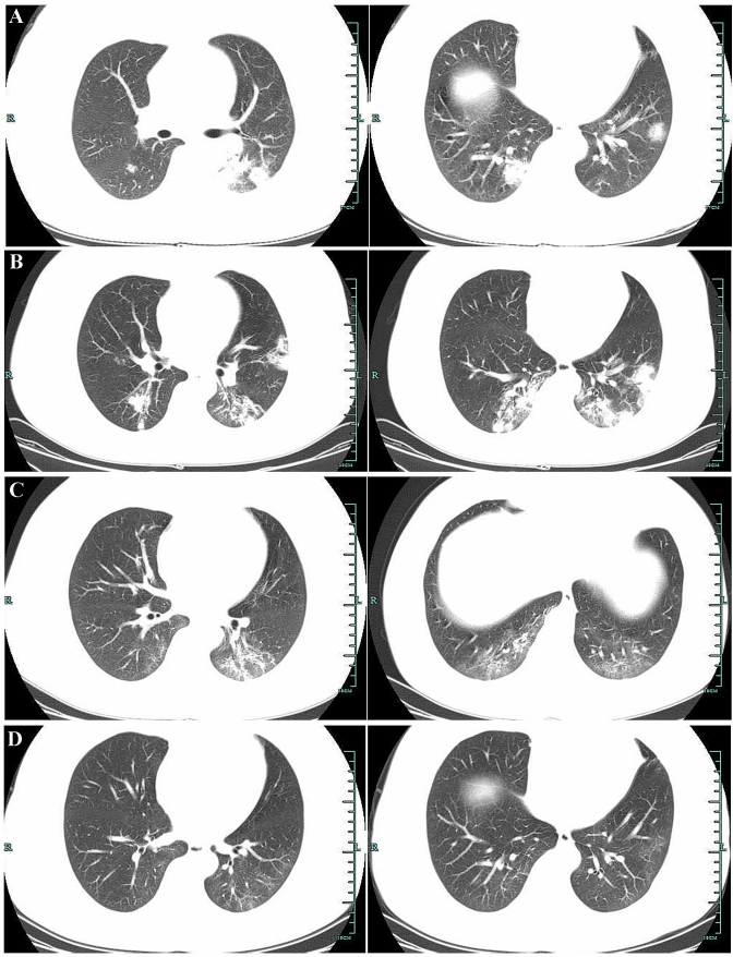
Fig. 1.
a Chest CT scans showed multiple flaky, consolidated lesions scattered throughout the right lower lobe, the left upper lobe apicoposterior segment, the lingula of the left lung, and the left lower lobe. b Chest CT showed some lesions had progressed. c Chest CT showed the bilateral lesions were significantly resolved when compared with the previous scans. d Chest CT showed the original focus was obviously absorbed

