Abstract
Purpose:
Beginning with the graduating class of 2018, the American Board of Surgery (ABS) requires that residents complete the ABS Flexible Endoscopy Curriculum, Fundamentals of Endoscopic Surgery (FES). This curriculum includes both didactic and simulator training. In the ideal setting residents gain proficiency using simulation prior to performing endoscopies in the clinical setting. This new requirement creates an increased demand for endoscopic simulators in all General Surgery residency programs. Due to the cost prohibitive nature of virtual reality simulators an economic alternative is needed.
Methods:
A mechanical simulator was created from inexpensive items easily acquired at a hardware store and in the hospital. Total cost of the simulator was approximately $120 USD. To validate the simulator, experienced endoscopists completed a training session with the device. A seven-question Likert scale survey (1 - strongly disagree to 5 - strongly agree) was completed after the session evaluated the simulated experience versus live upper endoscopies and the device’s ability to meet the goals of the FES curriculum.
Results:
Eight proficient endoscopists completed the training session and survey and agreed that the device closely replicated live colonoscopies and would meet all training requirements in the FES curriculum. Mean responses to all seven survey questions ranged from 3.8–4.4.
Conclusion:
This device is a cost-effective method for simulating live upper endoscopies and is appropriate for use in FES training.
Keywords: Endoscopy, Simulation, Residency, EGD simulator, General surgery residency
INTRODUCTION
Upper and lower endoscopies compose a vital portion of training for general surgical residents. The Accreditation Council of Graduate Medical Education requires that surgical residents complete 50 colonoscopies and 35 upper endoscopies prior to graduation.1 Proficiency on endoscopic simulators prior to live endoscopy has been studied in both upper and lower endoscopy. Though many different outcomes have been measured, residents who complete some form of simulator training routinely perform better than those without simulation when direct feedback is provided by proficient endoscopists at the time of training.2
Beginning with the graduating class of 2018 the American Board of Surgery (ABS) requires that all general surgery residents demonstrate proficiency in endoscopy, by completing the ABS Flexible Endoscopy Curriculum, to be eligible to sit for board examinations. This program includes the Society of American Gastrointestinal and Endoscopic Surgeons Fundamentals of Endoscopic Surgery Program (FES).1 A portion of the FES curriculum requires the use of realistic endoscopic simulators prior to live endoscopy. In the ideal setting residents demonstrate proficiency using simulation prior to beginning endoscopies in the clinical setting. Current simulator costs range from $6,000 for mechanical simulators to over $100,000 for virtual reality simulators.3,4
METHODS
In order to create a simulator that could be easily reproduced; all materials were sourced from the hospital itself or local stores. The total cost of all materials was $120.38 USD; recurring cost of $57.82 for each simulated mucosal layer and $62.56 in fixed costs for the head and base (Table 1). Time dedicated to assembly was less than 2 hours. Setup for each use is less than 10 minutes and the simulator mucosal layer is easily cleaned for use in multiple training sessions. Measurements from SEER training (Surveillance, Epidemiology, and End Results Program) were used to ensure realistic lengths for the simulator.
Table 1.
Simulator Cost Breakdown
| Fixed Costs | Recurring Costs | ||
|---|---|---|---|
| Foam Insulation | $32.52 | Transvaginal US Probe Cover | $5.10 |
| Gallon Jug | $0.99 | Foam Pads | Recycled |
| Size 5 LMA | $5.87 | Large Balloon | $1.99 |
| Small Plastic Ring | $2.99 | Breast Navigator Probe Cover | $50.73 |
| Flexible Discharge Tube | $11.65 | ||
| Endoscope and Tower | Hospital Owned | ||
| Rubber Bands | $2.89 | ||
| Elmer’s Glue Spots | $5.65 | ||
| Total fixed costs | $62.56 | Total recurring costs | $57.82 |
| Total cost | $120.38 | ||
The simulator was fashioned in three pieces, the base, the head, and the working portion or mucosal layer. The base and head were designed for repeated use and the mucosal layer was designed to be replaced as needed with wear and tear. A gallon milk jug was used for the head. The bottom of the jug was removed to allow access to the inside. The indented portion of the handle was cut on 3 sides leaving the portion closest to the cap intact. A small segment was cut from the threaded portion on top to permit the working portion, or mucosal layer, of the model to pass through. A mouth was cut opposite the handle to accommodate a bite block (Figure 1). Two small holes were created above the mouth and a string was passed through the handle and one end was brought out through either hole. This was used to tension the handle and create a palate for the endoscope to push against.
Figure 1.
Cut lines for gallon jug.
The mucosal layer was assembled in three portions: the esophagus, the stomach and the duodenum. The thin portion of a gamma probe cover, cut to 25 cm, with an appropriate diameter of 4.5 cm was used for the esophagus. A small plastic grommet with a 2 cm diameter was used as the upper esophageal sphincter (UES). One end of the cut probe cover was brought up through the gromet and secured around it with a small rubber band; this was taped to a number 5 laryngeal mask airway, which simulated the larynx and trachea. The simulated UES was approximately 18 cm from the incisors, the opening in the milk jug. When placed within the milk jug a space to simulate the piriform recess was created on either side of the esophagus (Figure 2).
Figure 2.
Top view of mucosal layer inserted into the gallon jug head.
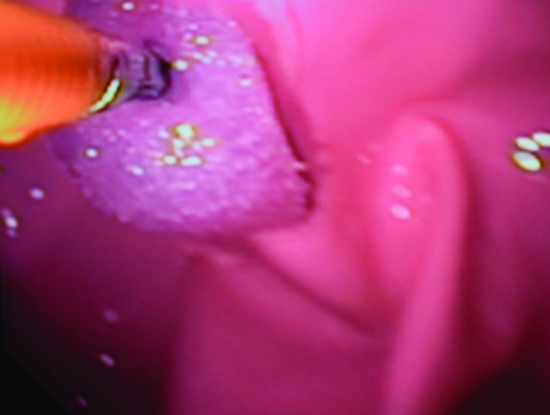
A small hole was cut at the top of a large party balloon (20 cm diameter) leaving an opening about 1.5 cm in diameter. The balloon was inverted, and the distal portion of the navigator probe cover was threaded through the hole cut in the top of the balloon. This was secured with rubber cement for an airtight seal. A rubber band just above the stomach served as a lower esophageal sphincter approximately 43 cm from the incisors. While the balloon was inverted small polyps fashioned from a foam pad were secured to the lumen of the stomach with Elmer’s glue spots. These would allow for instrumentation practice with biopsy forceps, injection needle, and snare biopsy (Figure 3). A vaginal ultrasound probe cover was secured to the end of the balloon with rubber cement after the balloon was returned to its original position. This served as the duodenal mucosa and the blind end of the ultrasound probe cover created an airtight system that would distend appropriately (Figure 4).
Figure 3.
Simulated polyp as seen during polypectomy.
Figure 4.
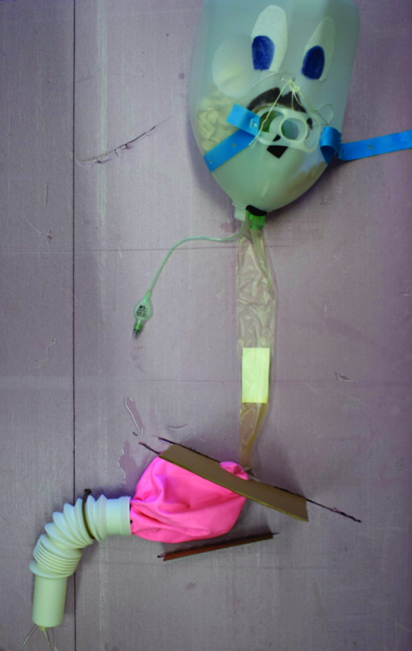
Assembled mucosal layer lying on assembled base and head.
A piece of 2” foam insulation was cut to 60 × 60 cm dimension and oriented in portrait fashion to be used as a base. The head was secured to the base through predrilled holes with heavy string in multiple locations to prevent movement. The mucosal layer was then passed through the top of the milk jug and out the bottom. The laryngeal mask airway and grommet at the UES were oriented appropriately and secured to the threaded portion of the milk jug with tape. A 2.5 cm hole was cut in a firm piece of cardboard and this was inserted into grooves in the base as a diaphragm. This could be moved inferiorly to simulate a hiatal hernia. The stomach was allowed to flop below the diaphragm; however, the duodenal mucosa was placed within flexible tubing that was secured to the base to simulate the fixed nature of the duodenum. Components were arranged on the base according to average anatomic distances according to the SEER database5 (Figure 5). To better simulate the gastrointestinal tract consistency, a slurry of lubricant and water was created in a cup and placed into the mucosal layer. The simulator was then used in conjunction with a hospital endoscope and tower.
Figure 5.
Assembled simulator ready for use.
A seven-question survey based on the FES curriculum was used to evaluate the effectiveness of the mechanical simulator as a teaching tool. Proficient endoscopists; including senior surgery residents, surgery attendings, gastroenterology fellows, and attendings completed a training session on the simulator. After the training session all endoscopists completed the seven-question Likert based survey (Figure 6). A mean and standard deviation was then obtained for each question.
Figure 6.
Survey.
RESULTS
Eight proficient endoscopists completed a training session on our model. On average the participants agree that our model replicates the skills necessary for upper endoscopy mean 3.8 (SD 0.66). Moreover, endoscopists agree the simulator is effective for teaching difficult maneuvers such as esophageal intubation mean 3.9 (SD .78), keeping a clear endoscopic view mean 4.4 (SD .70), and instrumentation techniques mean 4.1 (SD .78). Overall endoscopists strongly agree the model is useful for training mean 4 (SD .71) (Table 2).
Table 2.
Survey Responses
| Survey Question | Mean Response (Standard Deviation) |
|---|---|
| 1. This simulator closely replicates the skills necessary for live upper endoscopy. | 3.8 (.66) |
| 2. This simulator would be effective to teach residents/fellows esophageal intubation. | 3.9 (.78) |
| 3. This simulator would be effective to teach residents/fellows scope navigation including advancement/withdrawal, tip deflection and torque. | 4.0 (.71) |
| 4. This simulator would be effective to teach residents/fellows to keep a clear endoscopic field. | 4.4 (.70) |
| 5. This simulator would be effective to teach residents/fellows instrumentation. (i.e. biopsy and snare polypectomy, epi injection, etc.) | 4.1 (.78) |
| 6. This simulator could be used to effectively evaluate residents/fellows for efficiency and quality of examination during upper endoscopy. | 3.8 (.66) |
| 7. This simulator would be highly useful in the training of residents/fellows. | 4.0 (.71) |
DISCUSSION
Currently virtual reality simulators are becoming increasingly prevalent in the training of residents. Mechanical simulators are also available to residency programs. Both come at a significant cost both initially and in the maintenance of the simulators.6 Inexpensive lower endoscopy simulators and task specific simulators have been previously described in the literature; however, to our knowledge this is the first resident fabricated upper endoscopy simulator that has been described.7,8 While task specific simulators and lower endoscopy simulators can aid in training for basic scope control, certain tasks require a dedicated upper endoscopy simulator such as esophageal and duodenal intubation. This model will allow for upper endoscopy specific maneuvers. While there are simulators available or easily constructible to replicate the colon and to practice general scope manipulation, our model is important for training on upper endoscopy specific tasks.
CONCLUSION
Simulation prior to live procedures leads to better proficiency and patient comfort. Furthermore, the ACS now requires residents to show proficiency in endoscopy by completing the FES curriculum, which requires simulation prior to live endoscopy. With the price of simulators ranging from $6000 USD for mechanical models, with additional costs for consumables, to greater than $100,000 USD for virtual reality simulators there is a need for a high efficiency, low-cost model for community residency programs. This model is effective for teaching specialized maneuvers associated with upper endoscopy and is easily assembled by residents in less than 2 hours for a cost of less than $150 USD.
Contributor Information
Matthew J. Deere, Department of Surgery, Mclaren Greater Lansing, Lansing, Michigan..
Mark W. Jones, Department of Surgery, Mclaren Greater Lansing, Lansing, Michigan..
Leandra A. Jelinek, Department of Surgery, Mclaren Greater Lansing, Lansing, Michigan..
Werner H. Henning, Department of Surgery, Mclaren Greater Lansing, Lansing, Michigan..
References:
- 1.American Board of Surgery. The American Board of Surgery Booklet of Information. 2018. http://absurgery.org/xfer/BookletofInfo-Surgery.pdf. Accessed June 28, 2018.
- 2.Mahmood T, Scaffidi MA, Khan R, Grover SC. Virtual reality simulation in endoscopy training: current evidence and future directions. World J Gastroenterol. 2018;24(48):5439–5445. [DOI] [PMC free article] [PubMed] [Google Scholar]
- 3.Anatomy Warehouse. Koken EGD simulator. https://www.anatomywarehouse.com/koken-egd-simulator-a-105294. Accessed November 4, 2019.
- 4.Bittner JG, Marks JM, Dunkin BJ, Richards WO, Onders RP, Mellinger JD. Resident training in flexible gastrointestinal endoscopy: a review of current issues and options. J Surg Educ. 2007;64(6):399–409. [DOI] [PubMed] [Google Scholar]
- 5.National Cancer Institute. Anatomy of the Esophagus. https://training.seer.cancer.gov/ugi/anatomy/esophagus.html. Accessed November 4, 2019.
- 6.Blackburn SC, Griffin SJ. Role of simulation in training the next generation of endoscopists. World J Gastrointest Endosc. 2014;6(6):234–239. [DOI] [PMC free article] [PubMed] [Google Scholar]
- 7.Jones MW, Deere MJ, Harris JR, Chen AJ, Henning WH. Fabrication of an inexpensive but effective colonoscopic simulator. JSLS. 2017;21(2):e2017.00002. [DOI] [PMC free article] [PubMed] [Google Scholar]
- 8.King N, Kunac A, Johnsen E, Gallina G, Merchant AM. Design and validation of a cost-effective physical endoscopic simulator for fundamentals of endoscopic surgery training. Surg Endosc. 2016;30(11):4871–4879. [DOI] [PubMed] [Google Scholar]


