Abstract
An increasingly popular method for post-operative rehabilitation of an ACL reconstruction, as a substitute for traditional therapy, is blood flow restriction therapy (BFR). BFR therapy utilizes a pneumatic cuff to simulate strenuous exercise in an effort to stimulate muscle recruitment, mitigate atrophy, and promote hypertrophy in patients with load-bearing limitations. Because this is a relatively new form of therapy, there is a lack of established literature and protocol that is preventing widespread use of the therapy. This article will seek to confirm the value and validity of the utilization of BFR therapy. In order to validate the utilization of BFR, an evaluation of the science underlying BFR will be discussed as well as the technique and exercises preformed during therapy. Furthermore, analysis of other BFR literature will be utilized to lend further credence to the obtained conclusions. Based on the literature, BFR therapy mitigates atrophy through type II muscle recruitment while also stimulating hypertrophy in patients, supporting its use post-operatively. Moreover, positive results from BFR case series also lend credence to its value as a substitute for traditional therapy in patients who have weight-bearing limitations, specifically those who are recovering from anterior cruciate ligament reconstructions.
Key Words: ACL, lood flow restriction, econstruction, herapy
Introduction
Blood flow restriction therapy (BFR) has become an increasingly popular method of post-operative rehabilitation. Notably, this therapeutic method is growing more prolific particularly following anterior cruciate ligament (ACL) reconstruction. The American College of Sports Medicine recommends that, in order to maintain and increase optimal muscle strength, the muscle must be stressed with between 60 and 100 percent of the one repetition maximum (1).
However, load bearing is limited in post-operative knee reconstruction patients leading to muscular atrophy, a post-operative condition connected with pain and muscle weakness. BFR therapy allows for clinicians and patients to work in a low-load bearing environment while still being able to achieve the necessary musculoskeletal strengthening, avoiding the traditional muscle atrophy often prevalent following reconstructive knee surgeries. Because of this, BFR has become an increasingly attractive option. BFR utilizes the application of a pneumatic cuff which is placed at the most proximal location of the involved limb (2). The cuff is then set to a certain pressure to provide venous occlusion to the targeted muscle groups allowing for reduction of muscle atrophy (2). However, due to the limited evidence available, as well as a lack of established protocols, BFR has not been widely adapted. This article will seek to examine and reassert the value and validity of BFR therapy in patients recovering from ACL reconstructions.
Body Text
Historical Background
The first documented concept and practice of vascular occlusion moderation therapy occurred in the 1970s in Japan by Dr. Yoshiaki Soto. Soto’s discovery dubbed Kaatsu utilized bands and ropes to create a tourniquet in order to restrict muscular venous blood flow. Kaatsu, while unsophisticated, paved the way for the development of electric tourniquets as well as modern blood flow restriction therapy. The first study on BFR was published in 1998 with the development and utilization of electric tourniquets (3). In the early 2000’s, with the help of third generation tourniquets allowing BFR to be performed safely and accurately, blood flow restriction therapy was further investigated in patients who had restrictions with the amount of resistance they could withstand while exercising, specifically those who were elderly (4). These early investigations tried to distinguish BFR therapy as a tool for mitigating muscular atrophy in individuals with weight-bearing limitations. Recently, BFR has been utilized as a substitute for traditional postoperative therapy in patients recovering from a range of musculoskeletal injuries. BFR gained popular recognition through a published case series by Johnny Owens (5). The authors utilized BFR therapy to strengthen patients who experienced severe limb trauma causing lower extremity weakness with positive results in terms of muscle hypertrophy and recovery of range of motion (5). Consequently, both physicians and therapists have begun to recognize the potential of BFR therapy in treating musculoskeletal injuries and have started a shift towards widespread use of BFR in post-operative rehabilitation.
Mechanism of Action
Venous occlusion creates an anaerobic environment within the targeted muscles. At this lower oxygen level, the body utilizes muscle fibers that are almost exclusively reserved for strenuous activities, such as type II muscles (2). Stress induced on these muscle fibers leads to up-regulation of the muscle hypertrophy-signaling cascade by increased protein synthesis, proliferation of myogenic satellite cells, and activation of type II muscle fibers, mitigating the atrophic effects (6-9). As previously shown, many BFR exercises are done at 20% to 30% of the one repetition maximum. When doing these BFR exercises at 20/30% of the one repetition maximum, muscle change is observed equivalent to the muscle change observed in an individual exercising at 80% of their one repetition maximum (10-12)
Blood Flow Restriction Technique
In utilizing BFR therapy, a standard postoperative rehabilitation protocol was implemented following the guidelines provided by the Owens Recovery Science. For the therapy, there are three distinct phases: the protected phase (weeks one to six), the second phase (weeks six to twelve), and the third phase (three months and beyond). The pneumatic cuff should be placed more proximal than distal to mitigate neurovascular injury. In addition, for the traditional strengthening BFR exercises, four sets should be performed with reps of 30/15/15/15 including 30 second rest interval between each set with the BFR cuff still inflated. After the completion of exercise, the cuff is deflated for one minute to allow for reperfusion before continuing with the strengthening exercise (5). BFR exercises should be completed three times a week with the overall goal of mitigating atrophy while promoting hypertrophy.
Phase One
Phase one, also known as the protected phase, begins in the first week post-operatively after day three. The goals of this phase are pain control, reducing effusion within the joint, restoring range of motion, and maintaining muscular and aerobic endurance. BFR is utilized for a max of 25 minutes during the first phase. For phase one, the exercises are separated into three subsets: weeks one to two, weeks two to four, and weeks four to six.
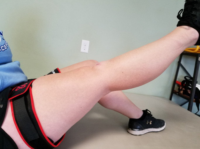
For weeks one to two, neuromuscular electrical stimulation at 10-20% maximal voluntary contraction is performed for ten minutes with the pad placement distal to the pneumatic cuff on the vastus laterals and the vastus medialis oblique. The exercise is run with a ten second contraction followed by 20 second rest. Furthermore, the leg is started in full extension before progressing to isometric holds with the leg in 60 to 90 degrees flexion. The second BFR exercise performed is hip adduction while the patients lies on their side. When starting, no weights are present but as the patient progresses through the week, cuff weights should be added. The third exercise performed is bent knee ankle plantar flexion with a light elastic band that is increased for more resistance as the patient progresses. Per Figure 1, the final BFR exercise of the first two weeks is straight leg raises which once again, begin with no weights with patient progression dictating when cuff weights are added.
Figure 1.
Unweighted straight leg raise with the pneumatic cuff. This is done as well with weights as the patient progresses
For weeks two-four, two new additional exercises are added in addition to the continuation of the exercises from weeks one through two. The exercises from the previous weeks are kept the same with no deviation from how they were performed. One additional exercise added is prone hip extension. This exercise is usually performed with no weights but as the patient progresses, cuff weights are added. Another additional exercise is lifting and straightening one leg to 30-90 degrees know as a long arc quad. This exercise should be performed with no anterior knee pain and no weights until week four.
During weeks four-six, straight leg raises, side-lying hip abduction, and long arc quad continue to be performed by the patient with the same protocol. Again, new BFR exercises are implemented. Bilateral bridging is performed with avoidance of joint line pain. Leg presses are also performed at 0 to 60 degrees with weight at 25% body weight. After the sixth week, progression is made to the second phase of BFR therapy.
Phase Two
Phase two places an emphasis on improving strength to progress to a normal, weight-bearing gait as well as achieving full range of motion. Many of the exercises are performed with weights approximately 30% of the one repetition maximum. These exercises mitigate atrophy while beginning the hypertrophic phase. Similar to phase one, phase two is separated into two subsets: weeks 6-10 and weeks 10-12.
For weeks 6-10, three BFR exercises are performed in order to strengthen the surrounding muscles. Leg presses at 25% of body weight or 30% of one repetition maximum are performed. Furthermore, resisted hamstring curls are also performed with weights approximately 30% of the one repetition maximum. The final BFR exercise for weeks 6-10, an exercise bike is utilized to build up endurance. The target for the exercise bike is 15 minutes at a resistance level three.
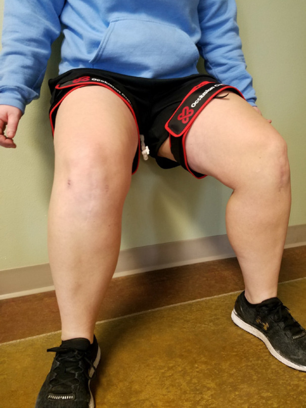
The final section of phase two, weeks 10-12, continues with the exercise bike in building the patient’s endurance. However, squatting is implemented into the exercise regimen. Per Figure 2, regular squatting as well as split squatting are performed at weights 30% of the estimated one repetition maximum. At this point, the patient has regained enough muscle to move onto phase three.
Figure 2.

Wall sit/squat where the patient is using the wall for support to ensure equa loading
Phase three
Phase three rehabilitation begins three months and beyond status post ACL reconstruction. This involves rotating from high interval intensity training (HIIT) to BFR training until the patient can do HIIT without pain or symptoms. At this point, one weans the patient off of BFR until HIIT is done exclusively.
Discussion
The goal of blood flow restriction therapy is to prevent muscle atrophy, regain endurance, and restore range of motion in patients recovering from ACL reconstructions. Whether in athletes or individuals performing their occupation, muscle strength must be achieved in order to complete extended periods of physical activity. As previously mentioned, the American College of Sports Medicine recommends, that in order to strengthen muscle and maintain muscular strength, resistance exercises should be performed at 60 to 100 percent of the one repetition maximum (1). By exercising at these resistances, individuals obtain type II muscle recruitment leading to increased muscle hypertrophy (6). In the absence of type II muscle recruitment, atrophy can result, causing stark limitations in the post-operative recovery. Atrophy results from a low load bearing state in which the down regulation of hypertrophic signaling cascade occurs as well as the activation of cascades that cause muscle protein degradation (2).
As many postoperative ACL reconstruction patients are unable to tolerate high load bearing states, it is paramount that type II muscle recruitment and muscle strengthening is achieved in a manner that benefits the patient. Multiple studies have confirmed the benefits of blood flow restriction therapy in a post-operative ACL reconstruction setting [able 1]. These studies assert that BFR therapy represents a suitable and positive alternative to traditional methods. BFR therapy is not hindered by traditional limitations. BFR therapy achieves hypertrophy in postoperative patients through venous occlusion of the affected extremity by a pneumatic cuff. As a result of BFR therapy, patients who are unable to tolerate heavier loads receive the strengthening necessary to avoid muscular atrophy, specifically those that are type II fiber specific, while also reducing pain and adverse joint loading. Owens et al, as well as Takarada et al, demonstrated and provided key examples of the success of BFR therapy in mitigating the effects of atrophy on patients with lower extremity trauma while positively stimulating muscle hypertrophy through proper implementation of rehabilitative BFR protocol (4, 5).
Table 1.
Previous Studies on Blood Flow Restriction Therapy and Its Use in Post-Operative Setting
| Title | Author | Date | Result |
|---|---|---|---|
| Low-load Resistance Muscular Training with Moderate Restriction of Blood Flow After Anterior Cruciate Ligament Reconstruction | H Ohta; H Kurosawa; H Ikeda; Y Iwase; N Satou; S Nakamura | 2003 | Findings from the study show that low-load resistance muscular training during moderate restriction of blood flow is an effective exercise for early muscular training after reconstruction of the anterior cruciate ligament. |
| Blood Flow Restriction Training in Clinical Musculoskeletal Rehabilitation: A Systematic Review and Meta-Analysis | L Hughes; B Paton; B Rosenblatt; C Gissane; SD Patterson | 2017 | Findings from the 20 eligible studies show that low-load BFR training is more effective, tolerable, and therefore a potential clinical rehabilitation tool. |
| Effect of Blood Flow Restriction Training on Quadriceps Muscle Strength, Morphology, Physiology, and Knee Biomechanics Before and After Anterior Cruciate Ligament Reconstruction: Protocol for a Randomized Clinical Trial | LN Erickson; KCH Lucas; KA Davis; CA Jacobs; KL Thompson; PA Hardy; AH Anderson; CS Fry; BW Noehren | 2017 | Findings from the study show that BFR therapy is a suitable option for improved targeted treatment for protracted quadriceps strength loss associated with ACL injury and reconstruction. |
| Blood Flow Restriction Therapy After Surgery: Indications, Safety Considerations, and Postoperative Protocol | N DePhillipo; M Kennedy; Z Aman, A Bernhardson; L O’Brien; R LaPrade | 2018 | Findings show that the current literature indicates that BFR is a safe intervention that may improve muscle strength and atrophy after knee surgery compared with traditional therapy. |
| Comparing the Effectiveness of Blood Flow Restriction and Traditional Heavy Load Resistance Training in the Post-Surgery Rehabilitation of Anterior Cruciate Ligament Reconstruction Patients: A UK National Health Service Randomized Controlled Trial | L Hughes; B Rosenblatt; F Haddad; C Gissane; D McCarthy; T Clarke; G Ferris; J Dawes; B Paton; SD Patterson | 2019 | Findings show that BFR resistance training can improve skeletal muscle hypertrophy and strength to a similar extent to traditional heavy load resistance training with a greater reduction in joint pain and effusion, leading to greater overall improvements in physical function. |
Furthermore, BFR therapy provides physicians and therapists with a process that contains very few side effects. Concerns associated with venal occlusion in postoperative ACL reconstructions revolve around blood clotting and the possibilities of embolisms. However, DePhillipo et al observed in their investigation of BFR a lack of potential side effects such as thromboembolism and ischemic changes in the extremities (14). Moreover, the principal limitation of BFR therapy application observed was a possible inadvertent increase in pain during treatment although this may be ascribed to cuff width (14). Given the lack of limitations and concerns associated with BFR, it becomes an increasingly viable method of rehabilitation.
Although adoption of blood flow restriction therapy on a widespread level has not occurred, it remains an increasingly viable and efficient method for both physicians and therapists to utilize in postoperative ACL reconstruction patients. With the increasing amount of literature being published on the topic with generally positive results, BFR is becoming increasingly adopted by the orthopedic community. Future research should be performed to obtain a universally acknowledged rehabilitation program for the application of BFR and limit any adverse side effects that occur from this form of therapy. Furthermore, it would be useful to have a randomized clinical trial to compare BFR to traditional therapy in order to further understand its impact on musculoskeletal patients. Additional research should also be done to test the validity of preoperative BFR training in patients prior to ACL reconstructions as an additional modality to increase muscle mass and strength, thereby potentially aiding recovery.
References
- 1.Ratamess NA, Alvar BA, Evetoch TE, Housh TJ, Ben Kibler W, Kraemer WJ, Triplett NT. Progression models in resistance training for healthy adults. Medicine and science in sports and exercise. 2009;41(3):687–708. doi: 10.1249/MSS.0b013e3181915670. [DOI] [PubMed] [Google Scholar]
- 2.Meyer RA. Does blood flow restriction enhance hypertrophic signaling in skeletal muscle? Journal of applied physiology. 2006;100(5):1443–4. doi: 10.1152/japplphysiol.01636.2005. [DOI] [PubMed] [Google Scholar]
- 3.Kouzaki M, Yoshihisa T, Fukunaga T. Efficacy of tourniquet ischemia for strength training with low resistance. European journal of applied physiology and occupational physiology. 1997;77(1-2):189–91. doi: 10.1007/s004210050319. [DOI] [PubMed] [Google Scholar]
- 4.Takarada Y, Takazawa H, Sato Y, Takebayashi S, Tanaka Y, Ishii N. Effects of resistance exercise combined with moderate vascular occlusion on muscular function in humans. Journal of applied physiology. 2000;88(6):2097–106. doi: 10.1152/jappl.2000.88.6.2097. [DOI] [PubMed] [Google Scholar]
- 5.Hylden C, Burns T, Stinner D, Owens J. Blood flow restriction rehabilitation for extremity weakness: a case series. J Spec Oper Med. 2015;15(1):50–6. [PubMed] [Google Scholar]
- 6.Abe T, Sakamaki M, Fujita S, Ozaki H, Sugaya M, Sato Y, et al. Effects of low-intensity walk training with restricted leg blood flow on muscle strength and aerobic capacity in older adults. Journal of geriatric physical therapy. 2010;33(1):34–40. [PubMed] [Google Scholar]
- 7.Adams GR, Cheng DC, Haddad F, Baldwin KM. Skeletal muscle hypertrophy in response to isometric, lengthening, and shortening training bouts of equivalent duration. Journal of Applied Physiology. 2004 doi: 10.1152/japplphysiol.01162.2003. [DOI] [PubMed] [Google Scholar]
- 8.Gundermann DM, Walker DK, Reidy PT, Borack MS, Dickinson JM, Volpi E, et al. Activation of mTORC1 signaling and protein synthesis in human muscle following blood flow restriction exercise is inhibited by rapamycin. American Journal of Physiology-Endocrinology and Metabolism. 2014;306(10):E1198–204. doi: 10.1152/ajpendo.00600.2013. [DOI] [PMC free article] [PubMed] [Google Scholar]
- 9.Nielsen JL, Aagaard P, Bech RD, Nygaard T, Hvid LG, Wernbom M, et al. Proliferation of myogenic stem cells in human skeletal muscle in response to low-load resistance training with blood flow restriction. The Journal of physiology. 2012;590(17):4351–61. doi: 10.1113/jphysiol.2012.237008. [DOI] [PMC free article] [PubMed] [Google Scholar]
- 10.Cook SB, Clark BC, Ploutz-Snyder LL. Effects of exercise load and blood-flow restriction on skeletal muscle function. Medicine and science in sports and exercise. 2007;39(10):1708–13. doi: 10.1249/mss.0b013e31812383d6. [DOI] [PubMed] [Google Scholar]
- 11.Takarada Y, Sato Y, Ishii N. Effects of resistance exercise combined with vascular occlusion on muscle function in athletes. European journal of applied physiology. 2002;86(4):308–14. doi: 10.1007/s00421-001-0561-5. [DOI] [PubMed] [Google Scholar]
- 12.Yamanaka T, Farley RS, Caputo JL. Occlusion training increases muscular strength in division IA football players. The Journal of Strength & Conditioning Research. 2012;26(9):2523–9. doi: 10.1519/JSC.0b013e31823f2b0e. [DOI] [PubMed] [Google Scholar]
- 13.Schoenfeld BJ. Is there a minimum intensity threshold for resistance training-induced hypertrophic adaptations? Sports Medicine. 2013;43(12):1279–88. doi: 10.1007/s40279-013-0088-z. [DOI] [PubMed] [Google Scholar]
- 14.DePhillipo NN, Kennedy MI, Aman ZS, Bernhardson AS, O’Brien LT, LaPrade RF. The role of blood flow restriction therapy following knee surgery: expert opinion. Arthroscopy: The Journal of Arthroscopic & Related Surgery. 2018;34(8):2506–10. doi: 10.1016/j.arthro.2018.05.038. [DOI] [PubMed] [Google Scholar]


