Abstract
TEA domain transcription factor 4 (TEAD4) is an important member of the TEAD family. As a downstream effector of the Hippo pathway, TEAD4 has essential roles in cell proliferation, cell survival, tissue regeneration, and stem cell maintenance. TEAD4 contains a TEA DNA binding domain that binds the promoters of target genes and a Yes-associated protein/transcriptional co-activator with PDZ-binding motif (YAP/TAZ) binding domain that associates with transcriptional cofactors. TEAD4 coordinates with YAP, TAZ, VGLL, and other transcription factors to regulate different cellular processes in cancer via its transcriptional output. Moreover, TEAD4 undergoes post-translational modifications and subcellular translocations, and both processes have been shown to shed new insights on how TEAD transcriptional activity can be modified. In summary, TEAD4 has important roles in cancer, including epithelial–mesenchymal transition (EMT), metastasis, cancer stem cell dynamics, and chemotherapeutic drug resistance, suggesting that TEAD4 may be a promising prognostic biomarker in cancer.
Keywords: TEAD4, Hippo pathway, YAP, cancer, targeted therapy
Introduction
TEAD transcription factors are downstream effectors of the Hippo pathway, and they regulate cell proliferation, tissue regeneration, and metastasis.1 In mammals, the TEAD family consists of TEAD1, TEAD2, TEAD3, and TEAD4. TEADs are widely expressed in different tissues, though each member also shows tissue-specific expression, indicating that they have both common functions (regulating cell proliferation and contact inhibition)1–3 and distinct functions (heart development,4 neural development,5 and trophectoderm lineage determination).6–8
TEAD4, also known as transcriptional enhancer factor-3 (TEF-3), is a key member of the TEAD family. TEAD4 is highly expressed in skeletal muscle, initial studies focused on its role in blastocyst formation6 and reported that TEAD4 is required for specification of the trophectoderm lineage in preimplantation embryos.6–8 Recently, TEAD4 was demonstrated to be a novel prognostic marker of gastric cancer,9 breast cancer,10 colorectal cancer,11 melanoma,12 laryngeal cancer,13 and head–neck squamous cell carcinoma.14 Apart from its role in the canonical Hippo–YAP/TAZ pathway, other studies have reported its participation in post-translational modifications,9,15 crosstalk between cancer-related signaling pathways,16 and cancer-causing mutations.17,18 In addition, targeting upstream molecules in the Hippo pathway may bring about unwanted side effects.3 Thus, TEAD4 is an ideal druggable target for cancer. This review highlights the important roles of TEAD4 in cancer biology and provides new insights into TEAD4 regulation in cancer.
Structure of TEAD4
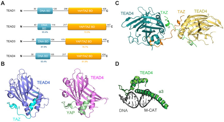
TEAD is a downstream transcriptional factor in the Hippo pathway. However, TEAD does not exhibit transcriptional activity in the absence of critical cofactors.19 The C-terminal YAP/TAZ binding domain of TEAD, which with high identity across all TEAD family members, associates with transcriptional cofactors such as YAP, TAZ, and VGLL.1 The structure of TEAD4 is shown in Figure 1A. The YAP/TAZ binding domain of TEAD4 contains an immunoglobulin-like fold, whereas the N-terminus of the coactivator YAP interacts with TEAD4 through two helixes and an intervening loop that contains the PXXΦP motif (X, any residue; Φ, hydrophobic residue) to trigger transcriptional activity (Figure 1B).18,20 Interestingly, the TAZ binding domain can associate with TEADs through two distinct mechanisms. In the first mechanism, TAZ binds to TEAD4, which is similar to the binding of YAP to TEAD4, and this binding involves helix α1, helix α2, and a loop in between (Figure 1B). In the second mechanism, two TAZ molecules straddle two TEAD molecules to form a heterotetramer (Figure 1C).21
Figure 1.
(A) The overall structure of TEADs. TEADs consist of a TEA DNA binding domain (blue) and a YAP/TAZ binding domain (yellow). The percent represents the identity for each domain of TEADs compared to that of TEAD1. (B) YAP binds to TEAD4 via two short helixes and an extended loop containing the PXXΦP motif. TAZ interacts with TEAD4 in a similar manner to the binding of TEAD4-YAP. (C) Two TAZ molecules straddle two TEAD molecules to form a heterotetramer. (D) The α3 helix of the TEA domain (green) binds to the M-CAT DNA duplex (grey).
Notes: Images Figure 1B and C are adapted from Kristal Kaan HY, Chan SW, Tan SKJet al Crystal structure of TAZ-TEAD complex reveals a distinct interaction mode from that of YAP-TEAD complex. Sci Rep. 2017;7(1):1–11. 10.1038/s41598-017-02219-9. Creative Commons license and disclaimer available from: http://creativecommons.org/licenses/by/4.0/legalcode.21Figure 1D reproduced from Holden JK, Cunningham CN. Targeting the hippo pathway and cancer through the TEAD family of transcription factors. Cancers (Basel). 2018;10 (3). 10.3390/cancers10030081. Creative Commons license and disclaimer available from: http://creativecommons.org/licenses/by/4.0/legalcode.20
The N-terminal DNA binding domain of TEADs is evolutionarily conserved.22 The DNA binding domain, which is responsible for TEAD–YAP/TAZ complex formation and transcriptional regulation, was initially reported to bind the M-CAT (5ʹ-CATTCCT-3ʹ) regulatory element of the simian virus 40 (SV40) enhancer and to activate transcription.23,24 The DNA recognition helix, namely, the α3 helix, determines the specificity of the TEA domain binding to a DNA sequence. By structure-guided biochemical analysis, two binding sites were identified at the interface of the TEAD4–DNA complex, with the sites binding to the minor and major grooves of DNA, respectively (Figure 1D).17
Regulatory Mechanism of TEAD4 in Cancer
Role of TEAD4 in the Hippo–YAP/TAZ Pathway
The Hippo pathway is evolutionarily conserved in mammals, where it regulates cell fate and tissue growth.25,26 YAP and TAZ are critical effectors in the Hippo pathway.25 When phosphorylated by upstream kinases LATS1 and LATS2, YAP and TAZ accumulate in the cytoplasm and cannot bind TEAD, which induces their ubiquitin-mediated proteolysis and autophagy-induced degradation.25,27 Upon dephosphorylation, YAP and TAZ translocate to the nucleus, where they interact with TEADs to drive the transcription of a variety of target genes involved in cell proliferation, metastasis, and apoptosis.1 Importantly, approximately 80% of TEAD4-bound promoters and enhancer regions were found to overlap with YAP and TAZ.28 Similar findings were also reported for their paralogs TEAD1 and TEAD2,28–30 indicating the important roles of YAP and TAZ as TEAD coactivators.
YAP and TAZ are paralogs that have similar domain structures and partially redundant functions.31 Specifically, they exhibit differences in their physiological functions and downstream targets.31,32 It was reported that YAP inhibition had a stronger influence than TAZ inhibition on a variety of cellular processes such as cell spreading, cell volume control, glucose uptake, cell proliferation, and cell migration.31 Furthermore, a recent study revealed that TAZ, but not YAP, formed biomolecular condensates through liquid–liquid phase separation to compartmentalize TEAD4, coactivators, and the elongation machinery, which included BRD4, MED1, and CDK9, to efficiently and specifically activate transcription.33
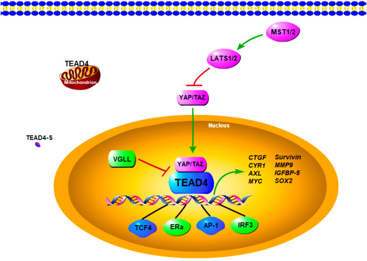
Downstream targets extend the roles of the TEAD–YAP/TAZ complex in the regulation of cancer stem cell renewal, tumorigenesis, cell metastasis, epithelial–mesenchymal transition (EMT), and tissue growth. In addition to well-studied TEAD target genes, such as CTGF, CYR61, and AXL,29 it was reported that survivin,34 vimentin,11 MYC,35 MMP9,36 IGFBP-5,37 and SOX238 are directly targeted by the TEAD4–YAP/TAZ complex (Figure 2).
Figure 2.
The regulatory mechanisms of TEAD in cancer biology. Upstream Hippo signaling pathway and downstream transcriptional outputs of TEAD4. TEAD4 associates with transcriptional factors such as TCF-4, ERα, AP-1, and IRF3 to enhance the stability of the transcriptional complex. The TEAD4 transcriptional outputs play critical roles in cancer progression, metastasis as well as stem cell maintenance. VGLL proteins directly compete with YAP for TEAD4 binding. Tead4 is the unique member of the TEAD family to localize to the mitochondrion and maintain mitochondrial energy homeostasis. TEAD4-S is a truncated isoform of TEAD4 spread in both the nucleus and cytoplasm, which inhibits proliferation and EMT in cancer cells.
Interaction with VGLL Proteins
The vestigial-like (VGLL) family consists of four members, VGLL1-4, all of which contain a TONDU domain that associates with TEADs.39 VGLL1 and VGLL4 were demonstrated to compete with YAP for direct binding to TEAD416,37,40,41 (Figure 2).
The structure of the VGLL1–TEAD4 complex has been determined. Although the primary sequence of VGLL1 is different from those of YAP and TAZ, VGLL1 similarly binds to TEAD4, interacting with the same region of TEAD4. In addition, the VGLL1–TEAD4 complex upregulates the expression of IGFBP-5, thereby facilitating anchorage-independent prostate cancer cell proliferation.37 In another study, the VGLL1–TEAD4 complex was reported to be regulated by PI3K/AKT/β-catenin signaling, which activated the transcription of MMP9, a key molecule that promotes gastric cancer cell proliferation and metastasis.36 Furthermore, VGLL4 inhibits the formation of the YAP–TEAD complex and negatively regulates its activity in lung carcinogenesis.42 TEAD4 was reported to directly bind to TCF4, a transcription factor in the Wnt/β-catenin pathway, thereby forming a complex with target genes, whereas VGLL4 was demonstrated to associate with the TEAD4–TCF4 complex to inhibit the transcriptional activation of TCF4 and TEAD4. Collectively, these findings reveal a novel mechanism for VGLL4 in the co-regulation of Hippo and Wnt pathways.16 In summary, VGLL1 functions as a TEAD4 coactivator and facilitates cell proliferation, as well as metastasis, whereas VGLL4 functions as a tumor suppressor and positively correlates with overall survival.
Cooperation with Transcription Factors
The results of recent high-throughput binding assays revealed that transcriptional factor cooperativity is a common cellular phenomenon,43 which has greatly enhanced our understanding of the interplay between transcription factors. As previously discussed, TEAD4 interacts with TCF4 to form a complex for cobind target genes.16 IRF3 is a member of the interferon regulatory transcription factor (IRF) family, which associates with the YAP–TEAD4 complex in the nucleus where it binds and regulates a variety of target genes such as CTGF, CYR61, and AXL.44 In addition, ERα is one of two main types of estrogen receptor, and it is also a key transcription factor in breast cancer.45 The YAP–TEAD4 complex co-regulates ERα for estrogen-regulated enhancer activation, indicating that a non-canonical TEAD4 mechanism is responsible for breast cancer growth.10 On the other hand, activator protein-1 (AP-1, a dimer of JUN and FOS) is a transcription factor and proto-oncogene.46 By ChIP-seq analysis, AP-1 was demonstrated to co-occupy chromatin with the TEAD–YAP/TAZ complex and to promote breast cancer cell proliferation.28 Further studies reported that the TEAD4–AP1 complex is widespread across a broad range of tumors including neuroblastoma, colon cancer, lung cancer, and endometrial cancer.47 The TEAD4–AP1 complex also regulates the activity of the Dock-Rac/CDC42 module and activates target genes that promote cell migration and invasion.47 In summary, TEAD4 can cooperatively bind DNA and transcription factors, which stabilizes the transcriptional complex (Figure 2).
Other Hippo-Independent Mechanisms
There is convincing evidence on the roles of YAP and TAZ as TEAD coactivators. However, there are few studies on the role of TEAD4 in cancer independent of YAP and TAZ. In breast cancer, for instance, glucocorticoids promote nuclear translocation and transcriptional activation of TEAD4. At the same time, glucocorticoids activate the glucocorticoid receptor, thereby forming a complex that is recruited to the TEAD4 promoter to upregulate its expression, indicating that glucocorticoid receptor signaling triggers the activation of TEAD4 in a Hippo-independent manner. In addition, the glucocorticoid receptor-TEAD4 complex mediates cell survival, metastasis, and chemotherapeutic drug resistance both in vitro and in vivo.48 Moreover, TEAD4 promotes EMT and the expression of vimentin, which enhances the role of TEAD4 in colorectal cancer cell shape and migration. However, YAP knockdown in colorectal cancer cells did not abolish the expression of vimentin, and YAP could not regulate EMT and metastasis.11 Taken together, these findings provide new insights into the roles of YAP in different types of cancers.
Molecular Mechanisms Determining TEAD4 Activity
Subcellular Localization
The nuclear accumulation of TEAD4 is essential for its ability to mediate transcriptional activation.1 Therefore, it is necessary to identify the factors involved in the nucleocytoplasmic shuttling of TEAD4. A variety of conditions can inhibit the activity and restrict the localization of YAP and TAZ to the cytoplasm such as serum starvation,49 energy stress,50,51 and PKA activation.49 However, these stimuli cannot affect the cellular localization of TEADs. By contrast, environmental stress, such as osmotic stress, cell detachment, and cell crowding, can drive the cytoplasmic translocation of TEADs. Importantly, environmental stress can induce the cytoplasmic translocation of TEADs through p38 MAPK in a Hippo-independent manner.52 Interestingly, a truncated isoform of TEAD4 (TEAD4-S), which lacks an N-terminal DNA binding domain, is regulated by RBM4 and located in the nucleus and cytoplasm with no obvious preference. In cancer cell lines, TEAD4-S can inhibit proliferation and EMT, whereas in vivo, it can improve overall survival.53 Another recent study reported that TEAD4 is a unique member that localizes to mitochondria where is functions in energy homeostasis54 (Figure 2). In cells in which TEAD4 has been knocked out, YAP activators, such as serum and LPA, cannot induce YAP nuclear accumulation and dephosphorylation. Therefore, a manipulation of localization of TEAD in cells with high YAP activity may serve as a therapeutic approach.52
Post-Translational and Epigenetic Modifications
Post-translational modifications (PTMs) are generally enzymatic and covalent modifications of proteins after biosynthesis.55 Examples of the different types of PTMs are phosphorylation, glycosylation, ubiquitination, acetylation, and lipidation, and are all involved in cancer development.56 However, the post-translational modifications of TEAD4 are poorly understood. TEAD palmitoylation has recently attracted significant attention as a novel type of modification.15,57,58 Palmitoylation involves the attachment of a fatty acid (palmitate) to cysteine residues. In TEADs, palmitoylation can regulate protein trafficking, membrane localization, and signaling.59 The palmitate moiety binds to a hydrophobic cavity known as the “central pocket”, and it can accommodate small molecules.58 TEAD1 palmitoylation is required for its association with YAP and TAZ,57 and TEAD2 palmitoylation is needed for its stability.15 Interestingly, TEAD4 palmitoylation was reported to enhance its stability, but it was not a prerequisite for its interaction with YAP and TAZ.58 TEAD depalmitoylation is regulated by enzymes, including APT2 and ABHD17A. Furthermore, TEAD4 depalmitoylation causes instability and subsequent degradation through the E3 ubiquitin ligase CHIP.60
Epigenetic modifications are stably inherited phenotype alterations in which the DNA sequence is unchanged. Examples of this modification are DNA methylation and histone modifications.61 Epigenetic modifications can silence tumor-related genes and induce cancer development and progression.62 In gastric cancer, TEAD4 was reported to be hypomethylated, and a correlation exists between the methylation level and the clinical overall survival.9 However, the precise mechanism of TEAD4 methylation is still unknown.
Functional Role of TEAD4 in Cancer
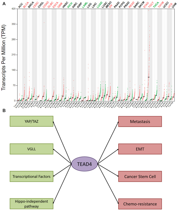
There is convincing evidence to indicate that TEAD4 has roles in cancer development. Figure 3A shows the TEAD4 expression profile across paired tumor and normal tissues from GEPIA (http://gepia.cancer-pku.cn). TEAD4 was upregulated in most tumor tissues, indicating that it is a potential prognostic biomarker. In addition, TEAD4 is involved in multiple stages in cancer (Figure 3B). EMT is a physiological process in which epithelial cells lose their adhesive ability, cell polarity is marked by decreased E-cadherin expression, and migration and invasion are triggered.63 Thus, EMT is a hallmark of metastasis.63,64 In colorectal cancer, TEAD4 overexpression affects vimentin expression, thereby promoting EMT and metastasis.11 In head–neck squamous cell carcinoma, decreased TEAD4 expression inhibited cell proliferation, migration, and invasion, as well as triggered cell apoptosis, whereas increased TEAD4 expression caused EMT. Furthermore, TEAD4 is required for TGF-β1-induced EMT,14 indicating that EMT is a prerequisite for metastasis. EMT can also affect the self-renewal and differentiation of cancer stem cells, thereby resulting in cancer development and progression.63 The TEAD4–TAZ complex can also bind to the promoter of SOX2 and modulate cancer stem cell self-renewal in head–neck squamous cell carcinoma.38 Consistent with the findings above, TEAD4-induced metastasis is common in gastric cancer,36 breast cancer,48 lung cancer,42 and colorectal cancer.11 This is plausible as cancer cells that undergo EMT detach, traverse the basement membrane, enter the bloodstream, and disseminate elsewhere for metastatic colonization.63 TEAD4 can also induce chemotherapeutic drug resistance.16 In summary, as the key effector of the Hippo pathway, TEAD4 and its transcriptional output promote cell proliferation and cancer development.
Figure 3.
(A) TEAD4 expression profile across all tumor samples and paired normal tissues. (B) Functional roles of TEAD4 in multiple stages of cancer progression. TEAD4 has essential functions in promoting metastasis, EMT, cancer stem cells, and chemo-resistance.
Note: Figure 3A is graphed by GEPIA (http://gepia.cancer-pku.cn).
Targeted Therapy
Targeting Upstream Kinases
As a promising prognostic marker, increased TEAD4 expression correlates with poor overall survival. Therefore, it is critical to develop anticancer therapies that target TEAD4. One potential strategy to suppress TEAD4 expression is to target upstream kinases. However, the design of small molecule kinase inhibitors is not straightforward as most kinases upstream of TEAD4, such as LATS1/2 and MST1/2, are tumor suppressors.25,65,66 Metformin is used to treat type 2 diabetes, but it has recently emerged as a magic bullet for cancer therapy. Metformin can suppress bladder cancer cell proliferation by upregulating AMPK and downregulating YAP–TEAD4 expression.67 Other studies have reported MST/LATS activation but TEAD–YAP/TAZ complex inhibition by small-molecule drugs.49,68,69 Furthermore, there is extensive crosstalk between different signaling pathways, and targeting upstream kinases can affect other pathways, which can cause side effects.3
Targeting the TEAD4–YAP/TAZ Complex
Presently, it is more feasible to target TEAD4 directly for cancer therapy.70 A structural study has reported the presence of a central palmitate-binding pocket in the YAP/TAZ binding domain of TEAD that is highly druggable due to its enclosure and hydrophobicity. A fragment screen revealed that the TEAD4 pocket can bind small-molecule inhibitors such as flufenamic acid and niflumic acid.71 Another study has reported the development of compounds with a conserved cysteine that can bind to the TEAD4 pocket, thereby resulting in allosteric inhibition of TEAD4.72 Similarly, VGLL4 is a YAP antagonist for TEAD4 binding in colorectal cancer,16 gastric cancer,41 and lung cancer.42 On the other hand, a mutation of the TEA domain has been demonstrated to abolish the association of TEAD4 with target genes, impairing TEAD4 transcriptional activity, indicating that the TEAD–DNA interplay is also a druggable target.17 Furthermore, verteporfin can suppress the progression of ovarian cancer73 and inhibit the tumorigenic characteristics of gastric cancer stem cells by suppressing the TEAD–YAP interaction.74 Amlexanox can target IRF3 to inhibit gastric cancer progression,44 but targeting the cooperative transcription factors of TEAD4 requires further investigation.
Targeting Non-Coding RNAs
Non-coding RNAs (ncRNAs) are RNAs that are not translated into proteins, including microRNAs (miRNAs), short interfering RNAs (siRNAs), long non-coding RNAs (lncRNA), and circular RNAs (circRNAs).75 Non-coding RNAs regulate the expression of oncogenes and tumor suppressors, making them novel targets for chemotherapy.76 Recently, studies have reported crosstalk between TEAD4 and ncRNAs. For instance, several miRNAs were identified to be involved in the inhibition of TEAD4 in gastric cancer and lung cancer.72–79 Additionally, TEAD4 can activate lncRNA MNX1–AS1 to promote gastric cancer progression through EZH2/BTG2 and miR-6785-5p/BCL2 axes.80 The greatest advantage of ncRNA-based therapies over other approaches is their ability to target multiple genes in a variety of pathways, thereby making them efficient regulators of several cellular processes.81 It might be feasible to target ncRNAs upstream of TEAD4 because the transcriptional output of oncogenes can be inhibited. Although the application of miRNA mimics is in Phase I clinical trials, there are several obstacles such as low-efficiency cellular uptake, off-target effects, and unexpected immune responses. Therefore, further studies are needed to overcome these challenges and to promote the clinical development of ncRNA-based therapies for cancer.76 It would be interesting to determine if ncRNA therapies can be combined with a targeted therapy involving the TEAD4–YAP/TAZ complex to optimize efficacy, reduce side effects, and avoid chemotherapeutic drug resistance.
Conclusions and Future Perspectives
In this review, we highlighted the structure, mechanism, and function of TEAD4 in various aspects of cancer development and progression such as EMT, drug resistance, and stem cell maintenance. Numerous studies have shown that TEAD4 interacts with the Hippo–YAP/TAZ pathway, VGLL proteins, and other transcription factors, thereby targeting downstream genes. However, YAP/TAZ-independent mechanisms that regulate TEAD4, such as glucocorticoids, post-translational modifications, and nucleocytoplasmic shuttling, require further studies.
It can be speculated that promoting the cytoplasmic translocation of TEAD4 might inhibit its transcriptional activity. Therefore, pharmacological agents are in need to prevent TEAD4 from entering the nucleus. Equally important, the identification of a hydrophobic palmitate-binding pocket in the YBD of TEAD4 has received much attention because it can be targeted by flufenamic acid and niflumic acid, resulting in allosteric inhibition of TEAD4. Apart from this, ncRNAs can serve as drug targets in cancers overexpressing TEAD4, but the main challenge is to improve the efficiency of cellular uptake and reduce side effects. Some oncogenic ncRNAs are also expressed in normal tissues to modulate other physiological functions, so specific delivery systems are required to reduce the dysregulation of biological processes. Moreover, there are not reliable carriers to enable ncRNA therapeutics to target hardly-accessible tissues such as solid tumors. To date, targeting the TEAD4–YAP/TAZ complex seems to be the most attractive therapeutic approach because targeting downstream molecules of the Hippo pathway does not affect upstream components. Hence, further studies are needed to understand the similarities and differences between YAP and TAZ in terms of their structure, localization, mechanisms, and downstream targets. It will be interesting to determine which would be the optimal therapy in specific conditions, or if combined approaches can promote overall survival.
Acknowledgments
This study was supported by grants from the Outstanding Leaders Training Program of Pudong Health Bureau of Shanghai (PWR12018-07), the Top-level Clinical Discipline Project of Shanghai Pudong (PWYgf2018-05), the Key Discipline Construction Project of Pudong Health Bureau of Shanghai (PWZxk2017-23).
Data Sharing Statement
The data that support the findings of this study are available from the corresponding author, Chunlong Zhong, upon reasonable request.
Disclosure
The authors declare no conflicts of interest.
References
- 1.Huh HD, Kim DH, Jeong HS, et al. Regulation of TEAD Transcription Factors in Cancer Biology. Cells. 2019;8(6):600. doi: 10.3390/cells8060600. [DOI] [PMC free article] [PubMed] [Google Scholar]
- 2.Lin KC, Park HW, Guan KL. Regulation of the Hippo pathway transcription factor TEAD. Trends Biochem Sci. 2017;42(11):862–872. doi: 10.1016/j.tibs.2017.09.003 [DOI] [PMC free article] [PubMed] [Google Scholar]
- 3.Landin-Malt A, Benhaddou AZA. An evolutionary, structural and functional overview of the mammalian TEAD1 and TEAD2 transcription factors. Gene. 2016;176(1):139–148. doi: 10.1016/j.physbeh.2017.03.040 [DOI] [PMC free article] [PubMed] [Google Scholar]
- 4.Chen Z, Friedrich GA, Soriano P. Transcriptional enhancer factor 1 disruption by a retroviral gene trap leads to heart defects and embryonic lethality in mice. Genes Dev. 1994;8(19):2293–2301. doi: 10.1101/gad.8.19.2293 [DOI] [PubMed] [Google Scholar]
- 5.Kaneko KJ, Kohn MJ, Liu CDM. Transcription factor TEAD2 Is involved in neural tube closure. Genesis. 2007;45(9):577–587. doi: 10.1002/dvg [DOI] [PMC free article] [PubMed] [Google Scholar]
- 6.Yagi R, Kohn MJ, Karavanova I, et al. Transcription factor TEAD4 specifies the trophectoderm lineage at the beginning of mammalian development. Development. 2007;134(21):3827–3836. doi: 10.1242/dev.010223 [DOI] [PubMed] [Google Scholar]
- 7.Nishioka N, Yamamoto S, Kiyonari H, et al. Tead4 is required for specification of trophectoderm in pre-implantation mouse embryos. Mech Dev. 2008;125(3–4):270–283. doi: 10.1016/j.mod.2007.11.002 [DOI] [PubMed] [Google Scholar]
- 8.Nishioka N, Ichi IK, Adachi K, et al. The Hippo signaling pathway components lats and yap pattern tead4 activity to distinguish mouse trophectoderm from inner cell mass. Dev Cell. 2009;16(3):398–410. doi: 10.1016/j.devcel.2009.02.003 [DOI] [PubMed] [Google Scholar]
- 9.Lim B, Park J, Kim H, et al. Integrative genomics analysis reveals the multilevel dysregulation and oncogenic characteristics of TEAD4 in gastric cancer. Carcinogenesis. 2014;35(5):1020–1027. doi: 10.1093/carcin/bgt409 [DOI] [PubMed] [Google Scholar]
- 10.Zhu C, Li L, Zhang Z, et al. A non-canonical role of YAP/TEAD is required for activation of estrogen-regulated enhancers in breast cancer. Mol Cell. 2019;75(4):791–806.e8. doi: 10.1016/j.molcel.2019.06.010 [DOI] [PMC free article] [PubMed] [Google Scholar]
- 11.Liu Y, Wang G, Yang Y, et al. Increased TEAD4 expression and nuclear localization in colorectal cancer promote epithelial–mesenchymal transition and metastasis in a YAP-independent manner. Oncogene. 2016;35(1665):2789–2800. doi: 10.1038/onc.2015.342 [DOI] [PubMed] [Google Scholar]
- 12.Yuan H, Liu H, Liu Z, et al. Genetic variants in Hippo pathway genes YAP1, TEAD1 and TEAD4 are associated with melanoma-specific survival. Int J Cancer. 2016;137(3):638–645. doi: 10.1002/ijc.29429.Genetic [DOI] [PMC free article] [PubMed] [Google Scholar]
- 13.Tsinias G, Nikou S, Mastronikolis N, et al. Expression and prognostic significance of YAP, TAZ, TEAD4 and p73 in human laryngeal cancer. Histol Histopathol. 2020;7:18228. doi: 10.14670/HH-18-228 [DOI] [PubMed] [Google Scholar]
- 14.Zhang W, Li J, Wu Y, et al. TEAD4 overexpression promotes epithelial-mesenchymal transition and associates with aggressiveness and adverse prognosis in head neck squamous cell carcinoma. Cancer Cell Int. 2018;18:178. doi: 10.1186/s12935-018-0675-z [DOI] [PMC free article] [PubMed] [Google Scholar]
- 15.Noland CL, Gierke S, Schnier PD, et al. Palmitoylation of TEAD transcription factors is required for their stability and function in hippo pathway signaling. Structure. 2016;24(1):179–186. doi: 10.1016/j.str.2015.11.005 [DOI] [PubMed] [Google Scholar]
- 16.Jiao S, Li C, Hao Q, et al. VGLL4 targets a TCF4–TEAD4 complex to coregulate Wnt and Hippo signalling in colorectal cancer. Nat Commun. 2017;8:14058. doi: 10.1038/ncomms14058 [DOI] [PMC free article] [PubMed] [Google Scholar]
- 17.Shi Z, He F, Chen M, et al. DNA-binding mechanism of the Hippo pathway transcription factor TEAD4. Oncogene. 2017;36(11):4362–4369. doi: 10.1038/onc.2017.24 [DOI] [PubMed] [Google Scholar]
- 18.Chen L, Chan SW, Zhang XQ, et al. Structural basis of YAP recognition by TEAD4 in the Hippo pathway. Genes Dev. 2010;24(3):290–300. doi: 10.1101/gad.1865310 [DOI] [PMC free article] [PubMed] [Google Scholar]
- 19.Xiao JH, Davidson I, Matthes H, et al. Cloning, expression, and transcriptional properties of the human enhancer factor TEF-1. Cell. 1991;65(4):551–568. doi: 10.1016/0092-8674(91)90088-G [DOI] [PubMed] [Google Scholar]
- 20.Holden JK, Cunningham CN. Targeting the hippo pathway and cancer through the TEAD family of transcription factors. Cancers (Basel). 2018;10:3. doi: 10.3390/cancers10030081 [DOI] [PMC free article] [PubMed] [Google Scholar]
- 21.Kristal Kaan HY, Chan SW, Tan SKJ, et al. Crystal structure of TAZ-TEAD complex reveals a distinct interaction mode from that of YAP-TEAD complex. Sci Rep. 2017;7(1):1–11. doi: 10.1038/s41598-017-02219-9 [DOI] [PMC free article] [PubMed] [Google Scholar]
- 22.Jacquemin P, Hwang JJ, Martial JA, et al. A novel family of developmentally regulated mammalian transcription factors containing the TEA/ATTS DNA binding domain. J Biol Chem. 1996;271(36):21775–21785. doi: 10.1074/jbc.271.36.21775 [DOI] [PubMed] [Google Scholar]
- 23.Jiang SW, Desai D, Khan S, et al. Cooperative binding of TEF-1 to repeated GGAATG-related consensus elements with restricted spatial separation and orientation. DNA Cell Biol. 2000;19(8):507–514. doi: 10.1089/10445490050128430 [DOI] [PubMed] [Google Scholar]
- 24.Anbanandam A, Albarado DC, Nguyen CT, et al. Insights into transcription enhancer factor 1 (TEF-1) activity from the solution structure of the TEA domain. Proc Natl Acad Sci U S A. 2006;103(46):17225–17230. doi: 10.1073/pnas.0607171103 [DOI] [PMC free article] [PubMed] [Google Scholar]
- 25.Harvey KF, Zhang X, Thomas DM. The Hippo pathway and human cancer. Nat Rev. 2013;13(4):246–257. doi: 10.1038/nrc3458 [DOI] [PubMed] [Google Scholar]
- 26.Wei C, Wang Y, Li X. The role of hippo signal pathway in breast cancer metastasis. Onco Targets Ther. 2018;11:2185–2193. doi: 10.2147/OTT.S157058 [DOI] [PMC free article] [PubMed] [Google Scholar]
- 27.Liang N, Zhang C, Dill P, et al. Regulation of YAP by mTOR and autophagy reveals a therapeutic target of tuberous sclerosis complex. J Exp Med. 2014;211(11):2249–2263. doi: 10.1084/jem.20140341 [DOI] [PMC free article] [PubMed] [Google Scholar]
- 28.Zanconato F, Forcato M, Battilana G, et al. Genome-wide association between YAP/TAZ/TEAD and AP-1 at enhancers drives oncogenic growth. Nat Cell Biol. 2015;17(9):1218–1227. doi: 10.1038/ncb3216 [DOI] [PMC free article] [PubMed] [Google Scholar]
- 29.Zhao B, Ye X, Yu J, et al. TEAD mediates YAP-dependent gene induction and growth control. Genes Dev. 2008;22(14):1962–1971. doi: 10.1101/gad.1664408 [DOI] [PMC free article] [PubMed] [Google Scholar]
- 30.Li X, Wang W, Wang J, et al. Proteomic analyses reveal distinct chromatin‐associated and soluble transcription factor complexes. Mol Syst Biol. 2015;11(1):775. doi: 10.15252/msb.20145504 [DOI] [PMC free article] [PubMed] [Google Scholar]
- 31.Plouffe SW, Lin KC, Moore JL, et al. The Hippo pathway effector proteins YAP and TAZ have both distinct and overlapping functions in the cell. J Biol Chem. 2018;293(28):11230–11240. doi: 10.1074/jbc.RA118.002715 [DOI] [PMC free article] [PubMed] [Google Scholar]
- 32.Bin Z, Karen T, Kun-Liang G. The Hippo pathway in organ size control, tissue regeneration and stem cell self-renewal. Nat Cell Biol. 2011;13(8):877–883. doi: 10.1038/jid.2014.371 [DOI] [PMC free article] [PubMed] [Google Scholar]
- 33.Lu Y, Wu T, Gutman O, et al. Phase separation of TAZ compartmentalizes the transcription machinery to promote gene expression. Nat Cell Biol. 2020;22(4):453–464. doi: 10.1038/s41556-020-0485-0 [DOI] [PMC free article] [PubMed] [Google Scholar]
- 34.Zhang J, Liu P, Tao J, et al. TEA domain transcription factor 4 is the major mediator of yes-associated protein oncogenic activity in mouse and human hepatoblastoma. Am J Pathol. 2019;189(5):1077–1090. doi: 10.1016/j.ajpath.2019.01.016 [DOI] [PMC free article] [PubMed] [Google Scholar]
- 35.Yu J, Rodriguez-barrueco R, Martinez D, et al. Cross-cohort analysis identifies a TEAD4 ↔ MYCN positive- feedback loop as the core regulatory element of high-risk neuroblastoma. Cancer Discov. 2018;8(5):582–599. doi: 10.1158/2159-8290.CD-16-0861. [DOI] [PMC free article] [PubMed] [Google Scholar]
- 36.Kim B, Cheong J, Im J, et al. PI3K/AKT/β-catenin signaling regulates vestigial-like 1 which predicts poor prognosis and enhances malignant phenotype in gastric cancer. Cancers (Basel). 2019;11(12):1923. doi: 10.3390/cancers11121923. [DOI] [PMC free article] [PubMed] [Google Scholar]
- 37.Pobbati AV, Chan SW, Lee I, et al. Structural and functional similarity between the Vgll1-TEAD and the YAP-TEAD complexes. Structure. 2012;20(7):1135–1140. doi: 10.1016/j.str.2012.04.004 [DOI] [PubMed] [Google Scholar]
- 38.Li J, Li Z, Wu Y, et al. The Hippo effector TAZ promotes cancer stemness by transcriptional activation of SOX2 in head neck squamous cell carcinoma. Cell Death Dis. 2019;10(8):603. doi: 10.1038/s41419-019-1838-0 [DOI] [PMC free article] [PubMed] [Google Scholar]
- 39.Vaudin P, Delanoue R, Davidson I, et al. TONDU (TDU), a novel human protein related to the product of vestigial (vg) gene of Drosophila melanogaster interacts with vertebrate TEF factors and substitutes for Vg function in wing formation. Development. 1999;126(21):4807–4816. [DOI] [PubMed] [Google Scholar]
- 40.Mesrouze Y, Hau JC, Erdmann D, et al. The surprising features of the TEAD4-Vgll1 protein-protein interaction. ChemBioChem. 2014;15(4):537–542. doi: 10.1002/cbic.201300715 [DOI] [PubMed] [Google Scholar]
- 41.Jiao S, Wang H, Shi Z, et al. A peptide mimicking VGLL4 function acts as a YAP antagonist therapy against gastric cancer. Cancer Cell. 2014;25(2):166–180. doi: 10.1016/j.ccr.2014.01.010 [DOI] [PubMed] [Google Scholar]
- 42.Zhang W, Gao Y, Li P, et al. VGLL4 functions as a new tumor suppressor in lung cancer by negatively regulating the YAP-TEAD transcriptional complex. Nat Publ Gr. 2014;24(3):331–343. doi: 10.1038/cr.2014.10 [DOI] [PMC free article] [PubMed] [Google Scholar]
- 43.Ibarra IL, Hollmann NM, Klaus B, et al. Mechanistic insights into transcription factor cooperativity and its impact on protein-phenotype interactions. Nat Commun. 2020;11(1):124. doi: 10.1038/s41467-019-13888-7 [DOI] [PMC free article] [PubMed] [Google Scholar]
- 44.Jiao S, Guan J, Chen M, et al. Targeting IRF3 as a YAP agonist therapy against gastric cancer. J Exp Med. 2018;215(2):699–718. doi: 10.1084/jem.20171116 [DOI] [PMC free article] [PubMed] [Google Scholar]
- 45.Moggs JG, Orphanides G. Estrogen receptors: orchestrators of pleiotropic cellular responses. EMBO Rep. 2001;2(9):775–781. doi: 10.1093/embo-reports/kve185. [DOI] [PMC free article] [PubMed] [Google Scholar]
- 46.Atsaves V, Leventaki V, Rassidakis GZ, et al. AP-1 transcription factors as regulators of immune responses in cancer. Cancers (Basel). 2019;11:7. doi: 10.3390/cancers11071037 [DOI] [PMC free article] [PubMed] [Google Scholar]
- 47.Liu X, Li H, Rajurkar M, et al. Tead and AP1 coordinate transcription and motility. Cell Rep. 2016;14(5):1169–1180. doi: 10.1016/j.celrep.2015.12.104. [DOI] [PMC free article] [PubMed] [Google Scholar]
- 48.He L, Yuan L, Sun Y, et al. Glucocorticoid receptor signaling activates TEAD4 to promote. Breast Cancer Prog. 2019;79. doi: 10.1158/0008-5472.CAN-19-0012 [DOI] [PubMed] [Google Scholar]
- 49.Yu FX, Zhao B, Panupinthu N, et al. Regulation of the Hippo-YAP pathway by G-protein-coupled receptor signaling. Cell. 2012;150(4):780–791. doi: 10.1016/j.cell.2012.06.037 [DOI] [PMC free article] [PubMed] [Google Scholar]
- 50.Mo JS, Meng Z, Kim YC, et al. Cellular energy stress induces AMPK-mediated regulation of YAP and the Hippo pathway. Nat Cell Biol. 2015;17(4):500–510. doi: 10.1038/ncb3111 [DOI] [PMC free article] [PubMed] [Google Scholar]
- 51.Wang W, Xiao ZD, Li X, et al. AMPK modulates Hippo pathway activity to regulate energy homeostasis. Nat Cell Biol. 2015;17(4):490–499. doi: 10.1038/ncb3113 [DOI] [PMC free article] [PubMed] [Google Scholar]
- 52.Lin KC, Moroishi T, Meng Z, et al. Regulation of Hippo pathway transcription factor TEAD by p38 MAPK-induced cytoplasmic translocation. Physiol Behav. 2017;176(5):139–148. doi: 10.1016/j.physbeh.2017.03.040 [DOI] [PMC free article] [PubMed] [Google Scholar]
- 53.Qi Y, Yu J, Han W, et al. A splicing isoform of TEAD4 attenuates the Hippo–YAP signalling to inhibit tumour proliferation. Nat Commun. 2016;7:ncomms11840. doi: 10.1038/ncomms11840 [DOI] [PMC free article] [PubMed] [Google Scholar]
- 54.Kaneko KJ, De Pamphilis ML. TEAD4 establishes the energy homeostasis essential for blastocoel formation. Development. 2013;140(17):3680–3690. doi: 10.1242/dev.093799 [DOI] [PMC free article] [PubMed] [Google Scholar]
- 55.Deribe YL, Pawson T, Dikic I. Post-translational modifications in signal integration. Nat Struct Mol Biol. 2010;17(6):666–672. doi: 10.1038/nsmb.1842 [DOI] [PubMed] [Google Scholar]
- 56.Duan G, Walther D. The roles of post-translational modifications in the context of protein interaction networks. PLoS Comput Biol. 2015;11(2):e1004049. doi: 10.1371/journal.pcbi.1004049 [DOI] [PMC free article] [PubMed] [Google Scholar]
- 57.Chan P, Han X, Zheng B, et al. Autopalmitoylation of TEAD proteins regulates transcriptional output of hippo pathway. Nat Chem Biol. 2016;12(4):282–289. doi: 10.1038/nchembio.2036.Autopalmitoylation [DOI] [PMC free article] [PubMed] [Google Scholar]
- 58.Mesrouze Y, Meyerhofer M, Bokhovchuk F, et al. Effect of the acylation of TEAD4 on its interaction with co-activators YAP and TAZ. Protein Sci. 2017;26(12):2399–2409. doi: 10.1002/pro.3312 [DOI] [PMC free article] [PubMed] [Google Scholar]
- 59.Resh MD. Palmitoylation of ligands, receptors, and intracellular signaling molecules. Sci STKE. 2006;2006(359):re14. doi: 10.1126/stke.3592006re14 [DOI] [PubMed] [Google Scholar]
- 60.Kim NG, Gumbiner BM. Cell contact and Nf2/Merlin-dependent regulation of TEAD palmitoylation and activity. Proc Natl Acad Sci U S A. 2019;116(20):9877–9882. doi: 10.1073/pnas.1819400116 [DOI] [PMC free article] [PubMed] [Google Scholar]
- 61.Dupont C, Armant DR, Brenner CA. Epigenetics: definition, mechanisms and clinical perspective. Semin Reprod Med. 2009;27(5):351–357. doi: 10.1055/s-0029-1237423 [DOI] [PMC free article] [PubMed] [Google Scholar]
- 62.Suvà ML, Riggi N, Bernstein BE. Epigenetic reprogramming in cancer. Science. 2013;339:6127. doi: 10.1126/science.1230184 [DOI] [PMC free article] [PubMed] [Google Scholar]
- 63.Shibue T, Weinberg RA. EMT, CSCs, and drug resistance: the mechanistic link and clinical implications. Nat Rev Clin Oncol. 2018;14(10):611–629. doi: 10.1038/nrclinonc.2017.44.EMT [DOI] [PMC free article] [PubMed] [Google Scholar]
- 64.Han Q, Kremerskothen J, Lin X, et al. WWC3 inhibits epithelial-mesenchymal transition of lung cancer by activating hippo-YAP signalling. Onco Targets Ther. 2018;11:2581–2591. doi: 10.2147/OTT.S162387 [DOI] [PMC free article] [PubMed] [Google Scholar]
- 65.Zhang J, Yang PL, Gray NS. Targeting cancer with small molecule kinase inhibitors. Nat Rev Cancer. 2009;9(1):28–39. doi: 10.1038/nrc2559 [DOI] [PubMed] [Google Scholar]
- 66.Gross S, Rahal R, Stransky N, et al. Targeting cancer with kinase inhibitors Find the latest version: targeting cancer with kinase inhibitors. J Clin Invest. 2015;125(5):1780–1789. doi: 10.1172/JCI76094.genomically [DOI] [PMC free article] [PubMed] [Google Scholar]
- 67.Wu Y, Zheng Q, Li Y, et al. Metformin targets a YAP1-TEAD4 complex via AMPKα to regulate CCNE1/2 in bladder cancer cells. J Exp Clin Cancer Res. 2019;38(1):376. doi: 10.1186/s13046-019-1346-1 [DOI] [PMC free article] [PubMed] [Google Scholar]
- 68.Bao Y, Nakagawa K, Yang Z, et al. A cell-based assay to screen stimulators of the Hippo pathway reveals the inhibitory effect of dobutamine on the YAP-dependent gene transcription. J Biochem. 2011;150(2):199–208. doi: 10.1093/jb/mvr063 [DOI] [PubMed] [Google Scholar]
- 69.Basu D, Lettan R, Damodaran K, et al. Identification, mechanism of action, and antitumor activity of a small molecule inhibitor of Hippo, TGF-β, and Wnt signaling pathways. Mol Cancer Ther. 2014;13(6):1457–1467. doi: 10.1158/1535-7163.MCT-13-0918 [DOI] [PubMed] [Google Scholar]
- 70.Gibault F, Sturbaut M, Bailly F, et al. Targeting transcriptional enhanced associate domains (TEADs). J Med Chem. 2018;61(12):5057–5072. doi: 10.1021/acs.jmedchem.7b00879 [DOI] [PubMed] [Google Scholar]
- 71.Pobbati AV, Han X, Hung AW, et al. Targeting the Central Pocket in human transcription factor TEAD as a potential cancer therapeutic strategy. Structure. 2015;23(11):2076–2086. doi: 10.1016/j.str.2015.09.009 [DOI] [PMC free article] [PubMed] [Google Scholar]
- 72.Bum-Erdene K, Zhou D, Gonzalez-Gutierrez G, et al. Small-molecule covalent modification of conserved cysteine leads to allosteric inhibition of the tead yap protein-protein interaction. Cell Chem Biol. 2019;26(3):378–389.e13. doi: 10.1016/j.chembiol.2018.11.010 [DOI] [PubMed] [Google Scholar]
- 73.Feng J, Gou J, Jia J, et al. Verteporfin, a suppressor of YAP–TEAD complex, presents promising antitumor properties on ovarian cancer. Onco Targets Ther. 2016;9:5371–5381. doi: 10.2147/OTT.S109979 [DOI] [PMC free article] [PubMed] [Google Scholar]
- 74.Giraud J, Molina-Castro S, Seeneevassen L, et al. Verteporfin targeting YAP1/TAZ-TEAD transcriptional activity inhibits the tumorigenic properties of gastric cancer stem cells. Int J Cancer. 2020;146(8):2255–2267. doi: 10.1002/ijc.32667 [DOI] [PubMed] [Google Scholar]
- 75.JS N. Non-coding RNAs: the architects of eukaryotic complexity. EMBO Rep. 2001;2(11):986–991. doi: 10.1093/embo-reports/kve230. [DOI] [PMC free article] [PubMed] [Google Scholar]
- 76.Arun G, Diermeier SD, Spector DL. Therapeutic targeting of long non-coding RNAs in cancer. Trends Mol Med. 2018;24(3):257–277. doi: 10.1016/j.molmed.2018.01.001 [DOI] [PMC free article] [PubMed] [Google Scholar]
- 77.Kim J, Bae D, Seo E, et al. Epigenetic silencing of miR-1271 enhances MEK1 and TEAD4 expression in gastric cancer. Cancer Med. 2018;7(7):3411–3424. doi: 10.1002/cam4.1605 [DOI] [PMC free article] [PubMed] [Google Scholar]
- 78.Zhang Q, Fan H, Zou Q, et al. TEAD4 exerts pro-metastatic effects and is negatively regulated by miR6839-3p in lung adenocarcinoma progression. J Cell Mol Med. 2018;22(7):3560–3571. doi: 10.1111/jcmm.13634 [DOI] [PMC free article] [PubMed] [Google Scholar]
- 79.Lu J, Tan T, Zhu L, et al. Hypomethylation Causes MIR21 Overexpression in Tumors. Mol Ther Oncol. 2020;18:47–57. doi: 10.1016/j.omto.2020.05.011 [DOI] [PMC free article] [PubMed] [Google Scholar]
- 80.Shuai Y, Ma Z, Liu W, et al. TEAD4 modulated LncRNA MNX1-AS1 contributes to gastric cancer progression partly through suppressing BTG2 and activating BCL2. Mol Cancer. 2020;19(1):6. doi: 10.1186/s12943-019-1104-1. [DOI] [PMC free article] [PubMed] [Google Scholar]
- 81.Garzon R, Marcucci G, Croce CM. Targeting microRNAs in cancer: rationale, strategies and challenges. Nat Rev Drug Discov. 2010;9(10):775–789. doi: 10.1038/nrd3179 [DOI] [PMC free article] [PubMed] [Google Scholar]


