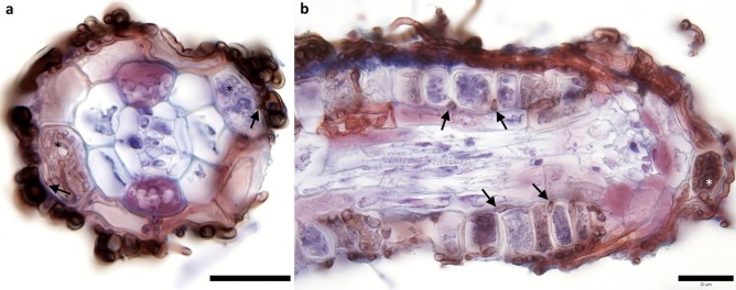
Fig. 5.
Transversal and longitudinal sections through hair roots of European blueberry. High magnification photos of longitudinal and transversal sections warrant the best illustration of the root colonization pattern. However, since the ericoid hair roots are too tiny for hand sectioning, paraffin thin sections (or an alternative) have to be obtained. a Thick dark brown surface hyphae that, upon entering the rhizodermis, dramatically change their diameter and give rise to intracellular hyphal coils morphologically corresponding to ericoid mycorrhiza (asterisks). Note hyphal connections between the surface hyphae and the intracellular hyphal coils (arrows). b Note the thick brownish intercellular hyphae (arrows) and an intracellular microsclerotium developed at the very tip of the hair root (asterisk). Both photos naturally colonized Vaccinium myrtillus hair roots collected in Northern Bohemia (Czechia), the sections were stained with acid fuchsin and trypan blue. Bars = 20 µm

