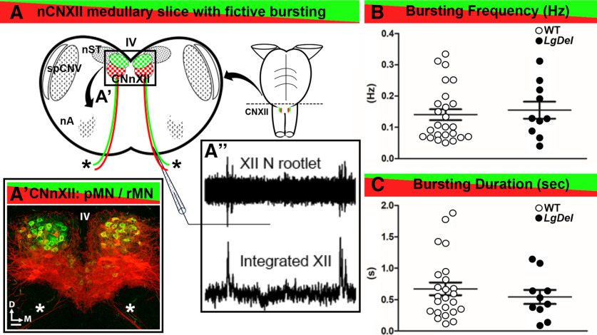
Figure 3.
Spontaneous fictive respiratory bursts in WT and LgDel CNXII slices. A, A schematic of a lower medulla section taken at the level of CNXII showing the nucleus (CNXII, box) ventral to the fourth ventricle (IV), segregated populations of pMNs (red) and rMNs (green), CNXII axons extending ventrally exit as CNXII rootlets (asterisks), and adjacent cranial nuclei including the nucleus of the solitary tract (nST), the spinal trigeminal nucleus (spCNV), and nucleus ambiguous (nA). A’, A confocal image of CNXII showing the segregation of the pMNs (red), rMNs (green), and CNXII axons extending ventrally (asterisks). Scale bar: 50 μm. D: dorsal, M: medial. A’’, Spontaneous inspiratory bursting activity recorded from hypoglossal nerve rootlets using a suction electrode (top), and represented as an integrated electrophysiological signal (right). B, Scatterplot of spontaneous inspiratory related bursting frequency recorded from WT and LgDel slices. C, Scatter plot of burst durations from WT and LgDel slices. Burst frequency and duration were not significantly different in the two genotypes; WT (n = 25) and LgDel (n = 10) animals, p > 0.05, unpaired two-tailed Student’s t test.

