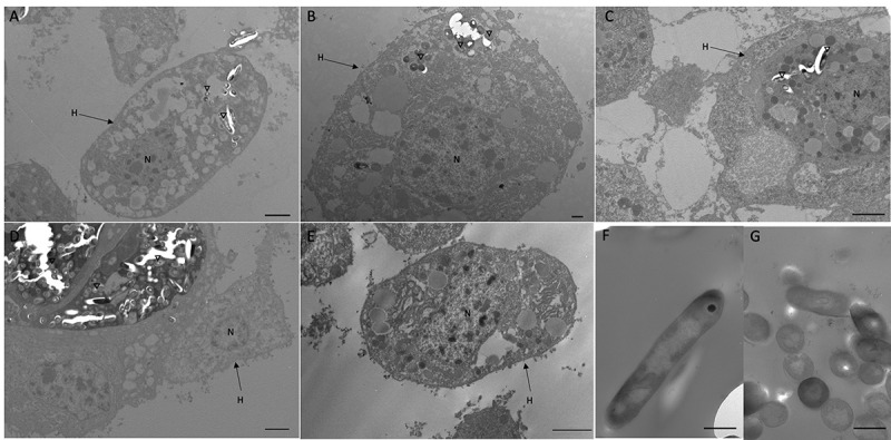
Figure 7.

Interaction between SAMTB lux and phagocytic hemocytes.
Transmission electron microscopy (TEM) was carried out on hemocytes extracted from G. mellonella larvae infected with SAMTB lux (2x10⁷ CFU) at: (a, b) 1 h (c) 24 h and (d) 168 h post infection. (e) shows healthy non-infected hemocytes. (f, g) show SAMTB lux bacilli in various geometric orientations. Internalization of SAMTB lux was observed as early as 1 h post-infection and presence of the bacilli persisted 24 h post-infection (A-C). By 168 h post-infection, aggregates of SAMTB lux bacilli, surrounded by hemocytes in an early granuloma-like structure, could be observed under TEM (D). H = hemocytes, N = nucleus, inverted triangles indicate SAMTB lux. Scale bars represent (a, c-e): 2 µm, (b, f, g): 500 nm.
