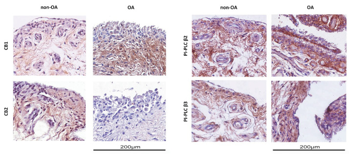
Figure 1.
Immunohistochemical analysis of synovial membranes from OA (osteoarthritis) and non-OA patients. Left panel: Slices were stained with anti-CB1 and anti-CB2 receptor antibodies. Right panel: Slices were stained with anti-PI-PLC β2 and anti-PI-PLC β3 antibodies. Slides were counterstained with hematoxylin and mounted with permanent mounting media. This figure shows representative images of different experiments (n = 5 non-OA and n = 6 OA).

