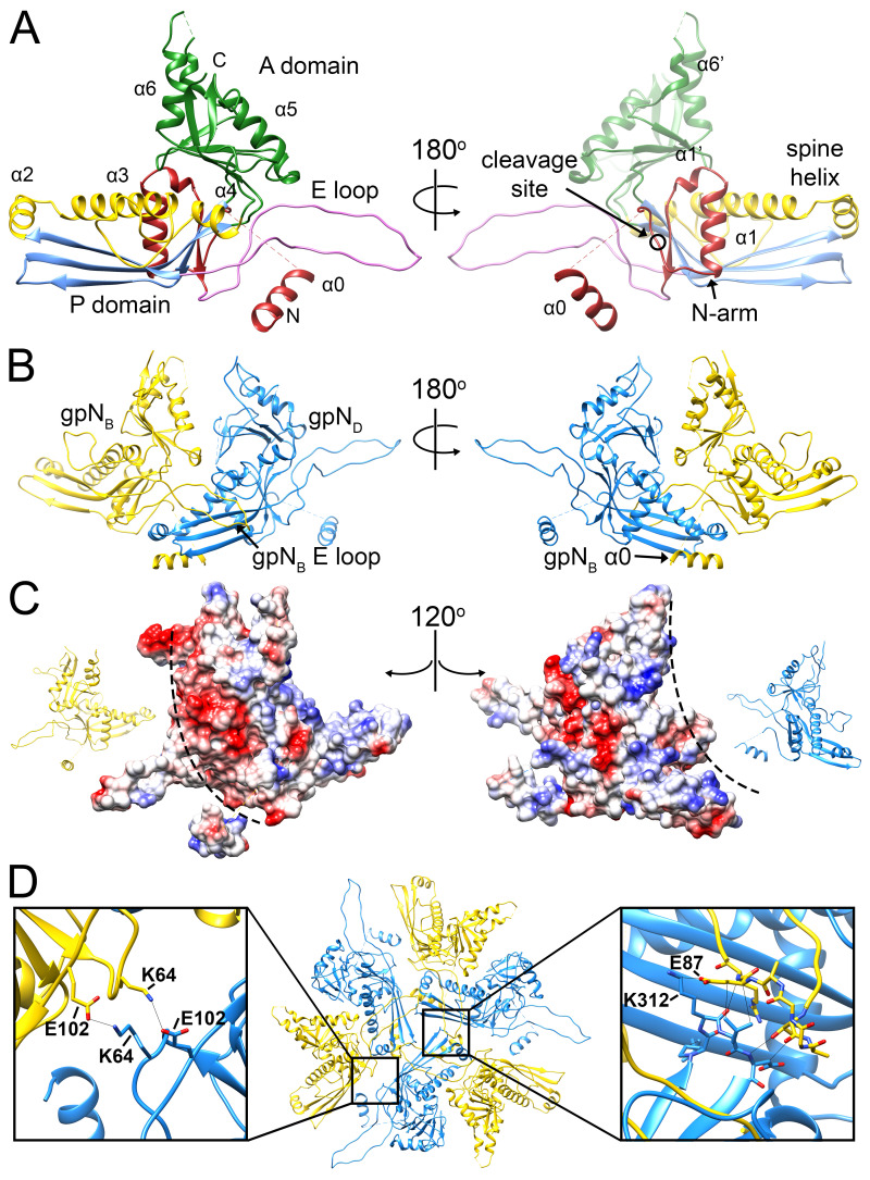
Figure 3.
The gpN capsid protein. (A) Ribbon diagram of gpNB (the most complete subunit), colored by structural feature: N-arm, red; E-loop, pink; P-domain β-sheet, blue; P-domain α-helices, yellow (α3 is the spine helix); A-domain, green. The right-hand panel is rotated by 180°. Key structural features are labeled, and the gpO cleavage site between residues 31 and 32 is indicated (circle). (B) Intra-capsomer interaction between the gpNB (yellow) and gpND (blue) subunits, showing how the gpNB E-loop overlays the spine helix of the neighboring (gpND) subunit. The right-hand panel is rotated by 180° and shows how the gpNB N-arm and α0 grasp the P-domain of gpND from underneath. (C) Electrostatic surfaces for the gpNB and gpND subunits from panel B, separated and rotated by 120° in opposite directions, as indicated by the insets. Interaction surfaces of opposite charge are indicated by the dashed lines. (D) Inter-capsomeric interaction between three gpND and their neighboring gpNB subunits related by the icosahedral threefold axis. The insets show details of the interactions between the gpND and gpNB subunits.

