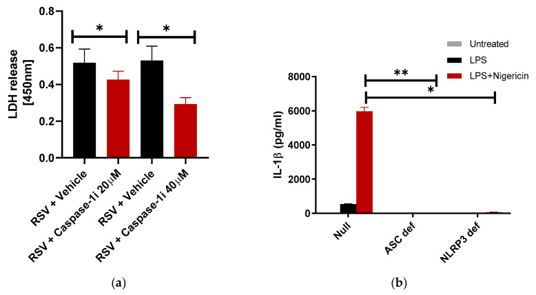
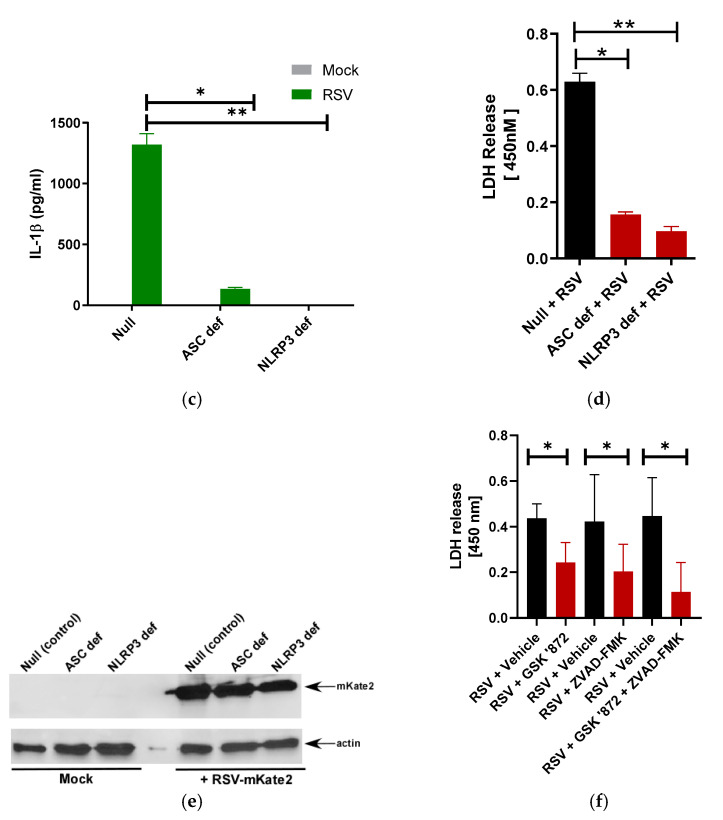
Figure 4.
Caspase-1 and ASC-NLRP3 inflammasome is required for pyroptotic cell death of macrophages during RSV infection. (a) Human THP-1 macrophages were infected with RSV (MOI = 1) in the presence of either vehicle (DMSO) or caspase-1 inhibitor (caspase-1i) Ac-YVAD-CHO (20 µM and 40 µM). LDH release was measured (at OD of 450 nm) at 16 h post-infection infection (n = 12 technical replicates from two independent experiments). * p ≤ 0.05 using a Student’s t-test. (b) Wild-type (null), ASC deficient (ASC def), and NLRP3 deficient (NLRP3 def) THP-1 macrophages were treated with LPS (100 ng/mL) for 4 h, followed by 30 min treatment with nigericin (15 µM). IL-1β release was measured by ELISA (n = 16 technical replicates from two independent experiments). * p and ** p ≤ 0.05 using a Student’s t-test. (c) Null, ASC def, and NLRP3 def THP-1 macrophages were infected with RSV (MOI = 1). At 16 h post-infection, IL-1β release was measured by ELISA (n = 16 technical replicates from two independent experiments). * p and ** p ≤ 0.05 using a Student’s t-test. (d) Null, ASC def, and NLRP3 def THP-1 macrophages were infected with RSV (MOI = 1). LDH release was measured (at OD of 450 nm) at 16 h post-infection infection (n = 16 technical replicates from two independent experiments). * p and ** p ≤ 0.05 using a Student’s t-test. (e) Null, ASC def, and NLRP3 def THP-1 macrophages were infected with RSV-mKate2 (MOI = 2) for 16 h. Equal levels of total protein from the cell lysates of Null, ASC def, and NLRP3 def cells were subjected to Western blotting with anti-RFP antibody. Western blot data shown is representative of three independent experiments with similar results. (f) Human THP-1 macrophages were infected with RSV (MOI = 1) the presence of DMSO (vehicle control), RIPK3-dependent necroptosis inhibitor GSK’872 (40 µM), Caspase-1 dependent pyroptosis inhibitor ZVAD-FMK (50 µM), or the combination of these inhibitors (GSK ‘872 + ZVAD-FMK). LDH release was measured (at OD of 450 nm) at 16 h post-infection (n = 14 technical replicates from two independent experiments). * p ≤ 0.05 using a Student’s t-test.


