Abstract
Simple Summary
Hepatitis E virus (HEV) is a lesser-known hepatitis virus, but its worldwide spread is undisputed and has increased in recent years. The zoonotic spread of HEV, mainly due to genotype (gt) 3, emerged in developed countries in the past decade. In addition, transmission via contaminated meat from pigs and boars was also established. Detailed analysis of viral dynamics and distribution is needed in order to identify associated risk factors. The aim of the current study is to present new and additional data on the HEV distribution among pigs, and for the first-time, also among the wild boar population in Bulgaria.
Abstract
(1) Background: Hepatitis E virus (HEV) is a causative agent of acute viral hepatitis, predominantly transmitted by the fecal–oral route. In developed countries, HEV is considered to be an emerging pathogen since the number of autochthonous cases is rising. Hepatitis E is a viral disease with a proven zoonotic potential for some of its genotypes. The main viral reservoirs are domestic pigs and wild boar. Consumption of undercooked meat, as well as occupational exposure, are key factors for the spread of HEV. In order to evaluate the risks of future viral evolution, a detailed examination of the ecology and distribution of the virus is needed. The aim of the present study is to investigate the prevalence of anti-HEV IgG Ab in domestic pigs and wild boar in Bulgaria; (2) Methods: In this study, during the period of three years between 2017 and 2019, 433 serum samples from 19 different pig farms and 1 slaughterhouse were collected and analyzed. In addition, 32 samples from wild boar were also collected and analyzed during the 2018–2019 hunting season. All samples were analyzed by commercial indirect ELISA; (3) Results: Overall, HEV seroprevalence was 60% (95% CI 42.7–77.1) in domestic pigs and 12.5% (4/32) in wild boar. The observed seroprevalence of the slaughter-aged pigs was 73.65% (95% Cl 58.7–87.3). Prevalence in domestic pigs was significantly higher in the samples collected during 2019 (98% (95% Cl 96.1–99.9)) compared to those collected during 2017 (45.33% (95% CI 2.7–87.3)) and 2018 (38.46% (95% CI 29.1–49.7.); (4) Conclusions: Our findings suggest that domesticated pigs and wild boar might be the reason for the increased HEV transmission across Bulgaria. The genotypic characterization of HEV found in pigs, wild boar and humans will give a more accurate view of the zoonotic transmission of this virus.
Keywords: hepatitis E virus, zoonotic transmission, foodborne infection, prevalence, boar, pigs
1. Introduction
Hepatitis E virus (HEV) is one of the leading causes of acute hepatitis worldwide. The hepatitis E infection is usually self-limiting in healthy human patients. However, immunocompromised, co-infected, and transplanted individuals may develop a fulminant infection with a high risk of developing a chronic disease. In addition, pregnant women infected with HEV have shown a higher mortality rate of up to 30% compared to 0.1–4% observed in the general population [1].
HEV is a small icosahedral virus from the Hepeviridae family, which is comprised of two genera Orthohepevirus and Piscihepevirus. The genus Orthohepevirus contains four species designated as Orthohepevirus A through D. Within the species Orthohepevirus A, 8 distinct mammalian genotypes (gt) have been identified so far (HEV 1–8) [2,3]. Gt 1 and gt 2 infect only humans and lead to epidemic outbreaks in developing countries located in Africa and Asia, as well as in Mexico. The HEV gt 1 and gt 2 are transmitted mainly through the fecal–oral route due to fecal contamination of drinking water [4]. In contrast, gt 3 and gt 4 have proven zoonotic potential and infect humans and several other animal species, such as pigs, wild boar, and deer [5,6,7]. Zoonotic transmission of gt 3 or gt 4 occurs through consumption of undercooked meat or contact with infected animals [8]. Gt 5 and gt 6 infect wild boar, while gt 7 infects dromedary camels. A recent study showed zoonotic transmission of gt 7 related to the consumption of camel meat. Gt 8 infects the Bactrian camel [9].
In Europe, HEV gt 3 infection is increasingly being recognized as an emerging disease [10]. Domestic pigs and wild boar are considered as the main animal reservoirs of HEV gt3. The reported anti-HEV IgG seroprevalence in swine herds ranges between 30% and 100% [11,12,13,14,15,16]. In Norway [12], 90% of the studied herds and 73% of the individual serum samples tested positive for specific IgG antibodies. Similar results were observed in many of the other European countries as well. The data from Italy shows that 97.43% of the tested herds and 50.21% of the serum samples were also positive for HEV specific IgG antibodies [13]. In Spain, 97.65% of the herds and 41.9% of the serum samples were positive as well [14].
HEV transmission within a herd occurs naturally through direct contact with HEV infected animals or with excreta from sick animals, or through contaminated water or food sources [17]. Animals can contract the HEV virus at various times during their growth period. Most studies show that pigs became infected between the age of 8 and 15 weeks, and some of them remain positive at slaughter [18,19,20,21]. In addition, it was shown that 2–15% of pigs were infected with HEV at slaughter [22]. The HEV viremia usually lasts up to 2 weeks with viral excretion, through feces, occurring usually between weeks 3 and 7 [20]. The infection in pigs causes the development of microscopic lesions in the liver, with asymptomatic clinical presentation [23]. Hepatitis E antibody seroconversion in pigs occurs between week 11 and week 13, following the waning in maternal antibodies, where IgM anti-HEV antibodies peak first, followed by the IgG anti-HEV antibodies [21].
Recent studies in Europe revealed molecular evidence for HEV infection in wild boar and high HEV seroprevalences [24,25,26,27,28]. The HEV disease-dynamic in wild boar is similar to domestic pigs; the infection frequency also increases with age as well as the possibility of viral chronification [29]. Viral transmission to humans is observed due to consumption of undercooked meat. Occupational exposure is a risk factor; veterinarians, slaughterers, and forest workers have shown higher anti-HEV antibody prevalence in comparison to people with other occupations [30]. Therefore, knowledge of the viral presence and dynamic in different hosts is crucial to establish prevention and healthcare approaches and measures.
Studies on the distribution of HEV in humans across Bulgaria show that the most acute hepatitis E cases in the country are caused by the HEV-3 subtypes 3e, 3f and 3c [31,32]. The first immunological study of pigs in Bulgaria from 2017 reported over 75% seropositivity. A noncommercial ELISA test based on the HEV ORF2 110–610 capsid protein was used to detect anti-HEV IgG antibodies in domestic pigs [33]. Another study reports overall HEV seroprevalence of 60.3%. More specifically, in weaners—25%, in fattening pigs—75.8%, and in sows—80% [34]. To date, there is no available information about HEV seroprevalence in Bulgarian wild boar. This study aims to provide further knowledge on the distribution of HEV in pigs across Bulgaria and to broaden perspectives of mapping the spread. Moreover, it provides the first data ever on HEV prevalence in the wild boar population within the Bulgarian territory.
2. Materials and Methods
2.1. Sample Collection and Selection Criteria
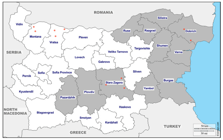
In total, 433 swine blood samples were collected from 19 different pig farms and 1 slaughterhouse, located in eleven districts across Bulgaria—Pazardzhik, Plovdiv, Stara Zagora, Yambol, Ruse, Razgrad, Silistra, Shumen, Varna, Dobrich, and Burgas district. The samples were collected during 2017, 2018, and 2019. Pig samples from 2017 (n = 75) were collected from two small pig farms (under 1000 pigs per farm) and a slaughterhouse, primarily used by local farmers. The age of swine tested was 5–6 months and older. Sera from 2018 (n = 158) were taken from industrial pig farms (over 1000 pigs per farm). Samples were taken from two age-groups, 6-month-old pigs that weighed around 80–100 kg (n = 78), and approximately 3-month-old pigs that weighed around 30 kg (n = 80). The 3-month-old pig samples were collected from four farms. Twenty samples were collected from each of the farms located in Shumen (2 farms), Dobrich and Burgas districts. Two of the farms (in Shumen and Dobrich districts) were also used for collection of 6-month-old pig samples. The 6-month-old pig samples were from five farms, located in four districts—Ruse (2 farms), Dobrich, Varna, and Shumen. The blood samples from 2019 (n = 200) were from ten farms from ten districts—Pazardzhik, Plovdiv, Stara Zagora, Yambol, Ruse, Razgrad, Silistra, Shumen, Varna and Dobrich (Figure 1). Twenty sera-samples from fattening farm pigs (5–6 months old) were collected from each farm. Information regarding their sex had not been recorded. The 6-month and the 3-month-old pig samples, collected during 2018, were chosen to examine the dynamics of the viral response through different pig ages.
Figure 1.
The map represents the origin of the farming pigs tested in grey. The origins of the wild boar’s testes are marked with red circles.
Geographically, districts Ruse, Razgrad, Silistra, Shumen, Dobrich and Varna are considered part of Northern Bulgaria. Districts Plovdiv, Stara Zagora, Pazardzhik, Yambol and Burgas are a part of Southern Bulgaria. The Stara Planina Mountain range located in the middle of the country, stretches from the eastern to the western borders of Bulgaria, acting as a natural divide, between Northern and Sothern Bulgaria.
In addition, 32 meat chunks from wild boar were collected for meat juice extraction. The samples were collected from animals hunted in Stara Zagora, Vratsa, Montana, and Dobrich districts during the official hunting season. Information regarding their sex and age was not recorded.
Eight samples were collected in district Stara Zagora (1–3 village Dalboki; 4–5 village Topolyane; 6–8 village Sladak Kladenec). Ten samples were collected from district Vratsa (1–6 village Butan; 7–8 village Leskovec; 9–10 village Dobrolevo). From district Montana, six samples were collected (1–2 village Medkovec; 3–6 village Staliyska Mahala), and from Dobrich district, eight (1–4 village Kozloduyci; 5–8 village Prilep). The origin given on the map indicates the closest place to the hunted area (Figure 1).
2.2. Ethics Statement
The authors report that all animals were treated following ethical principles approved by Bulgarian authorities. Sera samples were collected during routine health screenings of the pigs. Meat chunk samples from wild boar were obtained from hunted animals during the 2018/2019 hunting season. This study did not involve purposeful killing of animals.
2.3. Sample Processing
Blood samples from pigs (Sus scrofa domesticus) were taken by puncture of the sinus ophthalmicus. The blood samples were kept in plain vacutainers (without anticoagulant reagent) at room temperature (20 °C) until visible clot retraction. After centrifugation at 1500 g for 10 min, the sera were separated and kept at −20 °C until use.
Meat chunks from wild boar (Sus scrofa) were squeezed and meat juice samples were extracted. Approximately 200 mg of the meat was homogenized with a ceramic bead for 30 s at 4 speed (MP Biomedicals, Irvine, CA, USA). Samples were then squeezed through a layer of Miracloth and kept at −20 °C until further use.
2.4. Testing Assay
A commercial ELISA (PrioCHECK® HEV Ab porcine, Prionics AG, Schlieren, Switzerland) assay was used for the detection of HEV-specific antibodies. According to the manufacture protocol, dilutions of 1:100 for sera samples and 1:10 for meat juice samples were used. The test claims 91.0% sensitivity and 94.1% specificity. The cut-off value (Co) was calculated according to the manufacture protocol, as a means of optical density 450 (OD450) of the cut-off control multiplied by 1.2.
2.5. Data and Statistical Analysis
HEV prevalence was estimated from the ratio of positive samples to the total number of samples analyzed, with binomial confidence intervals of 95%. We calculated HEV prevalence during 2017, 2018 and 2019 in order to evaluate changes over time. Variables were expressed as percentages. Frequencies were compared using the chi-square test, and significance was set at a two-tailed p-value of less than 0.05. All data analysis was performed by Numbers 2019 (Apple Inc., Cupertino, CA, USA), Excel 2007 (Microsoft, Redmond, WA, USA) and SPSS Statistics 19.0 (IBM Corp., Armonk, New York, NY, USA).
3. Results
3.1. HEV Seroprevalence in Domestic Pigs
The overall HEV seroprevalence in domestic pigs was 60.05% (260 of 433 samples). There were no positive samples (0 of 80 samples) among the group of 3-month-old pigs (2018) originated from Shumen (2 farms), Dobrich (1 farm) and Burgas (1 farm) districts. Among the slaughter-aged pigs, the seroprevalence was 73.65% (260 of 353 samples). With respect to herd prevalence of anti-HEV antibodies, 17 out of 17 pig farms had HEV-positive pigs at slaughter age, demonstrating a 100% prevalence in the pig herds. The seropositivity in the slaughter-aged pigs varies based on the tested year and district (Table 1).
Table 1.
Anti-HEV IgG among slaughter-aged pigs (6-month-old) from different Bulgarian districts during 2017, 2018, and 2019. N designates the number of total samples; n, the number of positive samples; the districts are listed in alphabetical order.
| Year | Dobrich | Pazardzhik | Plovdiv | Razgrad | Ruse | Varna | Shumen | Silistra | Stara Zagora | Yambol | Total | ||
|---|---|---|---|---|---|---|---|---|---|---|---|---|---|
| 2017 | IgG+ | n | 34 | 34 | |||||||||
| % | 45.33 | 45.33 | |||||||||||
| Total | N | 75 | 75 | ||||||||||
| 2018 | IgG+ | n | 6 | 9 | 7 | 8 | 30 | ||||||
| % | 33.33 | 30 | 46.67 | 53.33 | 38.46 | ||||||||
| Total | N | 18 | 30 | 15 | 15 | 78 | |||||||
| 2019 | IgG+ | n | 19 | 20 | 20 | 18 | 20 | 20 | 20 | 20 | 20 | 19 | 196 |
| % | 95 | 100 | 100 | 90 | 100 | 100 | 100 | 100 | 100 | 100 | 98 | ||
| Total | N | 20 | 20 | 20 | 20 | 20 | 20 | 20 | 20 | 20 | 20 | 200 | |
| Total | IgG+ | n | 25 | 20 | 20 | 18 | 29 | 27 | 28 | 20 | 54 | 19 | 260 |
| % | 65.79 | 100 | 100 | 90 | 58 | 77.14 | 80 | 100 | 56.84 | 95 | 73.65 | ||
| Total | N | 38 | 20 | 20 | 20 | 50 | 35 | 35 | 20 | 95 | 20 | 353 | |
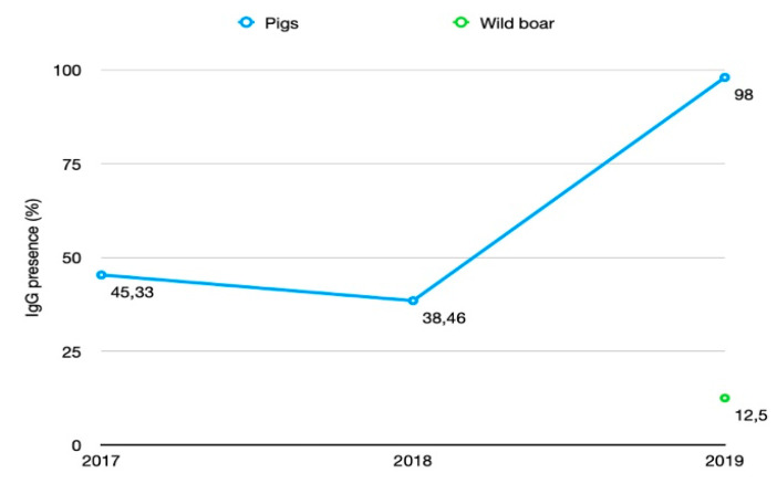
Samples collected during 2019 showed 98% (196 of 200 samples) seroprevalence. In 2017 and 2018, the HEV seroprevalence was 45.33% (34 of 75 samples) and 38.46% (30 of 78 samples) respectively (Figure 2).
Figure 2.
HEV IgG positive test results by year.
The observed high HEV seroprevalence during 2019 (98% (95% Cl 96.4–99.9)) compared to the previous two years, 2017 (45.33% (95% CI 2.7–87.3)) and 2018 (38.46% (95% CI 29.1–49.7)), is statistically significant, with a chi-square (χ2) value of 149.02 and a p-value <0.01 (Table 2).
Table 2.
Time factor of the HEV seroprevalence. N denotes the number of animals; OR is the odds ratio; 95% CI confidence interval; p—p-value and in the brackets [the chi-square statistic for each cell].
| Variable | N | HEV Positive | HEV Negative | OR (95% CI) | p |
|---|---|---|---|---|---|
| 2017 | 75 | 34 (55.67) [8.43] | 41 (19.33) [24.28] | 1.3 (95% CI: 0.69–2.5) | 0.3894 |
| 2018 | 78 | 30 (57.89) [13.44] | 48 (20.11) [38.69] | 0.75 (95% CI: 0.39–1.43) | 0.3894 |
| 2019 | 200 | 198 (148.44) [16.55] | 2 (51.56) [47.64] | 1.8984 (95% CI: 1.22–2.94) | 0.0044 |
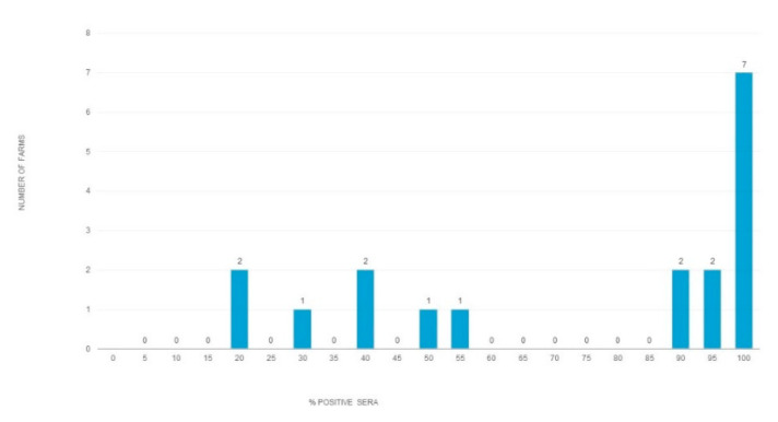
HEV seroprevalence did not differ significantly between 2017 and 2018 (χ2 = 0.742, df 1, p > 0.01). The seroprevalence in 2019 (98% (95% CI 96.4–99.9)) was significantly higher than the seroprevalence in 2017 and 2018 (χ2 = 146.2139, df 1, p < 0.00001). The HEV seroprevalence varied between the farms from 20% to 100% (73.05, (95% CI 58.2–88)) (Figure 3). The presence of specific IgG differed between farms from the same region. For example, in 2018, one of the two farms in Ruse district showed 20% positivity, while the other 40%. In Stara Zagora district, the variation between farms was even greater. In 2017, one of the two studied farms showed 20% positivity while the other, 88%.
Figure 3.
Variance between farms in HEV seroprevalence among slaughter-aged pigs. The table includes data collected from 17 farms and 1 slaughterhouse during the period of 2017–2020. The chart represents the number of farms (Y-axis) based on the percentage of positive sera per farm (X-axis).
HEV seroprevalence did not differ significantly between farms located in Southern and Northern Bulgaria (χ2 = 0.2997, and a p-value of 0.58) (Table 3).
Table 3.
The regional factor affecting HEV seroprevalence. N denotes the number of animals, p—p-value and in the brackets [the chi-square statistic for each cell].
| Region | Positive HEV IgG | Negative HEV IgG | Totals | p |
|---|---|---|---|---|
| Southern Bulgaria | 113 (110.88) [0.04] | 32 (34.12) [0.13] | 145 | 0.58 |
| Northern Bulgaria | 147 (149.12) [0.03] | 48 (45.88) [0.1] | 195 | 0.58 |
3.2. HEV Seroprevalence in Wild Boar
Twelve and half percent, or 4 of the 32 samples from wild boar, collected from four different regions during the 2018/2019 hunting season (Figure 1), were positive for anti-HEV IgG antibodies (Figure 2). Three of them originated from Dobrich district (2 near village Kozloduyci and 1 near village Prilep) and one from Stara Zagora district, near village Sladak Kladenec.
4. Discussion
The proven zoonotic potential of HEV in pigs, combined with the relatively high prevalence of HEV positive pigs in Europe, [21] and the presence of HEV infection in slaughter-aged pigs [35], may have a negative impact on the safety of pork products.
This research represents the first study on the HEV infection dynamics conducted during a three-year period in 19 different farms from eleven different Bulgarian districts. HEV seroprevalence was also shown in wild boar during the 2018/2019 hunting season. The overall seroprevalence obtained from domestic pigs was 60% (95% CI 42.7–77.1) and 12.5% (4/32) in wild boar. This rate is similar to the reported rate of HEV in pigs and wild boar in several European countries [15,16,29]. From the two studied age-groups of pigs, only the 6-month-old pigs were found to be seropositive. The 3-month-old pigs were not positive for anti-HEV IgG, confirming previous observations, showing that viremia most often occurs between the age of 2 and 4 months, and that anti-HEV IgG antibodies appear late during the period of viremia [36]. In addition, there are factors that can significantly delay HEV seroconversion, for example, co-infection with porcine reproductive and respiratory syndrome virus (PRRSV) and the presence of maternal antibodies. These factors may lead to HEV RNA presence at slaughter age, which appears to be a human health risk factor [37,38].
In this study, the HEV seroprevalence for anti-HEV IgG among the 6-month-old pigs, from different herds from ten Bulgarian districts (Table 1), ranged from 20% to 100%. The difference in seroprevalence suggests different transmission dynamics or exposure routes for HEV within the studied pig herds. A study from the Netherlands reported higher seropositivity in organic farms compared to conventional ones. The reason may be a greater contact frequency between pigs [39]. Other farming practices associated with HEV presence seem to include late weaning, mingling practices at the nursery stage, and poor hygiene measures [40].
The significantly higher seroprevalence observed in the sample cohort from 2019 compared to those from 2017 and 2018 suggests a rise in HEV infections in pigs, which may decrease or remain steadily high in the following years, which is concerning. Our findings complement the observations from previous studies done in Bulgaria and in other Balkan countries. Earlier studies on the distribution of HEV among Bulgarian pigs showed 40% overall seroprevalence (35/85) in pigs [41]. Another study showed 60.3% positive results for anti-HEV IgG in pigs from three Bulgarian districts [34]. A Romanian study showed 39% seropositivity among industrial pigs and 50% seroprevalence in backyard pigs [42,43]. In Serbia, a total of 315 serum samples from 3 to 4-month-old healthy backyard pigs were collected, of which 34.6% were positive for anti-HEV IgG [44].
A number of studies have shown that the overall seroprevalence in wild boar was significantly lower than the seroprevalence in domestic pigs [16,45,46]. Our study demonstrates that HEV is present among the wild boar population in Bulgaria, and it also confirms the observed lower seroprevalence among wild boar compared to domestic pigs. In our pilot investigation, only 12.5% of the wild boar samples were positive for HEV IgG antibodies. Differences seem to exist between the studied geographic areas (3 of the 4 positive samples were from one, Dobrich, of the four studied districts). However, it is difficult to draw meaningful conclusions with respect to the distribution of the virus due to the limited number of samples. Very similar results were reported in two studies across Romania, showing 10.29% and 9.61% HEV seroprevalence among the studied wild boar populations [47,48]. Interestingly, Dobrich is the only one of the four districts, where wild boar samples were collected, that shares a land border with Romania. Two studies, from France and the Netherlands, have also shown that HEV is endemic for these territories, with seropresence of 14% and 12%, respectively [49,50]. A research team from Spain reported a 23% presence of HEV RNA and suggested that the dynamics of the virus in wild boar may be seasonal, with the highest peak at the beginning of the hunting season, late October and November [26]. In addition, people with occupational exposure, such as hunters and forest workers, tend to show a higher presence of specific antibodies, which suggest an increased risk for HEV infection among them [30]. Although the researched pool is too small to be significant, this study is the first-ever proof for the spread of HEV among the wild boar population in Bulgaria. The results display the need for further studies to evaluate the degree of prevalence in this population and the associated risks of HEV prevalence in livers.
There are only a few immunological studies done in Bulgaria focused on HEV in humans. Two of them include only patients with clinical symptoms of acute hepatitis or liver failure. One of the records, collected between 2004 and 2012, showed 2.48% (20/806) presence of specific antibodies [51]. The other showed 13.2% (43/325) presence of anti-HEV IgM and 20.9% presence of anti-HEV IgG in patients studied between 2012 to 2016 [52]. Another report, from district Plovdiv, which includes 741 samples, collected between 2012 and 2013 from random patients, showed 9.04% presence of anti-HEV IgG and 1.48% of both anti-HEV IgG and IgM [53]. All of them showed a higher presence, which appears to increase with age. However, none of the studies investigated if there is a correlation between HEV in humans and pigs. In Bulgaria, two HEV phylogenetic studies confirmed the presence of HEV Subtypes 3e, 3f, 3c, 3i, and 3hi, and Subtype 1, in human samples [31,52]. Most of these subtypes refer to domestic pigs and wild boar, which reveals the probability of zoonotic transmission of HEV infection to humans. In addition, the higher frequency of detection of anti-HEV antibodies in veterinarians and pig farm workers strongly suggests the zoonotic potential of the disease [54,55]. Most likely, the transmission of the virus to the general public is predominantly a result of the consumption of undercooked pork and game meat [7,46,47].
This investigation has shown that domestic pigs and wild boar in Bulgaria are widely infected with HEV. There is a need for the production of HEV immunogenic proteins, which can be used for development of serological tests for detection of the HEV infection. In our previous study, we described a method for the production of recombinant HEV open reading frame (ORF) 2 protein in Nicotiana benthamiana plants and the successful use of this protein for serological detection of anti-HEV antibodies in both humans and swine [33,56]. Other studies have also demonstrated the efficacy of rORF2 gt 3 produced in various expression systems in the detection of anti-HEV serum Ab [57,58]. Ultimately, one of our main goals is to develop an HEV diagnostic kit based on the plant-derived rORF2 capsid protein. Such a kit would be cost-effective, and it would allow routine HEV testing, especially in developing countries.
5. Conclusions
For the first time, this study shows that HEV circulates in the Bulgarian wild boar population. The results of the HEV IgG seroprevalence in Bulgarian wild boar and domestic pigs further characterizes the presence of the Hepatitis E viral infection in Bulgaria, thus highlighting a significant concern for zoonotic virus transmission through consumption of raw or undercooked meat and direct contact with animals. Our results suggest that follow up studies, investigating the zoonotic transmission of this virus to humans, are needed, as well as measures for adequate meat product treatments, and thorough monitoring of the HEV disease distribution.
Acknowledgments
The authors want to thank Georgi Lukov M.D. (Faculty of Sciences, Brigham Young University–Hawaii) for his critical reading of the manuscript, Emilia Ivanova (National Diagnostic Science and Research Veterinary Medical Institute, Bulgarian Food Safety Agency) for collecting blood samples from pigs and Efstratios Kyriakakis (University of Plovdiv) for his support.
Author Contributions
Conceptualization, G.Z.; Funding acquisition, G.Z. and I.M., Investigation, K.T., Z.I. and T.K.; Project administration, V.T.; Supervision, G.Z. and V.T.; Writing, original draft preparation, K.T.; Writing review and editing, G.Z. All authors have read and agreed to the published version of the manuscript.
Funding
This research was funded by the European Union’s Horizon 2020 research and innovation programme, project PlantaSYST (SGA-CSA No. 739582 under FPA No. 664620), the European Regional Development Fund through the Bulgarian “Science and Education for Smart Growth” (project BG05M2OP001-1.003-001-C01) and by the Bulgarian National Science Fund, grant number DNTC/Russia 02/6.
Conflicts of Interest
The authors declare no conflict of interest.
References
- 1.Kmush B.L., Nelson K.E., Labrique A.B. Risk factors for hepatitis E virus infection and disease. Expert Rev. Anti Infect. Ther. 2015;13:41–53. doi: 10.1586/14787210.2015.981158. [DOI] [PubMed] [Google Scholar]
- 2.Purdy M.A., Harrison T.J., Jameel S., Meng X.-J., Okamoto H., Van der Poel W.H.M., Smith D.B. ICTV Virus Taxonomy Profile: Hepeviridae. J. Gen. Virol. 2017;98:2645–2646. doi: 10.1099/jgv.0.000940. [DOI] [PMC free article] [PubMed] [Google Scholar]
- 3.Smith D.B., Simmonds P., Jameel S., Emerson S.U., Harrison T.J., Meng X.J., Okamoto H., Van der Poel W.H., Purdy M.A., International Committee on the Taxonomy of Viruses Hepeviridae Study Group Consensus proposals for classification of the family Hepeviridae. J. Gen. Virol. 2014;95:2223–2232. doi: 10.1099/vir.0.068429-0. [DOI] [PMC free article] [PubMed] [Google Scholar]
- 4.Kamar N., Izopet J., Pavio N., Aggarwal R., Labrique A., Wedemeyer H., Dalton H.R. Hepatitis E virus infection. Nat. Rev. Dis. Primer. 2017;3:17086. doi: 10.1038/nrdp.2017.86. [DOI] [PubMed] [Google Scholar]
- 5.Anheyer-Behmenburg H.E., Szabo K., Schotte U., Binder A., Klein G., Johne R. Hepatitis E Virus in Wild Boars and Spillover Infection in Red and Roe Deer, Germany, 2013–2015. Emerg. Infect. Dis. 2017;23:130–133. doi: 10.3201/eid2301.161169. [DOI] [PMC free article] [PubMed] [Google Scholar]
- 6.Colson P., Borentain P., Queyriaux B., Kaba M., Moal V., Gallian P., Heyries L., Raoult D., Gerolami R. Pig liver sausage as a source of hepatitis E virus transmission to humans. J. Infect. Dis. 2010;202:825–834. doi: 10.1086/655898. [DOI] [PubMed] [Google Scholar]
- 7.Riveiro-Barciela M., Mínguez B., Gironés R., Rodriguez-Frías F., Quer J., Buti M. Phylogenetic demonstration of hepatitis E infection transmitted by pork meat ingestion. J. Clin. Gastroenterol. 2015;49:165–168. doi: 10.1097/MCG.0000000000000113. [DOI] [PubMed] [Google Scholar]
- 8.Drobeniuc J., Greene-Montfort T., Le N.-T., Mixson-Hayden T.R., Ganova-Raeva L., Dong C., Novak R.T., Sharapov U.M., Tohme R.A., Teshale E., et al. Laboratory-based Surveillance for Hepatitis E Virus Infection, United States, 2005–2012. Emerg. Infect. Dis. 2013;19:218–222. doi: 10.3201/eid1902.120961. [DOI] [PMC free article] [PubMed] [Google Scholar]
- 9.Woo P.C.Y., Lau S.K.P., Teng J.L.L., Cao K.-Y., Wernery U., Schountz T., Chiu T.H., Tsang A.K.L., Wong P.-C., Wong E.Y.M., et al. New Hepatitis E Virus Genotype in Bactrian Camels, Xinjiang, China, 2013. Emerg. Infect. Dis. 2016;22:2219–2221. doi: 10.3201/eid2212.160979. [DOI] [PMC free article] [PubMed] [Google Scholar]
- 10.Arends J.E., Ghisetti V., Irving W., Dalton H.R., Izopet J., Hoepelman A.I.M., Salmon D. Hepatitis E: An emerging infection in high income countries. J. Clin. Virol. Off. Publ. Pan Am. Soc. Clin. Virol. 2014;59:81–88. doi: 10.1016/j.jcv.2013.11.013. [DOI] [PubMed] [Google Scholar]
- 11.Salines M., Andraud M., Rose N. From the epidemiology of hepatitis E virus (HEV) within the swine reservoir to public health risk mitigation strategies: A comprehensive review. Vet. Res. 2017;48:31. doi: 10.1186/s13567-017-0436-3. [DOI] [PMC free article] [PubMed] [Google Scholar]
- 12.Lange H., Øverbø J., Borgen K., Dudman S., Hoddevik G., Urdahl A.M., Vold L., Sjurseth S.K. Hepatitis E in Norway: Seroprevalence in humans and swine. Epidemiol. Infect. 2017;145:181–186. doi: 10.1017/S0950268816002144. [DOI] [PMC free article] [PubMed] [Google Scholar]
- 13.Martinelli N., Luppi A., Cordioli P., Lombardi G., Lavazza A. Prevalence of hepatitis E virus antibodies in pigs in Northern Italy. Infect. Ecol. Epidemiol. 2011;1:7331. doi: 10.3402/iee.v1i0.7331. [DOI] [PMC free article] [PubMed] [Google Scholar]
- 14.Seminati C., Mateu E., Peralta B., de Deus N., Martin M. Distribution of hepatitis E virus infection and its prevalence in pigs on commercial farms in Spain. Vet. J. 2008;175:130–132. doi: 10.1016/j.tvjl.2006.11.018. [DOI] [PubMed] [Google Scholar]
- 15.Breum S.Ø., Hjulsager C.K., de Deus N., Segalés J., Larsen L.E. Hepatitis E virus is highly prevalent in the Danish pig population. Vet. Microbiol. 2010;146:144–149. doi: 10.1016/j.vetmic.2010.05.002. [DOI] [PubMed] [Google Scholar]
- 16.Ivanova A., Tefanova V., Reshetnjak I., Kuznetsova T., Geller J., Lundkvist Å., Janson M., Neare K., Velström K., Jokelainen P., et al. Hepatitis E Virus in Domestic Pigs, Wild Boars, Pig Farm Workers, and Hunters in Estonia. Food Environ. Virol. 2015;7:403–412. doi: 10.1007/s12560-015-9210-8. [DOI] [PubMed] [Google Scholar]
- 17.Bouwknegt M., Frankena K., Rutjes S.A., Wellenberg G.J., de Husman A.M.R., van der Poel W.H.M., de Jong M.C.M. Estimation of hepatitis E virus transmission among pigs due to contact-exposure. Vet. Res. 2008;39:1. doi: 10.1051/vetres:2008017. [DOI] [PubMed] [Google Scholar]
- 18.Casas M., Cortés R., Pina S., Peralta B., Allepuz A., Cortey M., Casal J., Martín M. Longitudinal study of hepatitis E virus infection in Spanish farrow-to-finish swine herds. Vet. Microbiol. 2011;148:27–34. doi: 10.1016/j.vetmic.2010.08.010. [DOI] [PubMed] [Google Scholar]
- 19.de Deus N., Casas M., Peralta B., Nofrarías M., Pina S., Martín M., Segalés J. Hepatitis E virus infection dynamics and organic distribution in naturally infected pigs in a farrow-to-finish farm. Vet. Microbiol. 2008;132:19–28. doi: 10.1016/j.vetmic.2008.04.036. [DOI] [PubMed] [Google Scholar]
- 20.Leblanc D., Ward P., Gagné M.-J., Poitras E., Müller P., Trottier Y.-L., Simard C., Houde A. Presence of hepatitis E virus in a naturally infected swine herd from nursery to slaughter. Int. J. Food Microbiol. 2007;117:160–166. doi: 10.1016/j.ijfoodmicro.2007.03.008. [DOI] [PubMed] [Google Scholar]
- 21.Krog J.S., Larsen L.E., Breum S.Ø. Tracing Hepatitis E Virus in Pigs From Birth to Slaughter. Front. Vet. Sci. 2019;6 doi: 10.3389/fvets.2019.00050. [DOI] [PMC free article] [PubMed] [Google Scholar]
- 22.Crossan C., Grierson S., Thomson J., Ward A., Nunez-Garcia J., Banks M., Scobie L. Prevalence of hepatitis E virus in slaughter-age pigs in Scotland. Epidemiol. Infect. 2015;143:2237–2240. doi: 10.1017/S0950268814003100. [DOI] [PMC free article] [PubMed] [Google Scholar]
- 23.Halbur P.G., Kasorndorkbua C., Gilbert C., Guenette D., Potters M.B., Purcell R.H., Emerson S.U., Toth T.E., Meng X.J. Comparative Pathogenesis of Infection of Pigs with Hepatitis E Viruses Recovered from a Pig and a Human. J. Clin. Microbiol. 2001;39:918–923. doi: 10.1128/JCM.39.3.918-923.2001. [DOI] [PMC free article] [PubMed] [Google Scholar]
- 24.Martinelli N., Pavoni E., Filogari D., Ferrari N., Chiari M., Canelli E., Lombardi G. Hepatitis E Virus in Wild Boar in the Central Northern Part of Italy. Transbound. Emerg. Dis. 2015;62:217–222. doi: 10.1111/tbed.12118. [DOI] [PubMed] [Google Scholar]
- 25.Porea D., Anita A., Demange A., Raileanu C., Oslobanu Ludu L., Anita D., Savuta G., Pavio N. Molecular detection of hepatitis E virus in wild boar population in eastern Romania. Transbound. Emerg. Dis. 2018;65:527–533. doi: 10.1111/tbed.12736. [DOI] [PubMed] [Google Scholar]
- 26.Rivero-Juarez A., Risalde M.A., Frias M., García-Bocanegra I., Lopez-Lopez P., Cano-Terriza D., Camacho A., Jimenez-Ruiz S., Gomez-Villamandos J.C., Rivero A. Prevalence of hepatitis E virus infection in wild boars from Spain: A possible seasonal pattern? BMC Vet. Res. 2018;14:54. doi: 10.1186/s12917-018-1377-4. [DOI] [PMC free article] [PubMed] [Google Scholar]
- 27.Spancerniene U., Grigas J., Buitkuviene J., Zymantiene J., Juozaitiene V., Stankeviciute M., Razukevicius D., Zienius D., Stankevicius A. Prevalence and phylogenetic analysis of hepatitis E virus in pigs, wild boars, roe deer, red deer and moose in Lithuania. Acta Vet. Scand. 2018;60:13. doi: 10.1186/s13028-018-0367-7. [DOI] [PMC free article] [PubMed] [Google Scholar]
- 28.Weigand K., Weigand K., Schemmerer M., Müller M., Wenzel J.J. Hepatitis E Seroprevalence and Genotyping in a Cohort of Wild Boars in Southern Germany and Eastern Alsace. Food Environ. Virol. 2018;10:167–175. doi: 10.1007/s12560-017-9329-x. [DOI] [PubMed] [Google Scholar]
- 29.Sonoda H., Abe M., Sugimoto T., Sato Y., Bando M., Fukui E., Mizuo H., Takahashi M., Nishizawa T., Okamoto H. Prevalence of Hepatitis E Virus (HEV) Infection in Wild Boars and Deer and Genetic Identification of a Genotype 3 HEV from a Boar in Japan. J. Clin. Microbiol. 2004;42:5371–5374. doi: 10.1128/JCM.42.11.5371-5374.2004. [DOI] [PMC free article] [PubMed] [Google Scholar]
- 30.Dremsek P., Wenzel J.J., Johne R., Ziller M., Hofmann J., Groschup M.H., Werdermann S., Mohn U., Dorn S., Motz M., et al. Seroprevalence study in forestry workers from eastern Germany using novel genotype 3- and rat hepatitis E virus-specific immunoglobulin G ELISAs. Med. Microbiol. Immunol. 2012;201:189–200. doi: 10.1007/s00430-011-0221-2. [DOI] [PubMed] [Google Scholar]
- 31.Bruni R., Villano U., Equestre M., Chionne P., Madonna E., Trandeva-Bankova D., Peleva-Pishmisheva M., Tenev T., Cella E., Ciccozzi M., et al. Hepatitis E virus genotypes and subgenotypes causing acute hepatitis, Bulgaria, 2013–2015. PLoS ONE. 2018;13:e0198045. doi: 10.1371/journal.pone.0198045. [DOI] [PMC free article] [PubMed] [Google Scholar]
- 32.Mrzljak A., Dinjar-Kujundzic P., Jemersic L., Prpic J., Barbic L., Savic V., Stevanovic V., Vilibic-Cavlek T. Epidemiology of hepatitis E in South-East Europe in the “One Health” concept. World J. Gastroenterol. 2019;25:3168–3182. doi: 10.3748/wjg.v25.i25.3168. [DOI] [PMC free article] [PubMed] [Google Scholar]
- 33.Mazalovska M., Varadinov N., Koynarski T., Minkov I., Teoharov P., Lomonossoff G.P., Zahmanova G. Detection of Serum Antibodies to Hepatitis E Virus Based on HEV Genotype 3 ORF2 Capsid Protein Expressed in Nicotiana benthamiana. Ann. Lab. Med. 2017;37:313–319. doi: 10.3343/alm.2017.37.4.313. [DOI] [PMC free article] [PubMed] [Google Scholar]
- 34.Tsachev I., Baymakova M., Ciccozzi M., Pepovich R., Kundurzhiev T., Marutsov P., Dimitrov K.K., Gospodinova K., Pishmisheva M., Pekova L. Seroprevalence of Hepatitis E Virus Infection in Pigs from Southern Bulgaria. Vector Borne Zoonotic Dis. Larchmt. N. 2019;19:767–772. doi: 10.1089/vbz.2018.2430. [DOI] [PubMed] [Google Scholar]
- 35.Di Bartolo I., Angeloni G., Ponterio E., Ostanello F., Ruggeri F.M. Detection of hepatitis E virus in pork liver sausages. Int. J. Food Microbiol. 2015;193:29–33. doi: 10.1016/j.ijfoodmicro.2014.10.005. [DOI] [PubMed] [Google Scholar]
- 36.Meng X.J., Halbur P.G., Haynes J.S., Tsareva T.S., Bruna J.D., Royer R.L., Purcell R.H., Emerson S.U. Experimental infection of pigs with the newly identified swine hepatitis E virus (swine HEV), but not with human strains of HEV. Arch. Virol. 1998;143:1405–1415. doi: 10.1007/s007050050384. [DOI] [PubMed] [Google Scholar]
- 37.Kanai Y., Tsujikawa M., Yunoki M., Nishiyama S., Ikuta K., Hagiwara K. Long-term shedding of hepatitis E virus in the feces of pigs infected naturally, born to sows with and without maternal antibodies. J. Med. Virol. 2010;82:69–76. doi: 10.1002/jmv.21647. [DOI] [PubMed] [Google Scholar]
- 38.Salines M., Barnaud E., Andraud M., Eono F., Renson P., Bourry O., Pavio N., Rose N. Hepatitis E virus chronic infection of swine co-infected with Porcine Reproductive and Respiratory Syndrome Virus. Vet. Res. 2015;46:55. doi: 10.1186/s13567-015-0207-y. [DOI] [PMC free article] [PubMed] [Google Scholar]
- 39.Rutjes S.A., Bouwknegt M., van der Giessen J.W., de Roda Husman A.M., Reusken C.B.E.M. Seroprevalence of Hepatitis E Virus in Pigs from Different Farming Systems in The Netherlands. J. Food Prot. 2014;77:640–642. doi: 10.4315/0362-028X.JFP-13-302. [DOI] [PubMed] [Google Scholar]
- 40.Walachowski S., Dorenlor V., Lefevre J., Lunazzi A., Eono F., Merbah T., Eveno E., Pavio N., Rose N. Risk factors associated with the presence of hepatitis E virus in livers and seroprevalence in slaughter-age pigs: A retrospective study of 90 swine farms in France. Epidemiol. Infect. 2014;142:1934–1944. doi: 10.1017/S0950268813003063. [DOI] [PMC free article] [PubMed] [Google Scholar]
- 41.Pishmisheva M., Baymakova M., Golkocheva-Markova E., Kundurzhiev T., Pepovich R., Popov G., Tsachev I. First Serological Study of Hepatitis E Virus Infection in Pigs in Bulgaria. Comptes Rendus Académie Bulg. Sci. Sci. Math. Nat. 2018;71:1001–1008. doi: 10.7546/CRABS.2018.07.18. [DOI] [Google Scholar]
- 42.Aniţă A., Gorgan L., Aniţă D., Oşlobanu L., Pavio N., Savuţa G. Evidence of hepatitis E infection in swine and humans in the East Region of Romania. Int. J. Infect. Dis. IJID Off. Publ. Int. Soc. Infect. Dis. 2014;29:232–237. doi: 10.1016/j.ijid.2014.10.018. [DOI] [PubMed] [Google Scholar]
- 43.Savuţa G., Adriana A., Aniţă D., Luanda L., Pavio N. Preliminary epidemiological investigations regarding hepatitis E virus infection in swine from the North-east of Romania. Bull. USAMV-CN. 2007;64:356–358. [Google Scholar]
- 44.Lupulovic D., Lazic S., Prodanov-Radulovic J., Oya N., Escribano-Romero E., Saiz J.-C., Petrovic T. First Serological Study of Hepatitis E Virus Infection in Backyard Pigs from Serbia. Food Environ. Virol. 2010;2:110–113. doi: 10.1007/s12560-010-9033-6. [DOI] [Google Scholar]
- 45.Burri C., Vial F., Ryser-Degiorgis M.-P., Schwermer H., Darling K., Reist M., Wu N., Beerli O., Schöning J., Cavassini M., et al. Seroprevalence of Hepatitis E Virus in Domestic Pigs and Wild Boars in Switzerland. Zoonoses Public Health. 2014;61:537–544. doi: 10.1111/zph.12103. [DOI] [PubMed] [Google Scholar]
- 46.Weiner M., Tokarska-Rodak M., Plewik D., Pańczuk A., Szepeluk A., Krajewska M. Preliminary study on the detection of hepatitis E virus (HEV) antibodies in pigs and wild boars in Poland. J. Vet. Res. 2016;60:385–389. doi: 10.1515/jvetres-2016-0057. [DOI] [Google Scholar]
- 47.Porea D., Anita A., Paslaru A., Savuta G. Serological evidence of wild boar hepatitis E infection in three counties from Eastern Romania. Lucr. Stiintifice Univ. Stiinte Agric. Banat. Timisoara Med. Vet. 2015;48:174–178. [Google Scholar]
- 48.Porea D., Anita A., Paslaru A., Savuta G. Wild Boar Hepatitis E Seroprevalence in Hunting Funds from Buzău and GalaÅ£i Counties from buzä‚ u and Galaèši Counties. Bull. Univ. Agric. Sci. Vet. Med. Cluj-Napoca Vet. Med. 2016;73:44–48. doi: 10.15835/buasvmcn-vm:11506. [DOI] [Google Scholar]
- 49.Carpentier A., Chaussade H., Rigaud E., Rodriguez J., Berthault C., Boué F., Tognon M., Touzé A., Garcia-Bonnet N., Choutet P., et al. High hepatitis E virus seroprevalence in forestry workers and in wild boars in France. J. Clin. Microbiol. 2012;50:2888–2893. doi: 10.1128/JCM.00989-12. [DOI] [PMC free article] [PubMed] [Google Scholar]
- 50.Rutjes S.A., Lodder-Verschoor F., Lodder W.J., van der Giessen J., Reesink H., Bouwknegt M., de Roda Husman A.M. Seroprevalence and molecular detection of hepatitis E virus in wild boar and red deer in The Netherlands. J. Virol. Methods. 2010;168:197–206. doi: 10.1016/j.jviromet.2010.05.014. [DOI] [PubMed] [Google Scholar]
- 51.Baymakova M., Sakem B., Plochev K., Popov G.T., Mihaylova-Garnizova R., Kovaleva V., Kundurdjiev T. Epidemiological characteristics and clinical manifestations of hepatitis E virus infection in Bulgaria: A report on 20 patients. Srp. Arh. Celok. Lek. 2016;144:63–68. doi: 10.2298/SARH1602063B. [DOI] [PubMed] [Google Scholar]
- 52.Stoykova Z., Ivanova L., Tsaneva-Damyanova D., Kostadinova T. Hepatitis E virus infection in Northeastern Bulgaria. Meditsinski Pregl. Med. Rev. 2017;53:30–34. [Google Scholar]
- 53.Teoharov P., Kevorkyan A., Raycheva R., Golkocheva-Markova E., Trandeva-Bankova D., Andonov A. Data on the prevalence of hepatitis E virus in Bulgaria. Comptes Rendus L’Academie Bulg. Sci. 2014;67:1427–1432. [Google Scholar]
- 54.Krumbholz A., Mohn U., Lange J., Motz M., Wenzel J.J., Jilg W., Walther M., Straube E., Wutzler P., Zell R. Prevalence of hepatitis E virus-specific antibodies in humans with occupational exposure to pigs. Med. Microbiol. Immunol. 2012;201:239–244. doi: 10.1007/s00430-011-0210-5. [DOI] [PubMed] [Google Scholar]
- 55.Meng X.J., Wiseman B., Elvinger F., Guenette D.K., Toth T.E., Engle R.E., Emerson S.U., Purcell R.H. Prevalence of antibodies to hepatitis E virus in veterinarians working with swine and in normal blood donors in the United States and other countries. J. Clin. Microbiol. 2002;40:117–122. doi: 10.1128/JCM.40.1.117-122.2002. [DOI] [PMC free article] [PubMed] [Google Scholar]
- 56.Zahmanova G.G., Mazalovska M., Takova K.H., Toneva V.T., Minkov I.N., Mardanova E.S., Ravin N.V., Lomonossoff G.P. Rapid High-Yield Transient Expression of Swine Hepatitis E ORF2 Capsid Proteins in Nicotiana benthamiana Plants and Production of Chimeric Hepatitis E Virus-Like Particles Bearing the M2e Influenza Epitope. Plants. 2020;9:29. doi: 10.3390/plants9010029. [DOI] [PMC free article] [PubMed] [Google Scholar]
- 57.Ponterio E., Di Bartolo I., Orrù G., Liciardi M., Ostanello F., Ruggeri F.M. Detection of serum antibodies to hepatitis E virus in domestic pigs in Italy using a recombinant swine HEV capsid protein. BMC Vet. Res. 2014;10:133. doi: 10.1186/1746-6148-10-133. [DOI] [PMC free article] [PubMed] [Google Scholar]
- 58.Jiménez de Oya N., Escribano-Romero E., Blázquez A.-B., Lorenzo M., Martín-Acebes M.A., Blasco R., Saiz J.-C. Characterization of Hepatitis E Virus Recombinant ORF2 Proteins Expressed by Vaccinia Viruses. J. Virol. 2012;86:7880–7886. doi: 10.1128/JVI.00610-12. [DOI] [PMC free article] [PubMed] [Google Scholar]


