Abstract
Extracellular matrix (ECM) proteins are crucial for skeletal muscle development and homeostasis. The stable knockdown of genes coding for ECM proteins in C2C12 myoblasts can be applied to study the role of these proteins in skeletal muscle development. Here, we describe a protocol to deplete the ECM protein ADAMTSL2 as an example, using small-hairpin (sh) RNA in C2C12 cells. Following transfection of shRNA plasmids, stable cells were batch-selected using puromycin. We further describe the maintenance of these cell lines and the phenotypic analysis via mRNA expression, protein expression, and C2C12 differentiation. The advantages of the method are the relatively fast generation of stable C2C12 knockdown cells and the reliable differentiation of C2C12 cells into multinucleated myotubes upon depletion of serum in the cell culture medium. Differentiation of C2C12 cells can be monitored by bright field microscopy and by measuring the expression levels of canonical marker genes, such as MyoD, myogenin, or myosin heavy chain (MyHC) indicating the progression of C2C12 myoblast differentiation into myotubes. In contrast to the transient knockdown of genes with small-interfering (si) RNA, genes that are expressed later during C2C12 differentiation or during myotube maturation can be targeted more efficiently by generating C2C12 cells that stably express shRNA. Limitations of the method are a variability in the knockdown efficiencies, depending on the specific shRNA that may be overcome by using gene knockout strategies based on CRISPR/Cas9, as well as potential off-target effects of the shRNA that should be considered.
Keywords: Genetics, Issue 156, ADAMTS proteases, ADAMTS-like proteins, C2C12 myoblasts, extracellular matrix, skeletal muscle differentiation, myogenesis, shRNA
Introduction
Extracellular matrix (ECM) proteins provide structural support for all tissues, mediate cell-cell communication, and determine cell fate. The formation and dynamic remodeling of ECM is thus critical to maintain tissue and organ homeostasis1,2. Pathological variants in several genes coding for ECM proteins give rise to musculoskeletal disorders with phenotypes ranging from muscular dystrophies to pseudomouscular build3,4. For example, pathogenic variants in ADAMTSL2 cause the extremely rare musculoskeletal disorder geleophysic dysplasia, which presents with pseudomuscular build, i.e., an apparent increase in skeletal muscle mass5. Together with gene expression data in mouse and humans, this suggests a role for ADAMTSL2 in skeletal muscle development or homeostasis6,7.
The protocol that we describe here was developed to study the mechanism by which ADAMTSL2 modulates skeletal muscle development and/or homeostasis in a cell culture setting. We stably knocked down ADAMTSL2 in the murine C2C12 myoblast cell line. C2C12 myoblasts and their differentiation into myotubes is a well-described and widely used cell culture model for skeletal muscle differentiation and skeletal muscle bioengineering8,9. C2C12 cells go through distinct differentiation steps after serum withdrawal, resulting in the formation of multinucleated myotubes after 3–10 days in culture. These differentiation steps can be reliably monitored by measuring mRNA levels of distinct marker genes, such as MyoD, myogenin, or myosin heavy chain (MyHC). One advantage of generating stable gene knockdowns in C2C12 cells is that genes that are expressed at later stages of C2C12 differentiation can be targeted more efficiently, compared to transient knockdown achieved by small-interfering (si) RNA, which typically lasts for 5–7 days after transfection, and is influenced by the transfection efficiency. A second advantage of the protocol as described here is the relatively fast generation of batches of C2C12 knockdown cells using puromycin selection. Alternatives, such as CRISPR/Cas9-mediated gene knockout or the isolation of primary skeletal muscle cell precursors from human or target-gene deficient mice are technically more challenging or require the availability of patient muscle biopsies or target-gene deficient mice, respectively. However, similar to other cell culture based approaches, there are limitations in the use of C2C12 cells as model for skeletal muscle cell differentiation, such as the two-dimensional (2D) nature of the cell culture set-up and the lack of the in vivo microenvironment that is critical to maintain undifferentiated skeletal muscle precursor cells10.
Protocol
1. Preparing the shRNA Plasmid DNA from Escherichia coli
- Generation of clonal bacterial colonies carrying the shRNA plasmids
-
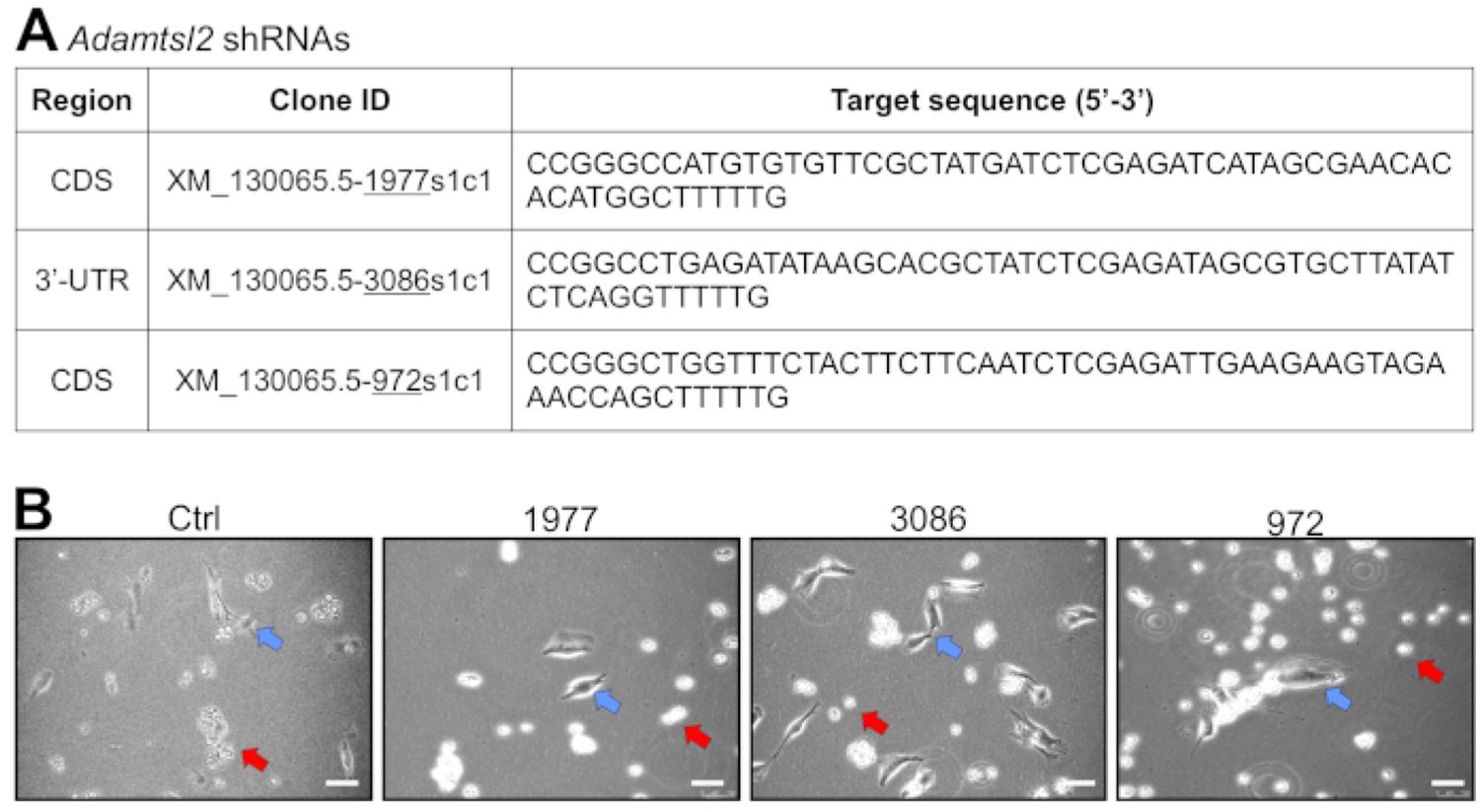
Obtain glycerol stocks of E. coli carrying target-specific shRNA plasmids and a control plasmid from commercial sources (Table of Materials).NOTE: Three different shRNA plasmids were used, targeting different regions of the murine Adamtsl2 mRNA. One shRNA was selected to target the 3’-untranslated region (3’UTR) of Adamtsl2 to facilitate rescue experiments with expression plasmids encoding recombinant full-length ADAMTSL2 or individual ADAMTSL2 protein domains. In addition, a scrambled shRNA plasmid was included as a negative control. Details of the shRNA sequences are provided in Figure 1A.
-
Thaw the shRNA bacterial glycerol stock at room temperature (RT). Transfer 10 µL of bacterial glycerol stock onto a Luria-Bertani (LB) agar plate supplemented with 100 µg/mL ampicillin (LB-Amp).NOTE: LB-Amp plates are prepared by autoclaving 1 L of LB medium (for 1 L: 10 g of tryptone, 5 g of yeast extract, 10 g of NaCl, adjust pH to 7.0) together with 12 g of agar. Let the medium cool down to ~50 °C and add ampicillin (stock solution: 50 mg/mL in sterile water) to a final concentration of µg/mL (LB-Amp agar). Immediately pour ~20 mL of LB-Amp agar in a 10 cm Petri dish and let the agar solidify before use. 1 L of LB-Amp agar is typically sufficient to pour about 40 10 cm Petri dishes. Petri dishes containing LB-Amp agar are stable for at least one month if stored sealed at 4 °C in the dark.
- Spread bacteria with sterile Drygalski spatula or any other appropriate method to achieve single bacterial colonies. Incubate bacterial plates upside down overnight at 37 °C.
-
- Plasmid preparation
- The next morning, remove the Petri dish with the individual bacterial colonies and store at 4 °C to avoid overgrowth of the bacterial colonies and the formation of satellite colonies. Seal the Petri dish if stored overnight or longer.
- In the afternoon, add 5 mL of LB-Amp medium into a polypropylene bacterial culture tube. Select a single bacterial colony from the Petri dish cultured overnight with a pipette tip and inoculate LB-Amp medium by ejecting the pipette tip in the bacterial culture tube containing the LB-Amp medium.
- Incubate bacterial culture overnight in a shaker at 250 rpm at 37 °C with the lid loosely attached to allow for aeration.
- Take out bacterial culture tube the next morning and keep at 4 °C to avoid bacterial overgrowth. In the afternoon, resuspend bacteria by vortexing and transfer 1 mL of the overnight bacterial culture in 50 mL of LB-Amp medium in a 250 mL conical flask. Incubate overnight in a shaker at 250 rpm at 37 °C.
-
On the next morning, transfer bacteria to a 50 mL disposable centrifuge tube and centrifuge bacteria at 6,000 x g at RT for 15 min. Remove the medium and continue with step 1.2.6.NOTE: If plasmid DNA cannot be isolated immediately, remove the LB-Amp medium after the centrifugation step and store the bacteria pellet at −20 °C.
-
Follow instructions for plasmid preparation kit (midi scale) to extract the plasmid DNA from the bacteria. Assess plasmid DNA quality by measuring the A260/A280 ratio using a spectrophotometer.NOTE: An A260/A280 ratio of >1.8 is desirable, indicating high purity of the plasmid DNA preparation. A lower A260/A280 ratio suggests contamination with protein or insufficient removal of extraction reagents. Additional plasmid purification steps may be required.
Materials
| Name | Company | Catalog Number | Comments |
|---|---|---|---|
| Acetone | Fisher Chemical | 191784 | |
| Agar | Fisher Bioreagents | BP1423 | |
| Ampicillin | Fisher Bioreagents | BP1760–5 | |
| Automated cell counter Countesse II | Invitrogen | A27977 | |
| Bradford Reagent | Thermo Scientific | P4205987 | |
| C2C12 cells | ATCC | CRL-1772 | |
| Chamber slides | Invitrogen | C10283 | |
| Chloroform | Fisher Chemical | 183172 | |
| DMEM | GIBCO | 11965–092 | |
| DMSO | Fisher Bioreagents | BP231–100 | |
| DNase I (Amplification Grade) | Invitrogen | 18068015 | |
| Fetal bovine serum | VWR | 97068–085 | |
| GAPDH | EMD Millipore | MAB374 | |
| Glycine | VWR Life Sciences | 19C2656013 | |
| Goat-anti-mouse secondary antibody (IRDYE 800CW) | Li-Cor | C90130–02 | |
| Goat-anti mouse secondary antibody (Rhodamine-red) | Jackson Immune Research | 133389 | |
| HCl | Fisher Chemical | A144S | |
| Incubator (Shaker) | Denville Scientific Corporation | 1704N205BC105 | |
| Mercaptoethanol | Amresco, VWR Life Sciences | 2707C122 | |
| Midiprep plasmid extraction kit | Qiagen | 12643 | |
| Myosin 4 (myosin heavy chain) | Invitrogen | 14–6503–82 | |
| Mounting medium | Invitrogen | 2086310 | |
| NaCl | VWR Life Sciences | 241 | |
| non-ionic surfactant/detergent | VWR Life Sciences | 18D1856500 | |
| Paraformaldehyde | MP | 199983 | |
| PBS | Fisher Bioreagents | BP399–4 | |
| PEI | Polysciences | 23966–1 | |
| Penicillin/streptomycin antibiotics | GIBCO | 15140–122 | |
| Petridishes | Corning | 353003 | |
| Polypropylene tubes | Fisherbrand | 149569C | |
| Protease inhibitor cocktail tablets | Roche | 33576300 | |
| Puromycin | Fisher Scientific | BP2956100 | |
| PCR (Real Time) | Applied Biosystems | 4359284 | |
| Reaction tubes | Eppendorf | 22364111 | |
| Reverse Transcription Master Mix | Applied Biosystems | 4368814 | |
| RIPA buffer | Thermo Scientific | TK274910 | |
| sh control plasmid | Sigma-Aldrich | 07201820MN | |
| sh 3086 plasmid | Sigma-Aldrich | TRCN0000092578 | |
| sh 972 plasmid | Sigma-Aldrich | TRCN0000092579 | |
| sh 1977 plasmid | Sigma-Aldrich | TRCN0000092582 | |
| Spectrophotometer (Nanodrop) | Thermo Scientific | NanoDrop One C | |
| SYBR Green Reagent Master Mix | Applied Biosystems | 743566 | |
| Trichloroacetic acid | Acros Organics | 30145369 | |
| Trizol reagent | Ambion | 254707 | |
| Trypan blue | GIBCO | 15250–061 | |
| Tryptone | Fisher Bioreagents | BP1421 | |
| Trypsin EDTA 0.25% | Gibco-Life Technology Corporation | 2085459 | |
| Water (DEPC treated and nuclease free) | Fisher Bioreagents | 186163 | |
| Western blotting apparatus | Biorad | Mini Protean Tetra Cell | |
| Yeast extract | Fisher Bioreagents | BP1422 |
Figure 1: Selection of stable C2C12 cells after transfection with shRNA-encoding plasmid DNA.

(A) Table showing the target region (CDS, coding sequence; 3’-UTR, 3’-untranslated region), clone ID (hereafter referred to as 1977, 3086, and 972), and sequence of the shRNAs used to target Adamtsl2. (B) The panels show the selection of C2C12 cells transfected with the control shRNA and the three Adamtsl2-targeting shRNAs. C2C12 cells were transfected with the shRNA plasmids and puromycin was added to the medium after 24 h. Puromycin-sensitive cells appear round and eventually detach during routine cell culture maintenance (red arrows). In contrast, puromycin-resistant cells harboring the integrated shRNA plasmids appear spindle-shaped, slightly elongated, attached, and viable (blue arrows). Scale bars = 100 µm.
2. Culturing and Transfection of C2C12 Cells and Puromycin Selection
- C2C12 cell culture
- Thaw C2C12 cells (Table of Materials) quickly in a 37 °C water bath and pour cells in sterile disposable 15 mL centrifuge tube containing 8 mL of Dulbecco’s modified Eagle medium (DMEM) medium supplemented with 100 units of penicillin and streptomycin antibiotics (serum-free DMEM) in a cell culture hood. Centrifuge cells for 3 min at 160 x g at RT.
- Aspirate supernatant and resuspend the cell pellet in 10 mL of serum-free DMEM supplemented with 10% fetal bovine serum (FBS) (complete DMEM). Transfer cells to 10 cm tissue culture treated plastic dishes. Incubate cells in a humidified incubator at 37 °C in a 5% CO2 atmosphere.
- On the next day, replace the medium with fresh complete DMEM.
- Expand low passage C2C12 cells on 3–4 10 cm dishes. When the C2C12 cells reach 50–60% confluence, aspirate the medium and rinse C2C12 cells with 10 mL of phosphate-buffered saline (PBS).
-
Add 1 mL of 0.25% trypsin-EDTA and incubate for 2 min at RT. Monitor cell detachment under a microscope and extend the incubation time if necessary, until most of the cells are detached.NOTE: Carefully tapping the dish can help to dislodge the cells.
- When most of the cells are detached, add 10 mL of complete DMEM to the dish, pipette the volume up and down several times, and transfer cell suspension to a sterile disposable 15 mL centrifuge tube. Centrifuge cells for 3 min at 160 x g at RT. Aspirate supernatant and resuspend the cell pellet in 2 mL of freezing medium (10% dimethyl sulfoxide [DMSO]/90% FBS).
-
Transfer two 1 mL aliquots per 10 cm dish in cryovials. Incubate in a cell-freezing container filled with RT isopropanol for at least 24 h in a −80 °C freezer resulting in a freezing rate of about −1 °C per min to avoid the formation of ice crystals. Store cells for long-term use in the vapor phase of liquid nitrogen.NOTE: The vendor recommends using C2C12 cells up to passage number 15. Authors’ experiences also showed that lower passage number cells showed more consistent and rapid differentiation into myotubes. It is essential to maintain C2C12 cells at low cell density (<50% confluence) to avoid premature onset of differentiation. Differentiating or differentiated C2C12 cells cannot be reverted into the original myoblast state and must be discarded.
- Preparing C2C12 cells for transfection
-
Culture undifferentiated C2C12 cells in a 10 cm dish until they reach 50–60% confluence. Aspirate the medium, rinse C2C12 cells with 10 mL of PBS, and add 1 mL of 0.25% trypsin-EDTA. Incubate for 2 min at RT, monitor cell detachment under a microscope, and extend the incubation time if necessary, until most of the cells are detached.NOTE: Carefully tapping the dish can help to dislodge the cells.
- When most of the cells are detached, add 10 mL of complete DMEM to the dish and transfer cell suspension to a sterile disposable 15 mL centrifuge tube. Centrifuge cells for 3 min at 160 x g at RT. Aspirate supernatant and resuspend the cell pellet in 4 mL of complete DMEM.
- Combine 10 µL of cell suspension with 10 µL of trypan blue in a 1.5 mL reaction tube, mix by pipetting up and down and carefully flicking the reaction tube, and transfer cells to a counting slide. Determine the cell number/mL using an automated cell counter.
- Dilute C2C12 cells to 50,000 cells/mL and seed 100,000 cells (2 mL) per well in a 6 well plate to achieve about 40–50% confluence after overnight incubation. Incubate cells in a humidified incubator at 37 °C in a 5% CO2 atmosphere.
-
- Transfection of C2C12 cells with the shRNA plasmid using polyethylenimine (PEI) and puromycin selection
- Preparation of PEI stock solution
- Dissolve 16 mg of PEI in 50 mL of sterile distilled water (concentration of stock solution: 0.32 mg/mL) in a 50 mL glass media bottle with a screw cap. Incubate the solution at 65 °C for 1 h and vortex the solution vigorously several times during the incubation period to completely dissolve the PEI.
-
Adjust to pH 8 by adding 15 µL of 1N HCl per 50 mL.CAUTION: HCl is corrosive. Concentrated HCl should be handled under the fume hood. Investigators should wear appropriate personal protective equipment.
- Freeze the PEI solution at −80 °C for 1 h with a loosely attached lid and rapidly thaw at 37 °C in a water bath to further enhance solubility. Repeat the freeze-thaw cycle 3x.
- Store PEI stock solution in 0.5 mL aliquots for one-time use at −20 °C.
- Transfection of C2C12 cells
- Combine 25.5 µL (8.5 µL/µg plasmid DNA) of the PEI stock solution with 100 µL of 25 mM NaCl in a 1.5 mL reaction tube and incubate for 5 min at 37 °C. Combine 3 µg of the plasmid DNA with 100 µL of 25 mM NaCl and incubate for 5 min at 37 °C.
- Combine the entire volume of diluted PEI reagent with the diluted plasmid and mix by gently pipetting up and down. Incubate for 25 min at 37 °C.
- During the incubation time, change the medium of the C2C12 cells intended for transfection to DMEM without FBS or antibiotics.
- Add the PEI/DNA transfection mix to C2C12 cells drop by drop and mix constantly by carefully moving the cell culture dish. Incubate in a humidified incubator at 37 °C in a 5% CO2 atmosphere. After 6 h, change the medium to complete DMEM.
- Puromycin selection
- 24 h after transfection, switch medium to selection medium (complete DMEM plus 5 µg/mL puromycin). Continue puromycin selection until puromycin-resistant C2C12 cells are obtained, which typically takes 10–14 days.
-
Expand puromycin resistant C2C12 cells at low cell density (<50–60% confluence), i.e., to maintain the undifferentiated state and cryopreserve of 6–10 vials for future experiments as described in steps 2.1.4–2.1.7.NOTE: Maintain puromycin-resistant C2C12 cells in the presence of 5 µg/mL puromycin for routine cell culture and expansion. However, puromycin was omitted in the differentiation experiments.
3. Phenotypic Analysis of C2C12 Differentiation
NOTE: The methods described below can easily be adapted for general phenotypic analysis of C2C12 myoblast differentiation into myotubes by varying the specific antibodies used in Western blotting or the gene specific primers used in the quantitative polymerase chain reaction (qPCR) analysis.
- C2C12 differentiation protocol and brightfield microscopy
- Seed 150,000 cells/well in a 12 well plate. Culture puromycin-resistant C2C12 stable cells in a 12 well plate in selection medium in a humidified incubator at 37 °C in a 5% CO2 atmosphere. Culture C2C12 cells in complete DMEM until they reach ~95% confluence.
-
To induce differentiation of C2C12 cells, replace complete DMEM with serum-free DMEM (day 0 of differentiation). Change medium every two days and follow C2C12 myotube formation using an inverted bright field microscope with camera.NOTE: Many protocols use 2% horse serum to differentiate C2C12 cells into myotubes. In authors’ hands, removing the serum entirely had a similar effect. Insulin is described as an additive to the cell culture medium to accelerate C2C12 differentiation. However, in the current protocol, C2C12 cells reliably differentiated into myotubes within 3–5 days after serum deprivation and addition of insulin was deemed unnecessary.
- Myosin heavy chain (MyHC) immunostaining to visualize myotubes
- Seed 50,000 puromycin-resistant C2C12 cells per chamber in an 8 well chamber slide in 500 µL of complete DMEM. Culture cells until they reach 95% confluence in complete DMEM (about 24 h).
-
To induce differentiation, switch from complete DMEM to 500 µL of serum-free DMEM (day 0 of differentiation). At the desired time point(s), aspirate the medium and rinse cells 3x with 0.5 mL of PBS.NOTE: Perform all following steps at RT.
-
Fix cells with 0.2 mL of 4% paraformaldehyde (PFA) diluted in PBS for 15 min. Rinse cells with 0.5 mL of PBS 3x for 5 min each.CAUTION: PFA is hazardous. Take appropriate precautions and discard paraformaldehyde solution as hazardous waste.
- Quench PFA with 0.2 mL of 0.5 M glycine in PBS for 5 min. Rinse cells with 0.5 mL of PBS 3x for 5 min each.
- Incubate cells with 0.2 mL of 0.1% non-ionic surfactant/detergent (Table of Materials) in PBS for 10 min to permeabilize the cell membrane. Block with 0.2 mL of 5% bovine serum albumin in PBS for 1 h. Rinse cells with 0.5 mL of PBS 3x for 5 min each.
- Incubate cells with 0.2 mL of MyHC antibody (1:200 in PBS) for 2 h. Rinse cells with 0.5 mL of PBS 3x for 5 min each.
- Incubate cells with 0.2 mL of goat-anti mouse rhodamine red-conjugated secondary antibody (1:200 in PBS). Rinse cells with 0.5 mL of PBS 3x for 5 min each.
- After removing PBS quantitatively, add one drop of mounting medium containing 4′,6-diamidino-2-phenylindole (DAPI) per chamber. Coverslip and cure mounting medium according to the manufacturer’s instructions. Seal with nail polish.
- Observe C2C12 cells using a fluorescence microscope using the appropriate filter set.
- Assessing knockdown efficiency by quantitative real-time polymerase chain reaction (qRT-PCR)
- Culture C2C12 cells under differentiation conditions in 12 well plates as described in section 3.1.
-
At the desired time point(s), remove the differentiation medium, rinse the cells once with 1 mL of PBS, and add 0.5 mL of RNA extraction reagent (Table of Materials) per well. Lyse the cells by carefully pipetting up and down and transfer cell lysate to a sterile 1.5 mL reaction tube. Isolate the RNA by carefully following the manufacturer’s protocol.CAUTION: The RNA extraction reagent contains phenol. It should be used under a fume hood. Collect the RNA extraction reagent waste and dispose of as hazardous waste. Investigators should wear appropriate personal protective equipment.
-
Dissolve the final RNA pellet in 20 µL of diethyl pyrocarbonate (DEPC)-treated water. Determine the quantity and quality of the RNA preparation using a spectrophotometer and determine the A260/A280 ratio as a quality measure.NOTE: A typical yield from 1 well of a 12 well plate is 5–7 µg of total RNA with an A260/A280 ratio of 1.8–2.
- To digest residual co-purified genomic DNA, dilute 1 µg of RNA in a total volume of 8 µL of DEPC-treated water in a PCR tube. Add 1 µL of 10x reaction buffer and 1 µL of DNase I (1 unit/µL) (Table of Materials) and incubate for 15 min at RT. Add 1 µL of stop solution (25 mM EDTA) and incubate at 70 °C for 10 min.
- To generate cDNA, combine 10 µL of DNase treated RNA with 10 µL of 2x reverse transcriptase master mix, containing the reverse transcriptase, random primers, 4 mM dNTP mix (1 mM each of dATP, dTTP, dGTP, and dCTP), and reverse transcriptase reaction buffer. Incubate the reaction in a thermocycler using the program as recommended by the manufacturer. Dilute cDNA with water in a 1:5 ratio.
- To prepare the qRT-PCR reaction, combine 5 µL of SYBR green qPCR master mix with 0.5 µL of gene-specific forward and reverse primers (stock: 10 µM), respectively, and 2 µL of DEPC-treated water per reaction. Pipette qPCR reaction in one well of a 96 well qRT-PCR plate and add 2 µL of diluted cDNA. Set up three technical replicates for each biological replicate and include a housekeeping gene such as Gapdh or Hprt1.
- Amplify the PCR product with a qRT-PCR thermocycler using the following program: 50 °C for 2 min (uracil-DNA glycosylase [UDG] activation), 95 °C for 2 min (dual-lock DNA polymerase activation), 40 cycles of 95 °C for 15 s, 60 °C for 30 s, and 72 °C for 1 min.
- Quantify qRT-PCR results using the DDCt method normalizing to a housekeeping gene, such as Gapdh or Hprt1.
- Assessing knockdown efficiency by Western blotting
- Seed 300,000 puromycin-resistant C2C12 cells per well in a 6 well plate for Western blot analysis. After 24 h, change medium to serum-free DMEM to initiate differentiation (day 0 of differentiation).
-
At the desired time point(s), collect two 1 mL of serum-free conditioned medium in a 1.5 mL reaction tube and centrifuge for 5 min at 500 x g at RT to remove detached cells and cell debris.NOTE: This step is necessary only if ECM proteins or other secreted proteins in conditioned medium are investigated. If the protein of interest is localized in the cytoplasm or is membrane bound, aspirate the cell culture medium and proceed with the cell lysis steps (3.4.7–3.4.12). Cell lysis should be performed in parallel to the protein precipitation from serum-free conditioned medium to minimize proteolytic degradation and other unintended consequences of prolonged storage of the cell layer.
-
After centrifugation, transfer 1 mL of conditioned medium in a new 1.5 mL reaction tube and precipitate proteins by adding 0.391 mL of a mixture of trichloroacetic acid (TCA) and non-ionic surfactant/detergent. Briefly vortex the mixture and incubate for 10 min on ice.NOTE: Prepare the protein precipitation mixture directly before use by combining 0.252 mL of 55% TCA and 0.139 mL of 1% non-ionic surfactant/detergent per mL of conditioned medium and vortex briefly. The solution becomes turbid.CAUTION: TCA is corrosive and must be handled appropriately.
-
Pellet the precipitated proteins at >16,000 x g for 10 min at 4 °C. Discard the supernatant.CAUTION: The supernatant contains TCA and should be collected separately and disposed of as hazardous material.
- Wash the protein pellet 3x with ice-cold acetone and centrifuge each time at >16,000 x g for 10 min at 4 °C.
-
Air-dry the protein pellet for 3–4 min at RT and dissolve in 50 µL of 1x sodium dodecyl sulfate polyacrylamide gel electrophoresis (SDS-PAGE) sample buffer (50 mM Tris pH 6.8, 2% SDS, 6% glycerol, and 0.004% bromophenol blue, supplemented with 5% β-mercaptoethanol). Boil the sample for 5 min at 95 °C and use for Western blotting to detect the desired target protein using standard procedures.CAUTION: β-Mercaptoethanol is odorous and toxic and must be handled in a fume hood.
- For intracellular and membrane bound proteins, rinse the cell layer once with 2 mL of PBS.
- Add 1 mL of PBS and dislodge cells by scraping them of the well using a cell scraper. Collect cells in a 1.5 mL reaction tube and centrifuge at 3,420 x g for 3 min at 4 °C. Wash the cell pellet 3x with 1 mL of PBS and centrifuge at 3,420 x g for 3 min at 4 °C each time.
- To lyse the cells, resuspend the cell pellet in 0.2 mL of lysis buffer (20 mM Tris-HCl pH 7.5, 150 mM NaCl, 1 mM Na2EDTA, 1 mM EGTA, 1% NP40, 1% sodium deoxycholate, 2.5 mM sodium pyrophosphate, 1 mM β-glycerophosphate, and 1 mM sodium orthovanadate) supplemented with 1x EDTA-free protease inhibitor cocktail reagent and incubate for 30 min on ice.
- Ultrasonicate for 15 s on ice with a power output setting of 10 at an operating frequency of 23 kHz.
- Remove cell debris by centrifugation at 13,680 x g for 20 min at 4 °C. Collect supernatant in a new 1.5 mL reaction tube and determine the protein concentration using a commercial Bradford assay or any other suitable method.
- For each sample, combine 100 µg of protein with 5x SDS-PAGE sample buffer in a total volume of 60 µL and boil for 5 min at 95 °C. Analyze samples by standard Western blotting procedures for the presence of the desired target protein.
Representative Results
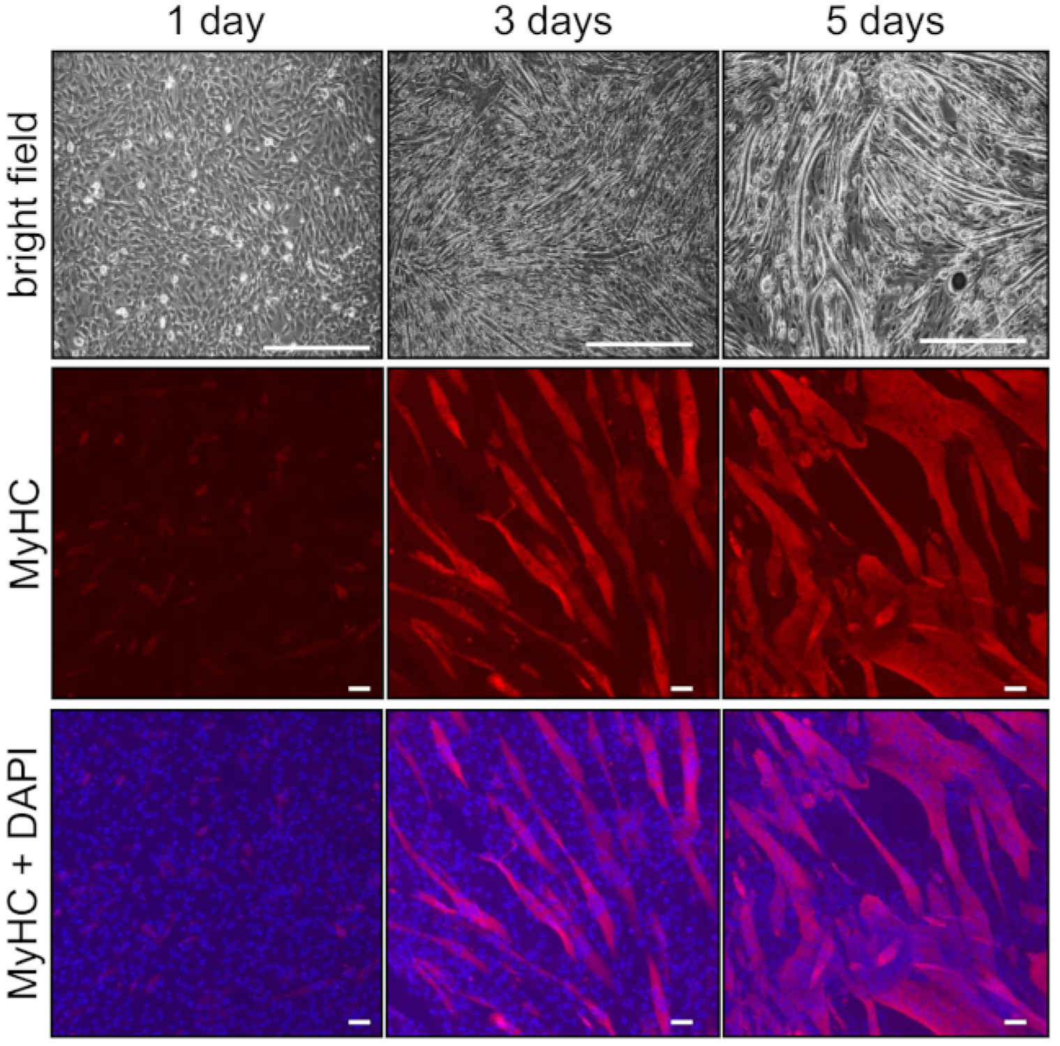
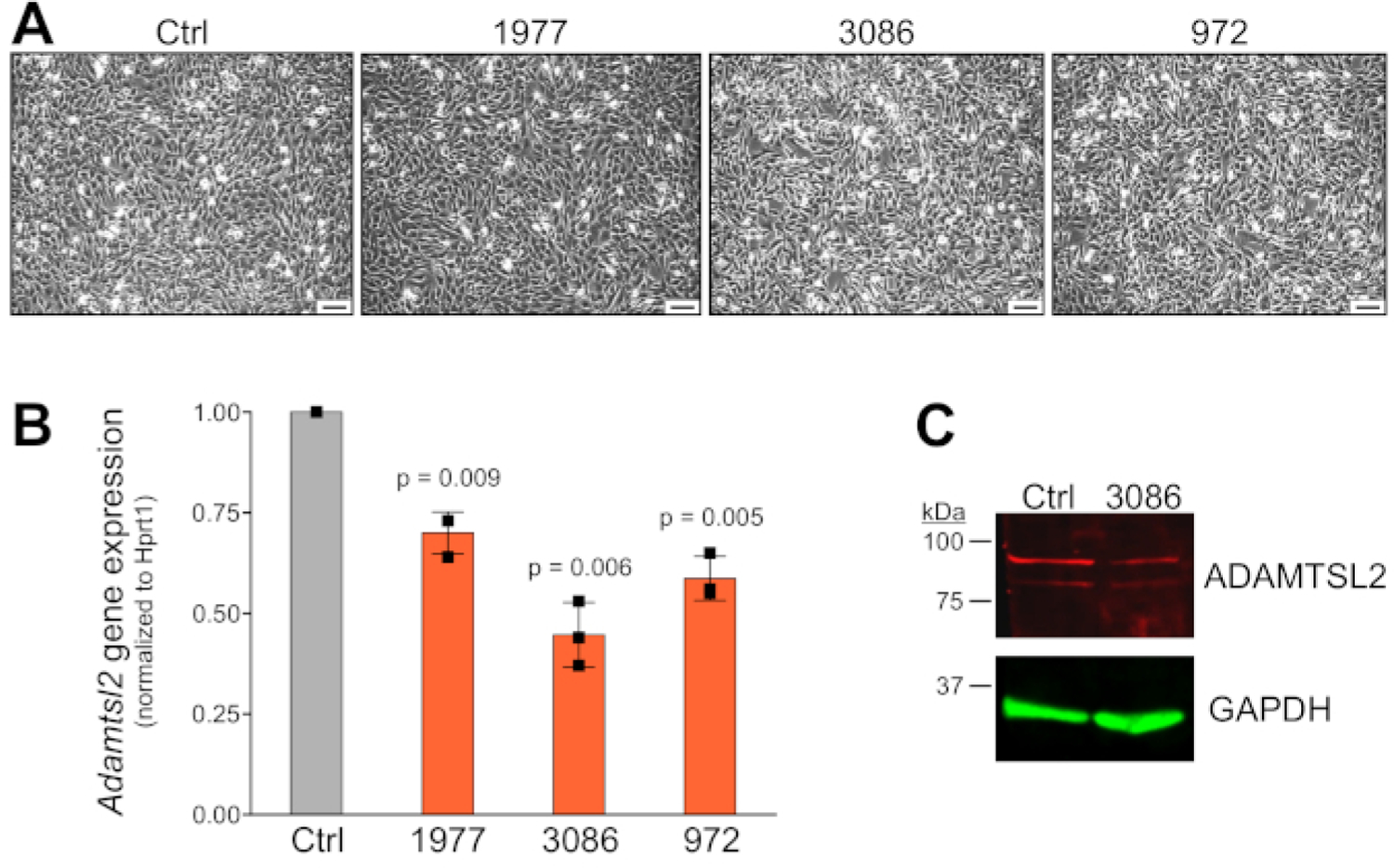
Selection of puromycin-resistant C2C12 can be achieved in 10–14 days after transfection due to efficient elimination of non-resistant, i.e., untransfected cells (Figure 1B). Typically, more than 80% of the cells detach from the cell culture dish and these cells are removed during routine cell maintenance. Puromycin-resistant C2C12 cells expressing the control (scrambled) shRNA retain the spindle-shape, elongated cell morphology at low cell density and the capability to differentiate into myotubes. C2C12 differentiation upon serum withdrawal can be monitored by bright field microscopy and by immunostaining for the myotube marker myosin heavy chain (MyHC) (Figure 2). MyHC-positive myotubes are observed between 3–5 days after differentiation initiation. Myotubes are multinucleated as shown by the presence of more than one DAPI-positive nucleus within the MyHC-positive cell boundaries. Figure 3A shows bright field images of stable C2C12 cells cultured in complete DMEM. The knockdown efficiency presented here ranges from 40–60% (Figure 3B). Since the mRNA was harvested in the proliferative state where little Adamtsl2 is expressed, the knockdown efficiency appears low, but the knockdown efficiency is expected to be larger at later time points during C2C12 differentiation, where endogenous Adamtsl2 is induced and thus expressed at much higher levels. Western blot analysis confirmed the successful knockdown of ADAMTSL2 in the cell lysate obtained from C2C12 cells stably expressing shRNA 3086 compared to control shRNA (Figure 3C).
Figure 2: C2C12 myoblast to myotube differentiation.

Bright field images of differentiating C2C12 cells show dense cobblestone appearance of the cells at the beginning of the differentiation (day 0–1) and multinucleated myotubes were observed after day 5 (upper row). Immunostaining of differentiating C2C12 cells with myosin heavy chain (MyHC, red), which is a marker for myotubes, is induced at day 3 of differentiation (middle panel). Nuclei were stained with DAPI and the merged image is shown in the lower panels. Scale bars = 50 µm.
Figure 3: Validation of stable knockdown in proliferating C2C12 cells.

(A) Bright field images of stable C2C12 cells cultured in complete DMEM. Scale bars = 300 µm. (B) qRT-PCR analysis of Adamtsl2 mRNA expression in stable C2C12 cells. Ct values were normalized to the housekeeping gene Hprt1. mRNA was harvested before the onset of differentiation. Error bars represent standard deviation. (C) Western blot analysis showing reduced ADAMTSL2 protein in the cell lysate/ECM fraction from C2C12 cells stably expressing shRNA 3086. Endogenous ADAMTSL2 was detected using a custom-made polyclonal peptide antibody (available upon request).
Discussion
We describe here a protocol for the stable knockdown of ECM proteins in C2C12 myoblasts and for phenotypic analysis of the differentiation of C2C12 myoblasts into myotubes. Several factors determine the outcome of the experiment and need to be considered carefully. Maintaining C2C12 cells in the proliferating phase is a critical step to keep the C2C12 cells in the myoblast precursor state. Retaining the capability of C2C12 cells to consistently differentiate into myotubes depends on i) the passage number of the cells, ii) the density of the cultured cells during routine maintenance, and iii) nutrient availability, requiring frequent and regular replenishment of the cell culture medium11,12,13. Due to some unknown mechanisms, higher passage number C2C12 cells also lose the potential for further myoblast fusion11. The instructions from the provider of the C2C12 cells suggest maintaining these cells up to passage number 15. Thereafter, the differentiation potential may be reduced and experiments with such cells may result in less consistent myotube formation. On the other hand, the cell density during maintenance can result in similar effects9. Reaching confluent C2C12 cell densities during routine cell culture promote initiation of C2C12 myoblast differentiation and thus may negatively influence the differentiation potential of the cell population. Therefore, it is of critical importance to prevent C2C12 cells from reaching high cell densities during routine C2C12 cell maintenance. This can be achieved by already sub-culturing C2C12 cells at low cell densities (<50–60% confluence). Serum starvation is used to induce C2C12 cell differentiation into myotubes. Therefore, maintaining cells for longer times without replenishing medium severely exhaust the nutrient and serum levels. Replenishing with fresh serum containing medium at least every two days can prevent the onset of unwanted premature differentiation due to nutrient and serum deprivation.
C2C12 differentiation is typically induced by serum starvation. The percentage of serum used to induce C2C12 differentiation can greatly influence the results, specifically the time it takes to form MyHC positive myotubes14,15. Several protocols show successful induction of differentiation under various serum concentrations. Use of 2–10% FBS or horse serum and complete serum deprivation has been reported and all conditions result in C2C12 myotube formation. The serum percentage or change in the serum lot can significantly alter markers for differentiation. In addition, the source of the serum may affect the experimental outcome, because the country of origin may or may not allow certain additives during bovine serum production8. The serum level could be adjusted to achieve the differentiation rate according to specific experimental requirements. Insulin can be added to the culture medium of C2C12 cells to accelerate differentiation and myotube formation16.
The ability to deliver plasmids encoding shRNA or recombinant proteins into C2C12 cells via transfection is an attractive feature of C2C12 cells. Several commercial liposome-based transfection reagents have been used previously to deliver plasmid DNA into C2C12 cells with variable reported transfection efficiencies17,18,19,20,21. PEI also shows reasonable transfection efficiency and is a cost-effective alternative to liposome-based transfection reagents. A comparison between transfection with a commercial liposome-based transfection reagent and PEI showed no considerable change in the transfection efficiency and slightly higher efficiency was found with PEI22. In our hands, transfection efficiency with PEI was sufficient to generate stable puromycin-resistant C2C12 cells. However, a critical step in this protocol is to keep the incubation time of the transfection reagent short. As mentioned above, C2C12 cells are sensitive to serum withdrawal and prolonged exposure to low serum conditions during transfection may favor C2C12 cell differentiation. Since transfection is performed in the absence of serum, the incubation time after transfection was kept short to resupply quickly serum-containing medium to prevent premature differentiation. Puromycin was added to the culture medium 24 h after transfection to initiate the selection of puromycin-resistant C2C12 cells. Methods for introducing plasmid DNA into differentiated myotubes include transfection (up to 85% efficiency reported), electroporation, or a biolistic approach23,24,25. Alternatively, stable C2C12 cells harboring an inducible shRNA plasmid could be generated to knock down genes at later stages of myotube differentiation or during myotube maturation26.
The method described here resulted in stable knockdown of ADAMTSL2 in C2C12 cells where its function during differentiation into myotubes can now be determined. The generation of stable knockdown cells may be especially important to study the function of proteins that are induced during myotube formation or maturation. Transient transfection with siRNA for example would not be sufficient to efficiently knockdown these genes, since the transient knockdown effect of siRNA may wean off after 5–7 days. Alternatively, the knockout of an ECM protein using CRISPR/Cas9 is technically more challenging and individual clones need to be selected and sequenced to ensure knockout of the desired gene. However, the evolving line of reagents may render CRISPR/Cas9 the method of choice for loss-of-function experiments in the future.
Acknowledgments
D.H. is supported by the National Institutes of Health (National Institute for Arthritis and Musculoskeletal and Skin Diseases, NIAMS, grant number AR070748) and seed funding from the Leni & Peter W. May Department of Orthopedics, Icahn School of Medicine at Mt. Sinai.
Footnotes
Video Link
The video component of this article can be found at https://www.jove.com/video/60824/
Disclosures
The authors have nothing to disclose.
References
- 1.Tanzer ML Current concepts of extracellular matrix. Journal of Orthopaedic Science: Official Journal of the Japanese Orthopaedic Association. 11 (3), 326–331 (2006). [DOI] [PMC free article] [PubMed] [Google Scholar]
- 2.Hubmacher D, Apte SS The biology of the extracellular matrix: novel insights. Current Opinion in Rheumatology. 25 (1), 65–70 (2013). [DOI] [PMC free article] [PubMed] [Google Scholar]
- 3.Sakai LY, Keene DR Fibrillin protein pleiotropy: Acromelic dysplasias. Matrix Biology. 80, 6–13 (2019). [DOI] [PubMed] [Google Scholar]
- 4.Iozzo RV, Gubbiotti MA Extracellular matrix: The driving force of mammalian diseases. Matrix Biology. 71–72, 1–9 (2018). [DOI] [PMC free article] [PubMed] [Google Scholar]
- 5.Le Goff C et al. ADAMTSL2 mutations in geleophysic dysplasia demonstrate a role for ADAMTS-like proteins in TGF-beta bioavailability regulation. Nature Genetics. 40 (9), 1119–1123 (2008). [DOI] [PMC free article] [PubMed] [Google Scholar]
- 6.Dubail J, Apte SS Insights on ADAMTS proteases and ADAMTS-like proteins from mammalian genetics. Matrix Biology. 44–46, 24–37 (2015). [DOI] [PubMed] [Google Scholar]
- 7.Koo BH et al. ADAMTS-like 2 (ADAMTSL2) is a secreted glycoprotein that is widely expressed during mouse embryogenesis and is regulated during skeletal myogenesis. Matrix Biology. 26 (6), 431–441 (2007). [DOI] [PubMed] [Google Scholar]
- 8.Khodabukus A, Baar K The effect of serum origin on tissue engineered skeletal muscle function. Journal of Cellular Biochemistry. 115 (12), 2198–2207 (2014). [DOI] [PubMed] [Google Scholar]
- 9.Bajaj P et al. Patterning the differentiation of C2C12 skeletal myoblasts. Integrative Biology. 3 (9), 897–909 (2011). [DOI] [PubMed] [Google Scholar]
- 10.Mashinchian O, Pisconti A, Le Moal E, Bentzinger CF The Muscle Stem Cell Niche in Health and Disease. Current Topics in Developmental Biology. 126, 23–65 (2018). [DOI] [PubMed] [Google Scholar]
- 11.Hindi L, McMillan JD, Afroze D, Hindi SM, Kumar A Isolation, Culturing, and Differentiation of Primary Myoblasts from Skeletal Muscle of Adult Mice. Bio-protocol. 7 (9), e2248 (2017). [DOI] [PMC free article] [PubMed] [Google Scholar]
- 12.Krauss RS, Joseph GA, Goel AJ Keep Your Friends Close: Cell-Cell Contact and Skeletal Myogenesis. Cold Spring Harbor Perspectives in Biology. 9 (2), a029298 (2017). [DOI] [PMC free article] [PubMed] [Google Scholar]
- 13.Lawson MA, Purslow PP Differentiation of myoblasts in serum-free media: effects of modified media are cell line-specific. Cells Tissues Organs. 167 (2–3), 130–137 (2000). [DOI] [PubMed] [Google Scholar]
- 14.Fujita H, Endo A, Shimizu K, Nagamori E Evaluation of serum-free differentiation conditions for C2C12 myoblast cells assessed as to active tension generation capability. Biotechnology and Bioengineering. 107 (5), 894–901 (2010). [DOI] [PubMed] [Google Scholar]
- 15.Cheng CS et al. Conditions that promote primary human skeletal myoblast culture and muscle differentiation in vitro. American Journal of Physiology-Cell Physiology. 306 (4), C385–395 (2014). [DOI] [PMC free article] [PubMed] [Google Scholar]
- 16.Conejo R, Valverde AM, Benito M, Lorenzo M Insulin produces myogenesis in C2C12 myoblasts by induction of NF-kappaB and downregulation of AP-1 activities. Journal of Cellular Physiology. 186 (1), 82–94 (2001). [DOI] [PubMed] [Google Scholar]
- 17.Dodds E, Dunckley MG, Naujoks K, Michaelis U, Dickson G Lipofection of cultured mouse muscle cells: a direct comparison of Lipofectamine and DOSPER. Gene Therapy. 5 (4), 542–551 (1998). [DOI] [PubMed] [Google Scholar]
- 18.Balcı B, Dinçer P Efficient transfection of mouse-derived C2C12 myoblasts using a matrigel basement membrane matrix. Biotechnology Journal. 4 (7), 1042–1045 (2009). [DOI] [PubMed] [Google Scholar]
- 19.Xia D et al. Overexpression of chemokine-like factor 2 promotes the proliferation and survival of C2C12 skeletal muscle cells. Biochimica et Biophysica Acta (BBA)-Molecular Cell Research. 1591 (1), 163–173 (2002). [DOI] [PubMed] [Google Scholar]
- 20.Tapia O, Gerace L Analysis of Nuclear Lamina Proteins in Myoblast Differentiation by Functional Complementation. Methods in Molecular Biology. 1411, 177–194 (2016). [DOI] [PMC free article] [PubMed] [Google Scholar]
- 21.Yamano S, Dai J, Moursi AM Comparison of transfection efficiency of nonviral gene transfer reagents. Molecular Biotechnology. 46 (3), 287–300 (2010). [DOI] [PubMed] [Google Scholar]
- 22.Luo J et al. An efficient method for in vitro gene delivery via regulation of cellular endocytosis pathway. International Journal of Nanomedicine. 10, 1667–1678 (2015). [DOI] [PMC free article] [PubMed] [Google Scholar]
- 23.Sandri M, Bortoloso E, Nori A, Volpe P Electrotransfer in differentiated myotubes: a novel, efficient procedure for functional gene transfer. Experimental Cell Research. 286 (1), 87–95 (2003). [DOI] [PubMed] [Google Scholar]
- 24.Yi CE, Bekker JM, Miller G, Hill KL, Crosbie RH Specific and potent RNA interference in terminally differentiated myotubes. Journal of Biological Chemistry. 278 (2), 934–939 (2003). [DOI] [PubMed] [Google Scholar]
- 25.Antolik C, De Deyne PG, Bloch RJ Biolistic transfection of cultured myotubes. Science’s STKE. 2003 (192), PL11 (2003). [DOI] [PubMed] [Google Scholar]
- 26.Shintaku J et al. MyoD Regulates Skeletal Muscle Oxidative Metabolism Cooperatively with Alternative NF-kappaB. Cell Reports. 17 (2), 514–526 (2016). [DOI] [PMC free article] [PubMed] [Google Scholar]


