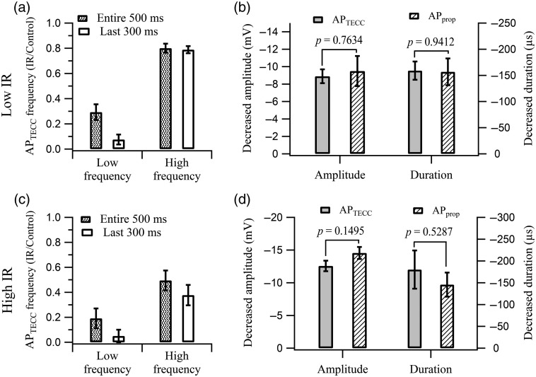
Fig. 2.
IR light pulses suppressed the firing and inhibited the AP amplitude and duration. (a) IR light pulses with 7.1-mW power significantly reduced the firing at low frequencies (10 to 20 Hz) to () while only decreasing the firing at high frequencies (40 to 60 Hz) to (). The ratios of firing frequency calculated from the 500-ms IR light illumination period are presented as the patterned bars. The open bars represent the ratios calculated based on the firing frequency during the last 300 ms of the IR light illumination period, when the temperature rise on the surface of axons had reached steady-state (see Fig. S1 in the Supplementary Material). In this case, the inhibition ratio was () for low frequencies and () for high frequencies. (b) IR light with 7.1-mW power suppressed the amplitude and duration of () and the () to comparable levels. (c) Reduction in firing frequency () at 13.1-mW IR light pulses illumination. The firing at low frequencies and high frequencies were reduced to () and (), respectively, by IR light pulses during the entire 500-ms illumination period. When the firing frequency of during the last 300 ms of the illumination period was evaluated, the inhibition was further reduced to () for low frequencies and () for high frequencies. (d) Suppression in amplitude and duration between () and () was similar when 13.1-mW IR light pulses were used.

