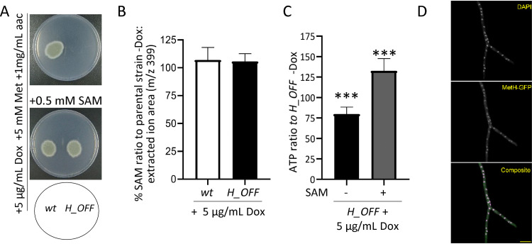
FIG 4.
External SAM reconstitutes ATP levels and growth. (A) The addition of SAM to the medium reconstituted growth of the H_OFF strain in restrictive conditions. The phenotypic analysis was repeated in two independent experiments. Representative plates are shown. (B) The levels of SAM did not significantly decrease upon Dox administration (5 μg/ml) in wild-type or H_OFF strains. Graphs depict the means and SD of three biological and two technical replicates. Data were analyzed by using one-way analysis of variance with a Bonferroni posttest adjustment. (C) The presence of SAM in the medium (0.5 mM) prevented the decrease in ATP levels observed in the H_OFF strain upon Dox addition. In fact, ATP levels were increased compared to the minus Dox condition. Graphs depict the means and SD of two biological and four technical replicates. (D) Expression of MetH-GFP in the H_OFF background showed that the protein localized in both the cytoplasm and the nucleus. Singular channels are shown in gray scale. In the composite image, magenta indicates DAPI and green indicates GFP. Scale bar, 10 μm.

