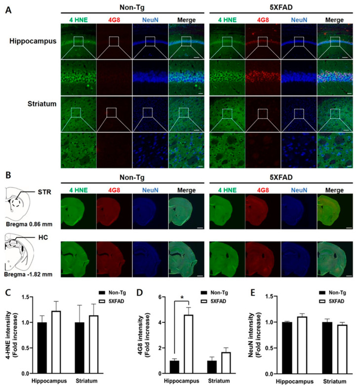
Figure 4.
Triple immunofluorescence staining of the hippocampus and striatum of the 4-month-old non-Tg control and 5XFAD mice. (A) Confocal microscopic images of triple immunofluorescence with 4-HNE, 4G8, and NeuN antibodies in the hippocampus (HC) and striatum (STR). (B) Schematic diagram of the analyzed brain regions and half-brain immunofluorescence images. The dashed circle line in the diagrams indicates the region of the interest. (C) No differences between the two groups in the 4-HNE levels of the hippocampus and striatum were found. (D) Levels of 4G8-positive signals were significantly higher in the hippocampus of 5XFAD than that in control mice, but not in the striatum. (E) No differences in intensities of NeuN-positive signals were found between the two groups. Top row: scale bar indicates 100 μm; bottom row: scale bar indicates 20 μm. Non-Tg, n = 4; 5XFAD, n = 6. * p < 0.05.

