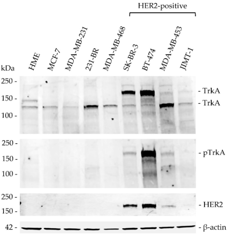
Figure 3.
TrkA, phospho-TrkA and HER2 protein expression in a panel of breast cancer cell lines. Western blotting for TrkA was performed with cellular proteins extracted from HME (non-tumourigenic human mammary epithelial) cells and the following breast cancer cell lines: MCF-7 (luminal A), MDA-MB-231 (triple-negative) and 231-BR (brain metastatic derivative), MDA-MB-468 (triple-negative); as well as the following the HER2-positive cell lines: SK-BR-3, BT-474, MDA-MB-453 and JIMT-1. TrkA was detected as a 140 kDa band in all cell lines. In addition, a second band at 180 kDa was observed in SK-BR-3 and BT-474 cell lines. Another band at 150 kDa was detected only in HME cells. The intensity of TrkA immunoreactive bands was higher in the HER2-positive cell lines SK-BR-3, BT-474 and MDA-MB-453. Phospho-TrkA (p-TrkA) immunoreactive bands were observed at 180 kDa in SK-BR-3, BT-474 and MDA-MB-453 cell lines. HER2 protein expression was confirmed across all HER2-positive breast cancer cell lines and β-actin protein expression was used as the equal loading control.

