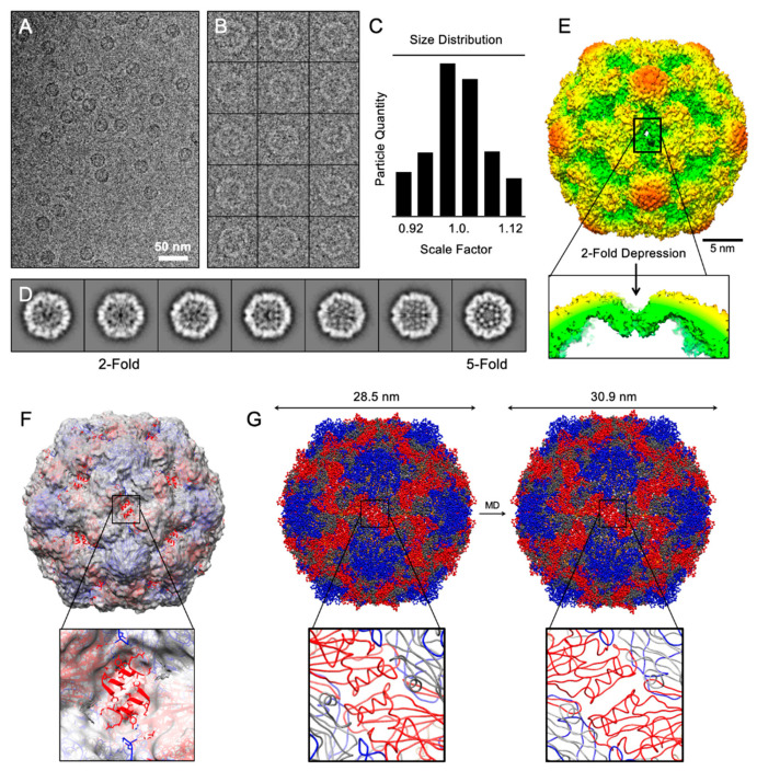
Figure 2.
Structural analysis of CVB3 virus-like particle. (A) Field view of cryo-EM micrographs of frozen-hydrated CVB3-VLPs. (B) A gallery of selected particles used towards structure determination (inverted). (C) Particle sorting by scale factor using Polar Fourier Transform method. Particles within ±3% of the average were used to calculate 2D-referece-free class averages shown in (D). (E) 3D surface rendering 2-fold view of CVB3-VLP colored by radial distance from cold to warm. A depression at 2-fold axis is pointed out. (F,G) Rigid-body fitting and dynamic flexible fitting of CVA6-VLP into cryo-EM density revealing the expanded size as a result of 2-fold depression.

