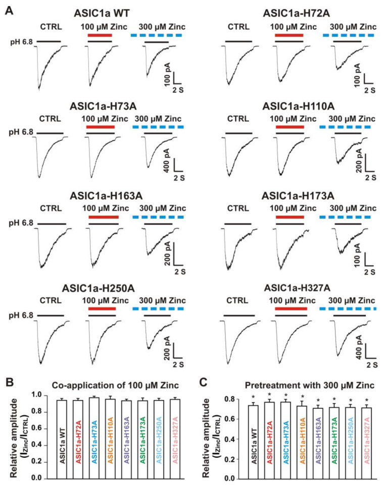
Figure 8.
Zinc effects on currents from ASIC1a WT and ASIC1a mutants of histidine residues in the extracellular domain of ASIC1a. (A) Representative traces show that co-application of zinc at a concentration of 100 µM had no effect on peak amplitude of the currents from ASIC1a WT, ASIC1a-H72A, ASIC1a-H73A, ASIC1a-H110A, ASIC1a-H163A, ASIC1a-H173A, ASIC1a-H250A, and ASIC1a-H327A; whereas pretreatment with zinc at a concentration of 300 µM displayed an inhibitory effect on peak amplitude of the currents from ASIC1a WT, ASIC1a-H72A, ASIC1a-H73A, ASIC1a-H110A, ASIC1a-H163A, ASIC1a-H173A, ASIC1a-H250A, and ASIC1a-H327A. The solid red and black lines represent 100 µM zinc and pH 6.8 application, respectively (each recoding with 7 s of zinc and pH 6.8 application). The dashed blue line represents pretreatment with 300 µM zinc in the pH 7.4 solution with a duration of 2 min. (B,C) Statistical bar graphs show relative peak amplitudes of the ASIC1a/3 currents by co-application of zinc (B, n = 8 to 10) and pretreatment with zinc (C, n = 6 to 10). There was no significant difference for zinc inhibition among ASIC1a WT and their histidine mutants (p > 0.05, ANOVA). For ASIC1a mutation, histidine residues in the extracellular domain of ASIC1a were replaced with alanine. Whole-cell patch-clap recording was performed and currents from homomeric ASIC1a WT and its histidine mutants were activated by a drop in pH from 7.4 to 6.8. Data are presented as mean ± SEM. CTRL, control. * p < 0.05 (t-test).

