Abstract
This study compared the ability of the lichen Evernia prunastri, chitin and chitosan to take up Cu2+ and Zn2+. It was hypothesized that chitin and chitosan have an accumulation capacity comparable to the lichen, so that these biopolymers could replace the use of E. prunastri for effective biomonitoring of Cu and Zn air pollution. Samples of the lichen E. prunastri, as well as chitin (from shrimps) and chitosan (from crabs), were incubated with Cu and Zn solutions at concentrations of 0 (control), 10, 25, 50, 75, and 100 µM and analyzed by Inductively Coupled Plasma Mass Spectrometry (ICP-MS). Metal concentrations accumulated by lichen, chitin and chitosan samples were strongly and linearly correlated with the concentrations in the treatment solutions. The lichen always showed significantly higher accumulation values compared to chitin and chitosan, which showed similar accumulation features. The outcomes of this study confirmed the great effectiveness of the lichen Evernia prunastri for environmental biomonitoring and showed that chitin and chitosan have a lower accumulation capacity, thus suggesting that although these biopolymers have the potential for replacing E. prunastri in polluted areas, their suitability may be limited in areas with intermediate or low pollution levels.
Keywords: bioaccumulation, biopolymers, biosorption, Cu, ion exchange, Zn
1. Introduction
The use of lichens for biomonitoring of trace metal air pollution is well established [1] and accepted to the point that these organisms are also used in environmental forensics [2,3] and decision-making processes [4]. Lichens do not have a root system and their mineral nutrition depends mainly on atmospheric inputs; they lack protective structure such as a cuticle and stomata, and trace metals can accumulate in their thalli up to levels well in excess of metabolic requirements [5]. Excluding particle interception, metal accumulation by lichens involves dynamic processes of uptake and release until an equilibrium with the surrounding environment is reached [6]. Accumulation of metal ions at extracellular binding sites on the cell wall by passive mechanisms is a well-documented, reversible process, largely dependent on the nature of the exchange sites and the affinity of the metal ions for these sites [7,8].
The major mechanisms of metal accumulation in lichens are particle deposition and extracellular ion exchange, which may account for up to 95% of the total [9]. Therefore, biomonitoring of airborne metal pollution by lichens mostly relies on these two mechanisms. This is also confirmed by: (i) the positive correlation between the elemental contents in the bulk deposition and in the thalli [10] and (ii) the similarity in the metal accumulation capacity of living and dead (devitalized) thalli in some transplant experiments [11]. Intracellular accumulation is commonly very limited, since excessive concentration of metals into the cell may cause severe damage [5]. The partitioning of extra- and intracellular metal in lichen thalli depends on the species and on the element [12,13]. For example, lichens demonstrate the ability to accumulate high concentrations of Cu and Zn extracellularly [14,15,16]. Both of these metals are of great environmental interest, being associated with atmospheric emissions from vehicular traffic [17]. For this reason, Cu and Zn have been commonly included in biomonitoring studies with lichens aimed at identifying and characterizing pollution sources in urban environments [18,19,20,21]. The opportunity and the convenience of environmental biomonitoring is seldom disputed with regard to some controversial issues, such as the definition of proper background values [22], the identification of the detection limit for a specific effect [23] and, above all, the large data variability [24], which is a possible cause of uncertainty in the results [25]. In addition, an ethical issue arise because, in large-scale and/or repeated surveys, a large amount of lichen material is required, which may drive a remarkable decrease in the lichen vegetation. This is also likely when using lichen transplants, where samples are collected from an unpolluted site and exposed elsewhere, with the risk of causing a dramatic reduction in the abundance of the selected species. Replacement of living organisms by passive samplers such as cellulose or quartz filters for the interception of airborne particles or cation exchange filters for the interception of elements in ionic form is simple and relatively inexpensive, although often not as effective as lichens [26].
Chitin (N-acetyl-D-glucosamine), after cellulose, is the second most abundant biopolymer worldwide, with an annual estimated biosynthesis of 100 million tons [27]. Together with its deacetylated form, chitosan, glucosamine-derived biopolymers are important components of the cell wall of fungi [28], as well as of several parts of crustaceans, insects, other arthropods, some bacteria, and mollusks [29]. Chitin and chitosan have a high adsorption capacity and, because of this, they have been widely used for the removal of toxic metals from wastewaters [30,31,32]. Fränzle [33] suggested using chitin for environmental biomonitoring and biogeochemical studies, and demonstrated the effectiveness of both the exoskeleton of living insects (cricket, Gryllus assimilis) and isolated chitin grafted on glass at taking up metals from different environmental media. Grafted chitin and chitin surfaces can be analyzed with minimal harm to organisms [33,34] because chitin can be dissolved in a mixture (solubility: 25–30 g chitin/L) of dimethylformamide (DMF), other carboxamides, and lactams, providing sizable amounts of lithium salts (e.g., chloride, nitrate, perchlorate, but not acetate). By adding water to the dissolving mixture, chitin re-precipitates as fibers or films without chemical changes. The Li+ solution in DMF is then put to the chitin surface and a thin layer is plainly removed, without producing cracks, holes, or craters. This extraction procedure should be repeated at least eight times [35].
While lichens have been tentatively used as biosorbents for wastewater remediation [36], the use of chitin and chitosan for monitoring airborne metals in ionic form remains unexplored. The aim of this study was to compare the ability of the lichen Evernia prunastri, chitin and chitosan to take up Cu2+ and Zn2+. The lichen species Evernia prunastri (L.) Ach., being widely used in field and laboratory studies [37,38,39,40], was chosen for the experiment because of its documented ability to bioaccumulate great amounts of trace elements and to reflect atmospheric bulk deposition [10,39,41]. In addition, the fruticose (shrubby) habitus of this epiphytic (tree-inhabiting) lichen allows easy handling of the thalli. We hypothesized that chitin and chitosan have an accumulation capacity comparable to the lichen, so that these biopolymers could replace the use of E. prunastri for effective biomonitoring of Cu and Zn air pollution.
2. Materials and Methods
2.1. Experimental Approach
Apparently healthy thalli of E. prunastri were harvested from a remote area of Tuscany, central Italy (43°11′60″ N, 11°21′33″ E, 310 m a.s.l.), far from any local source of air pollution. In the laboratory, extraneous material such as bark, insects, and other lichen species, were removed from the lichens with plastic tweezers and left to acclimate for 24 h in a climatic chamber at 15 ℃, RH = 65%, 40 µmol/m2/s photons PAR. Commercial chitin (from shrimps) and chitosan (from crabs) were used (Sigma-Aldrich, St. Louis, MO, USA).
Samples (ca. 200 mg) of the lichen and flakes of chitin and chitosan were separately put inside small nylon bags of ca. 8 cm3 (mesh 10 µm, side ca. 2 cm), that were first abundantly sprayed with deionized water to remove particles deposited onto the surface. The bags were then soaked by stirring for 1 h with individual Cu2+ and Zn2+ solutions provided as CuCl2 and ZnCl2 respectively, maintaining a constant 200/50 w/v ratio, at concentrations of 0 (control), 10, 25, 50, 75, 100 µM. These concentrations were selected by taking into account previous studies on element uptake in lichens [12] and are within the ranges of ecologically relevant levels found in polluted environments, such as urban and industrial areas [42]. The pH of the solutions was adjusted to 5.5 to maximize metal uptake [43]. After treatments, samples were rinsed three times for 5 s in deionized water to remove unbound ions simply deposited over the thallus surface. Samples were then allowed to air-dry on absorbing paper for 24 h in a climatic chamber, as described above, to allow possible later uptake [7]. The experiment was replicated independently three times.
2.2. Chemical Analysis
The total Cu and Zn content of whole lichen thalli, chitin and chitosan was measured by acid digestion in a microwave digestion system (Ethos 900, Milestone) using 3 mL of 70% HNO3, 0.2 mL of 60% HF and 0.5 mL of H2O2, and subsequent quantification by ICP-MS (Sciex Elan 6100, Perkin-Elmer, Waltham, MA, USA). One procedural blank and one sample of the certified material IAEA-336 ‘lichen’ were always included in the analysis. Recoveries were in the range 98–102% and precision of analysis, expressed as coefficient of variation of five replicates, was within 5% for all elements. Results are expressed on a dry weight basis.
2.3. Statistical Analysis
Linear regressions were run between metal concentrations in the treatment solutions and those accumulated by the lichen, chitin and chitosan to check for linear accumulation trends. Significance of differences (p < 0.05) between accumulated values in the three matrices were checked with the student t test, correcting for multiple comparisons according to Benjamini and Hochberg [44]. Data normality was checked with the Shapiro–Wilk test and equality of variances with the Levene test. All calculations were run using the free software R [45].
3. Results
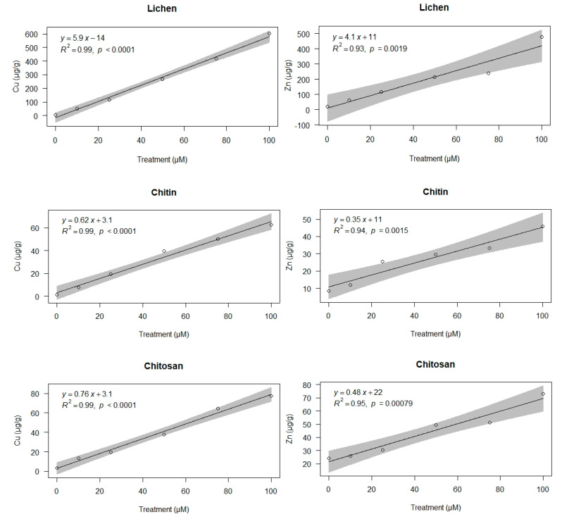
Metal concentrations accumulated by lichen, chitin and chitosan samples after incubation with Cu and Zn solutions were strongly and linearly correlated with the concentrations in the treatment solutions (Figure 1). All regression models showed residuals with normal distributions and homogeneous variances. The slopes of the regression lines for lichen samples were always ca. 10-fold higher than those of chitin and chitosan, which were similar.
Figure 1.
Copper and zinc concentrations (µg/g) in lichen, chitin, and chitosan samples after incubation with Cu (left) and Zn (right) solutions at the concentration 0 (control), 10, 25, 50, 75, and 100 µM. The grey area indicates the IC95 confidence interval.
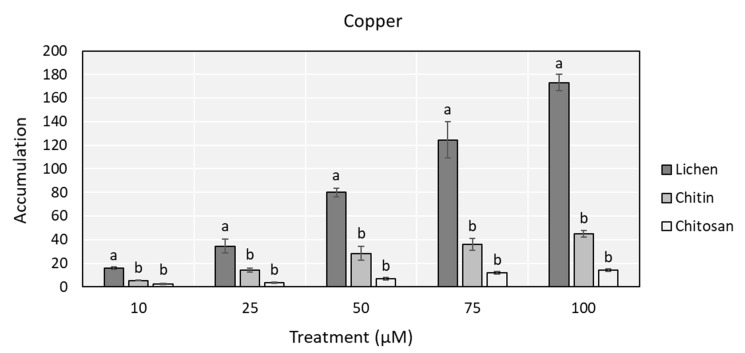
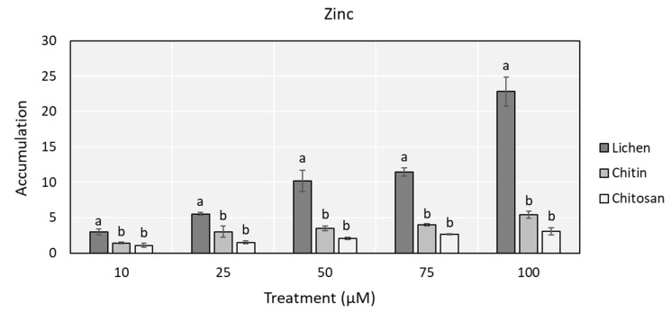
Control samples of lichen and chitosan showed similar concentrations for both Cu and Zn, while those of chitin were significantly lower (Table 1). For this reason, although differences in absolute concentrations were striking, to normalize for different starting concentrations, comparisons were made on data expressed as ratios to control values (Figure 2). Lichens always showed significantly higher accumulation values than chitin and chitosan, which presented similar accumulation features.
Table 1.
Metal concentrations (mean ± SE, µg/g·dw) in control samples. Different letters (a, b) indicate statistically significant (p < 0.05) differences among matrices.
| Element | Lichen | Chitin | Chitosan |
|---|---|---|---|
| Cu | 3.4 ± 0.1 a | 1.4 ± 0.4 b | 3.2 ± 0.3 a |
| Zn | 21 ± 2.5 a | 8.4 ± 2.2 b | 24 ± 2.1 a |
Figure 2.
Copper and zinc accumulation (ratio between treated and control value ± SE) in lichen, chitin, and chitosan samples after incubation with Cu (up) and Zn (down) solutions at the concentration of 10, 25, 50, 75, and 100 µM. Different letters (a, b) indicate statistical significant differences between matrices (p < 0.05).
4. Discussion
Papers reporting on lichen biomonitoring surveys almost invariably state that the peculiarity of lichens to accumulate and tolerate huge amounts of toxic metals is especially related to their ability to trap airborne particles. As an example, Nieboer et al. [6] stated that “trapping of particulates contributes significantly to the elemental levels found in lichens”. Nevertheless, here we have clearly documented that accumulation in ionic form is also very important: when elements are provided in ionic form only, they are nicely and quickly accumulated proportionally to the amount available in the medium. On the contrary, there is plenty of literature reporting metal adsorption by chitin and chitosan from aqueous solutions (see e.g., [30,46]), but very little is known about their ability to intercept and trap metal-rich particles from dry deposition. Comparative data are available for some elements (Mn, Ni, La, Ce, Sm, Eu, Dy, and Yb) accumulated in chitin from the peel of Pandalus borealis shrimp and grafted chitin from the same source and particle attachment was observed in Ni solid phases of different kinds [47,48]. Dust particles can be retained by chitin, while also transferring ions: we studied this concerning nickel for both ground minerals (Millerite NiS, Nickelin NiAs) and insoluble Ni salts (oxalate, hexacyanoferrate(II), carbonate), and found sizable retention of Ni from these sources [47]. At normal pH, that is pH >> pzzp (point of zero zeta potential ≈ 3.5 for chitin), chitin is negatively charged; many particles found in the environment are positively charged and thus may stick to chitin. Whereas ion exchange normally does not produce fractionation, adsorption tendencies to chitin do vary with metal and oxidation state [49].
The binding capacity of chitin is about 40 µmol/g irrespective of the element [30,49,50]. The cation exchange capacity of E. prunastri is known to be ca. 150 µmol/g [51], roughly corresponding to ca. 40 µmol/g of lichen chitin, based on a weight share of chitin around 25–30%. Similar conclusions were reached by Puckett et al. [52], which showed that Cu saturation in lichens occurs at ca. 40 µmol/g. Thus, the binding capacity of lichens and chitin/chitosan is similar. Moreover, the differential metal affinity for Cu and Zn is also similar in lichens and chitin/chitosan, with Cu being much higher than Zn [6,53].
In lichens from unpolluted areas, it has been shown that the extracellular fraction of Cu and Zn, which are micronutrients, is as low as 5% and 1%, respectively [12]. The cellular fractionation may account for great differences in the starting condition for metal adsorption: in chitin and chitosan all Cu and Zn is adsorbed, while in lichens the amounts bound to the cell wall may be as low as 0.17 and 0.21 µg/g·dw, thus leaving a very high potential for metal binding. In addition, in living organisms, intracellular uptake may surely play an important role in sequestering important amounts of the provided elements, especially when they are soluble micronutrients [6]. In the lichen Xanthoria parietina, intracellular concentrations can represent up to 15–30% of total Cu and Zn, respectively [12]. Moreover, in addition to free chitin and chitosan, the cell wall of the lichen mycobiont is known to contain chitin– and chitosan–glucan complexes [54], which are the main structural polysaccharides of the cell walls of fungi [55,56]. Lastly, it is generally accepted that ion exchange is the dominant mode of uptake in lichens, while electrolyte sorption plays only a minor role [52].
All of the above reasons may account for a larger accumulation of Cu and Zn in lichens than in chitin and chitosan, as we have found in the present study. Metal uptake by chitin and chitosan is reported as determined by adsorption processes [30], with adsorption depending on several factors such as chitin/chitosan biomass and exposure time [31]. A biomass of 2–5 g/L of chitin was found to be optimal for maximum divalent metal ion removal [53], and our experimental conditions of 4 g/L are consistent with these values. The adsorption of metals to chitin is very fast, and it has been shown that full equilibrium is already reached after 30 min [53], and in some cases there is evidence that 10 minutes are enough for chitin-adsorption equilibrium except for a few elements, like Fe [35,48]. Our experimental time of 60 min was thus enough to warrant full adsorption equilibrium with metal ions in the treatment solutions.
5. Conclusions
This study confirmed that the lichen Evernia prunastri effectively accumulated Cu2+ and Zn2+ over a wide range of concentrations, further supporting the use of this lichen species for environmental biomonitoring. The accumulation capacity of chitin and chitosan was comparably lower, thereby suggesting that although these two biopolymers may, in fact, replace E. prunastri, this may be appropriate in areas with intermediate or low pollution levels.
Author Contributions
S.L. and A.V. conceived and designed the experiments; A.V. and D.D. performed the experiments; S.L. and A.V. analyzed the data; F.M., F.B. and M.E. contributed analysis tools; S.L. wrote the paper, S.F. supervised the text. All authors have read and agreed to the published version of the manuscript.
Funding
This research received no external funding.
Conflicts of Interest
The authors declare no conflict of interest.
References
- 1.Loppi S. Lichens as sentinels for air pollution at remote alpine areas (Italy) Environ. Sci. Pollut. R. 2014;21:2563–2571. doi: 10.1007/s11356-013-2181-0. [DOI] [PubMed] [Google Scholar]
- 2.Loppi S. May the diversity of epiphytic lichens be used in environmental forensics? Diversity. 2019;11:36. doi: 10.3390/d11030036. [DOI] [Google Scholar]
- 3.Purvis O.W., Williamson B.J., Spiro B., Udachin V., Mikhailova I.N., Dolgopolova A. Lichen monitoring as a potential tool in environmental forensics: Case study of the Cu smelter and former mining town of Karabash, Russia. Geol. Soc. Spec. Publ. 2013;384:133–146. doi: 10.1144/SP384.6. [DOI] [Google Scholar]
- 4.Cecconi E., Fortuna L., Benesperi R., Bianchi E., Brunialti G., Contardo T., Nuzzo L.D., Frati L., Monaci F., Munzi S., et al. New interpretative scales for lichen bioaccumulation data: The italian proposal. Atmosphere. 2019;10:136. doi: 10.3390/atmos10030136. [DOI] [Google Scholar]
- 5.Bačkor M., Loppi S. Interactions of lichens with heavy metals—A review. Biol. Plant. 2009;53:214–222. doi: 10.1007/s10535-009-0042-y. [DOI] [Google Scholar]
- 6.Nieboer E., Richardson D.H.S., Tomassini F.D. Mineral uptake and release by lichens: An overview. Bryologist. 1978;81:226–246. doi: 10.2307/3242185. [DOI] [Google Scholar]
- 7.Brown D.H., Beckett R.P. The role of the cell wall in the intracellular uptake of cations by lichens. In: Brown D.H., editor. Lichen Physiology and Cell Biology. Springer; Boston, MA, USA: 1985. pp. 247–258. [DOI] [Google Scholar]
- 8.Loppi S., Di Lucia A., Vannini A., Ancora S., Monaci F., Paoli L. Uptake and release of copper ions in epiphytic lichens. Biologia. 2020 doi: 10.2478/s11756-020-00522-x. [DOI] [Google Scholar]
- 9.Branquinho C., Brown D.H., Catarino F. The cellular location of Cu in lichens and its effects on membrane integrity and chlorophyll fluorescence. Environ. Exp. Bot. 1997;38:165–179. doi: 10.1016/S0098-8472(97)00015-4. [DOI] [Google Scholar]
- 10.Loppi S., Paoli L. Comparison of the trace element content in transplants of the lichen Evernia prunastri and in bulk atmospheric deposition: A case study from a low polluted environment (C Italy) Biologia. 2015;70:460–466. doi: 10.1515/biolog-2015-0053. [DOI] [Google Scholar]
- 11.Adamo P., Bargagli R., Giordano S., Modenesi P., Monaci F., Pittao E., Spagnuolo V., Tretiach M. Natural and pre-treatments induced variability in the chemical composition and morphology of lichens and mosses selected for active monitoring of airborne elements. Environ. Pollut. 2008;152:11–19. doi: 10.1016/j.envpol.2007.06.008. [DOI] [PubMed] [Google Scholar]
- 12.Paoli L., Vannini A., Monaci F., Loppi S. Competition between heavy metal ions for binding sites in lichens: Implications for biomonitoring studies. Chemosphere. 2018;199:655–660. doi: 10.1016/j.chemosphere.2018.02.066. [DOI] [PubMed] [Google Scholar]
- 13.Osyczka P., Rola K. Integrity of lichen cell membranes as an indicator of heavy-metal pollution levels in soil. Ecotoxicol. Environ. Saf. 2019;174:26–34. doi: 10.1016/j.ecoenv.2019.02.054. [DOI] [PubMed] [Google Scholar]
- 14.Rola K. Insight into the pattern of heavy-metal accumulation in lichen thalli. J. Trace Elem. Med. Biol. 2020;61:126512. doi: 10.1016/j.jtemb.2020.126512. [DOI] [PubMed] [Google Scholar]
- 15.Vantová I., Bačkor M., Klejdus B., Bačkorová M., Kováčik J. Copper uptake and copper-induced physiological changes in the epiphytic lichen Evernia prunastri. Plant Growth Regul. 2013;69:1–9. doi: 10.1007/s10725-012-9741-z. [DOI] [Google Scholar]
- 16.Bačkor M., Péli E.R., Vantová I. Copper tolerance in the macrolichens Cladonia furcata and Cladina arbuscula subsp. mitis is constitutive rather than inducible. Chemosphere. 2011;85:106–113. doi: 10.1016/j.chemosphere.2011.05.035. [DOI] [PubMed] [Google Scholar]
- 17.Nawrot N., Wojciechowska E., Rezania S., Walkusz-Miotk J., Pazdro K. The effects of urban vehicle traffic on heavy metal contamination in road sweeping waste and bottom sediments of retention tanks. Sci. Total Environ. 2020;749:141511. doi: 10.1016/j.scitotenv.2020.141511. [DOI] [PubMed] [Google Scholar]
- 18.Parviainen A., Casares-Porcel M., Marchesi C., Garrido C.J. Lichens as a spatial record of metal air pollution in the industrialized city of Huelva (SW Spain) Environ. Pollut. 2019;253:918–929. doi: 10.1016/j.envpol.2019.07.086. [DOI] [PubMed] [Google Scholar]
- 19.Vannini A., Paoli L., Russo A., Loppi S. Contribution of submicronic (PM1) and coarse (PM > 1) particulate matter deposition to the heavy metal load of lichens transplanted along a busy road. Chemosphere. 2019;231:121–125. doi: 10.1016/j.chemosphere.2019.05.085. [DOI] [PubMed] [Google Scholar]
- 20.Conti M.E., Tudino M., Stripeikis J., Cecchetti G. Heavy metal accumulation in the Lichen Evernia prunastri transplanted at urban, rural and industrial sites in Central Italy. J. Atmos. Chem. 2004;49:83–94. doi: 10.1007/s10874-004-1216-9. [DOI] [Google Scholar]
- 21.Monaci F., Bargagli R., Gasparo D. Air pollution monitoring by lichens in a small medieval town of central Italy. Acta Bot. Neerl. 1997;46:403–412. doi: 10.1111/plb.1997.46.4.403. [DOI] [Google Scholar]
- 22.Bergamaschi L., Rizzio E., Giaveri G., Profumo A., Loppi S., Gallorini M. Determination of baseline element composition of lichens using samples from high elevations. Chemosphere. 2004;55:933–939. doi: 10.1016/j.chemosphere.2003.12.010. [DOI] [PubMed] [Google Scholar]
- 23.Klumpp A., Ansel W., Klumpp G., Belluzzo N., Calatayud V., Chaplin N., Garrec J.P., Gutsche H.J., Hayes M., Hentze H.W., et al. EuroBionet: A Pan-European biomonitoring network for urban air quality assessment. Environ. Sci. Potlut. Res. 2002;9:199–203. doi: 10.1007/BF02987489. [DOI] [PubMed] [Google Scholar]
- 24.Frati L., Brunialti G., Loppi S. Problems related to lichen transplants to monitor trace element deposition in repeated surveys: A case study from central Italy. J. Atmos. Chem. 2005;52:221–230. doi: 10.1007/s10874-005-3483-5. [DOI] [Google Scholar]
- 25.Loppi S., Ravera S., Paoli L. Coping with uncertainty in the assessment of atmospheric pollution with lichen transplants. Environ. Forensics. 2019;20:1–6. doi: 10.1080/15275922.2019.1627615. [DOI] [Google Scholar]
- 26.Giordano S., Adamo P., Spagnuolo V., Tretiach M., Bargagli R. Accumulation of airborne trace elements in mosses, lichens and synthetic materials exposed at urban monitoring stations: Towards a harmonisation of the moss-bag technique. Chemosphere. 2013;90:292–299. doi: 10.1016/j.chemosphere.2012.07.006. [DOI] [PubMed] [Google Scholar]
- 27.Tharanathan R.N., Kittur F.S. Chitin—the undisputed biomolecule of great potential. Crit. Rev. Food Sci. Nutr. 2003;43:61–87. doi: 10.1080/10408690390826455. [DOI] [PubMed] [Google Scholar]
- 28.Abo Elsoud M.M., El Kady E.M. Current trends in fungal biosynthesis of chitin and chitosan. Bull. Nat. Res. Cent. 2019;43:59. doi: 10.1186/s42269-019-0105-y. [DOI] [Google Scholar]
- 29.Yeul V.S., Rayalu S.S. Unprecedented chitin and chitosan: A chemical overview. J. Polym. Environ. 2013;21:606–614. doi: 10.1007/s10924-012-0458-x. [DOI] [Google Scholar]
- 30.Anastopoulos I., Bhatnagar A., Bikiaris D.N., Kyzas G.Z. Chitin adsorbents for toxic metals: A review. Int. J. Mol. Sci. 2017;18:114. doi: 10.3390/ijms18010114. [DOI] [PMC free article] [PubMed] [Google Scholar]
- 31.Franco L.D.O., Maia R.D.C.C., Porto A.L.F., Messias A.S., Fukushima K., Campos-Takaki G.M.D. Heavy metal biosorption by chitin and chitosan isolated from Cunninghamella elegans (IFM 46109) Braz. J. Microbiol. 2004;35:243–247. doi: 10.1590/S1517-83822004000200013. [DOI] [Google Scholar]
- 32.Pinto P.X., Al-Abed S.R., Reisman D.J. Biosorption of heavy metals from mining influenced water onto chitin products. Chem. Eng. J. 2011;166:1002–1009. doi: 10.1016/j.cej.2010.11.091. [DOI] [Google Scholar]
- 33.Fränzle S. Adsorption to chitin—A viable and organism-protecting method for biomonitoring metals present in different environmental compartments getting contacted with arthropods. Ann. Bot. 2015;118:579–585. doi: 10.4462/annbotrm-13065. [DOI] [Google Scholar]
- 34.Fränzle S., Erler M., Blind F., Ariuntsetseg L., Narangarvuu D. Chitin adsorption in environmental monitoring: Not an alternative to moss monitoring but a method providing (lots of) bonus information. J. Sci. Arts. 2019;3:659–674. [Google Scholar]
- 35.Gebauer T. Master’s Thesis. IHI Zittau TU Dresden; Dresden, Germany: 2016. Method Optimization Concerning Transfer of Metal Ions from Models of Environmental Samples Towards Chitin for Purposes of Analytical Biomonitoring and Investigation of Diffusion/Spreading of Analytes within Chitin Samples (in German) [Google Scholar]
- 36.Cansaran-Duman D., Aras S. Lichens as an alternative biosorbent: A Review. In: Ansari A., Gill S., Gill R., Lanza G., Newman L., editors. Phytoremediation. Springer; Cham, Switzerland: 2015. [DOI] [Google Scholar]
- 37.Loppi S., Pacioni G., Olivieri N., Di Giacomo F. Accumulation of trace metals in the lichen Evernia prunastri transplanted at biomonitoring sites in central Italy. Bryologist. 1998;101:451–454. doi: 10.1639/0007-2745(1998)101[451:AOTMIT]2.0.CO;2. [DOI] [Google Scholar]
- 38.Munzi S., Paoli L., Fiorini E., Loppi S. Physiological response of the lichen Evernia prunastri (L.) Ach. to ecologically relevant nitrogen concentrations. Environ. Pollut. 2012;171:25–29. doi: 10.1016/j.envpol.2012.07.001. [DOI] [PubMed] [Google Scholar]
- 39.Vannini A., Paoli L., Nicolardi V., Loppi S. Seasonal variation of intracellular trace elements content and physiological parameters in the lichen Evernia prunastri transplanted to an urban environment. Acta Bot. Croat. 2017;76:171–176. doi: 10.1515/botcro-2017-0007. [DOI] [Google Scholar]
- 40.Paoli L., Fačkovcová Z., Maccelli C., Guttová A., Kresáňová K., Loppi S. Evernia goes to school: Bioaccumulation of heavy metals and photosynthetic performance in lichen transplants exposed indoors and outdoors in public and private environments. Plants. 2019;8:125. doi: 10.3390/plants8050125. [DOI] [PMC free article] [PubMed] [Google Scholar]
- 41.Contardo T., Vannini A., Sharma K., Giordani P., Loppi S. Disentangling sources of trace element air pollution in complex urban areas by lichen biomonitoring. A case study in Milan (Italy) Chemosphere. 2020;256:127155. doi: 10.1016/j.chemosphere.2020.127155. [DOI] [PubMed] [Google Scholar]
- 42.Chettri M.K., Cook C.M., Vardaka E., Sawidis T., Lanaras T. The effect of Cu, Zn and Pb on the chlorophyll content of the lichens Cladonia convoluta and Cladonia rangiformis. Environ. Exp. Bot. 1998;39:1–10. doi: 10.1016/S0098-8472(97)00024-5. [DOI] [Google Scholar]
- 43.Chettri M.K., Sawidis T., Zachariadis G.A., Stratis J.A. Uptake of heavy metals by living and dead Cladonia thalli. Environ. Exp. Bot. 1997;37:39–52. doi: 10.1016/S0098-8472(96)01023-4. [DOI] [Google Scholar]
- 44.Benjamini Y., Hochberg Y. Controlling the false discovery rate: A practical and powerful approach to multiple testing. J. R. Stat. Soc. B. 1995;57:289–300. doi: 10.1111/j.2517-6161.1995.tb02031.x. [DOI] [Google Scholar]
- 45.R Core Team R: A Language and Environment for Statistical Computing R Foundation for Statistical Computing, Vienna, Austria 2020. [(accessed on 23 August 2020)]; Available online: https://www.R-project.org/
- 46.Zhang L., Zeng Y., Cheng Z. Removal of heavy metal ions using chitosan and modified chitosan: A review. J. Mol. Liq. 2016;214:175–191. doi: 10.1016/j.molliq.2015.12.013. [DOI] [Google Scholar]
- 47.Retschke D. Master’ Thesis. IHI Zittau TU Dresden; Dresden, Germany: 2017. Preliminary Investigation on Adsorption of Heavy Metals (Nickel) to Chitin at Sites Where There Are Selected Complex Ligands and in Areas of Potential and Actual Methanogenesis (in German) [Google Scholar]
- 48.Erler M. Master’ Thesis. IHI Zittau TU Dresden; Dresden, Germany: 2020. Studies of Binding of Selected Elements and Their Complexes Pertinent to Soil to Chitin (in German) [Google Scholar]
- 49.Kuppusamy V., Balasubramanian R. Single and binary biosorption of cerium and europium onto crab shell particles. Chem. Eng. J. 2010;163:337–343. doi: 10.1016/j.cej.2010.08.012. [DOI] [Google Scholar]
- 50.Bauer A. Master’ Thesis. IHI Zittau TU Dresden; Dresden, Germany: 2014. Preliminary Investigations on Binding of Metal Ions to Chitin and on Use of Arthropods Depending on this for Determining Environmental Metal Ion Concentrations (in German) [Google Scholar]
- 51.Branquinho C. Lichens. In: Prasad M.N.V., editor. Metals in the Environment. Analysis by Biodiversity. Marcel Dekker Inc.; New York, NY, USA: 2001. pp. 117–158. [Google Scholar]
- 52.Puckett K.J., Nieboer E., Gorzynski M.J., Richardson D.H.S. The uptake of metal ions by lichens: A modified ion-exchange process. New Phytol. 1973;72:329–342. doi: 10.1111/j.1469-8137.1973.tb02040.x. [DOI] [Google Scholar]
- 53.Boulaiche W., Hamdi B., Trari M. Removal of heavy metals by chitin: Equilibrium, kinetic and thermodynamic studies. Appl. Water Sci. 2019;9:39. doi: 10.1007/s13201-019-0926-8. [DOI] [Google Scholar]
- 54.Meichik N.R., Vorob’ev D.V. Chitin-glucan complex in cell walls of the Peltigera aphthosa lichen. Appl. Biochem. Micro. 2012;48:307–311. doi: 10.1134/S0003683812030088. [DOI] [PubMed] [Google Scholar]
- 55.Beran K., Holan Z., Baldrián J. The chitin-glucan complex in Saccharomyces cerevisiae. Folia Microbiol. 1972;17:322–330. doi: 10.1007/BF02884098. [DOI] [PubMed] [Google Scholar]
- 56.New N., Stevens W.F., Tokura S., Tamura H. Characterization of chitosan and chitosan–glucan complex extracted from the cell wall of fungus Gongronella butleri USDB 0201 by enzymatic method. Enzyme Microb. Technol. 2008;42:242–251. doi: 10.1016/j.enzmictec.2007.10.001. [DOI] [Google Scholar]


