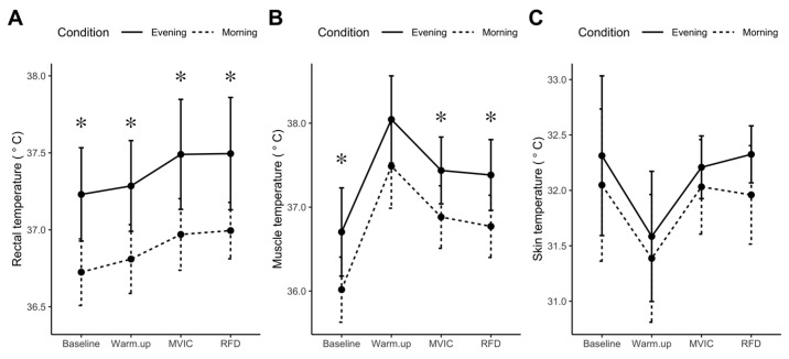
Figure 3.
Diurnal differences in core, muscle, and skin temperature. Core (rectal) temperature (A), muscle temperature (B), and skin temperature (C), were recorded during the last 5 min of baseline and immediately after cycle ergometry warm-up, maximal voluntary isometric contraction (MVIC), or peak rate of force development (RFD) protocols. Data are presented as mean, standard deviation (n = 10), experimental condition is identified by morning (dashed line) and evening (solid line). * p < 0.05 statistical difference between morning and evening condition.

