Severe acute respiratory syndrome coronavirus-2 (SARS-CoV-2) first emerged in Wuhan, China, and subsequently spread globally resulting in the current coronavirus disease-2019 (COVID-19) pandemic.1 There have been reports of myocardial damage and cardiovascular complications2; however, there is limited literature on incidence of cardiac arrhythmias. In this study, we aimed to characterize electrocardiographic characteristics and incidence of arrhythmias in patients admitted with SARS-CoV-2.
Patients ≥18 years old with a confirmed diagnosis of COVID-19 hospitalized at Vidant Medical Center between March 1 to April 26, 2020 were retrospectively identified after Institutional Review Board approval (no. UMCIRB 20-001064 and no. UMCIRB 20-000825). Data on patient demographics, comorbidities, and medications were collected. Baseline, admission, and subsequent serial 12-lead EKGs and telemetry strips were reviewed by 2 independent study investigators to identify rhythm abnormalities and compute baseline and maximum PR interval, QRS duration, and corrected QT interval (QTc) using Bazett formula. In presence of atrial fibrillation, QT measurement was performed using an average of 10 consecutive beats. Differences between mean EKG intervals from admission to maximum value were evaluated using paired t test. Subgroup analysis was performed by gender, race, intensive care unit (ICU) admission, troponin-I levels, and SARS-CoV-2 specific therapy (hydroxychloroquine, azithromycin, tocilizumab). Additional analysis for PR interval was performed in those who received atrioventricular nodal blocking agents (beta-blockers, calcium channel blockers, amiodarone, or digoxin). The data that support findings of this study are available from the corresponding author upon reasonable request.
As of April 26, 2020, there were 107 patients hospitalized with COVID-19. Mean age was 60.0±16.4 years with a predominantly female (58.9%) and Black (69.2%) population. Forty-nine (45.8%) patients required ICU care. Fifty-seven (53.3%) patients were treated with a combination of hydroxychloroquine and azithromycin, 18 (16.8%) patients received only hydroxychloroquine, and 3 (2.8%) patients received only azithromycin. Predominant arrhythmia was sinus tachycardia (30.9%). Tachyarrhythmias were observed in 13 (12.2%) patients. First-degree atrioventricular block was seen in 20 (18.7%) patients and 1 (0.9%) patient developed transient Mobitz II atrioventricular block. There were no cases of torsades de pointes.
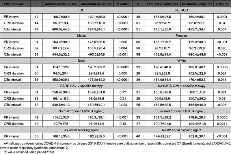
PR interval significantly prolonged from 158.7±33.2 ms at the time of admission to 173.9±34.3 ms (P<0.001) during hospitalization. Similarly, QRS duration increased significantly from 95.5±21.0 to 99.9±20.1 ms (P=0.0003) and QTc increased significantly from 452.0±40.1 to 473.8±45.4 ms (P<0.001). PR interval was prolonged regardless of whether patients received atrioventricular nodal blocking agents (Table, last row). Admission and peak PR were longer in Whites (176.4±40.9 versus 185.9±40.1, P=0.009) compared to Black (154.1±27.8 versus 170.7±32.2, P<0.001). QRS duration significantly increased in ICU patients (95.9±18.4 versus 103.7±18.3, P<0.001); but remained unchanged in non-ICU patients (P=0.34; Table). Patients had significant lengthening of their QTc intervals during hospitalization regardless of whether they received SARS-CoV-2 specific therapy. PR interval was prolonged regardless of the troponin value. QRS duration was increased only in patients with elevated troponin-I (100.7±21.6 to 109.4±21.8 ms, P=0.001; Table).
Table.
PR Interval, QRS Duration, and QTc Interval (Mean±SD) in Different Subgroups of Patients Hospitalized With COVID-19

Ours is the first report of electrocardiographic changes in patients hospitalized with SARS-CoV-2 showing significant prolongation of PR, QRS, and QTc intervals.
PR interval prolonged significantly in all patients admitted with SARS-CoV-2 infection regardless of medication status or troponin elevation. This suggests involvement of cardiac conduction system with SARS-CoV-2 infection which occurs early (ie, before clinically detectable myocardial injury) and is independent of myocardial inflammation. We also observed racial differences in PR intervals, suggesting that Whites may be more susceptible to cardiac conduction system involvement with SARS-CoV-2.
Critically ill patients with SARS-CoV-2 admitted to ICU and those with elevated troponin-I levels had a significantly wider QRS at the time of admission and during the hospital course. We hypothesize that QRS widening is a sign of myocardial injury and may help risk-stratify COVID-19 patients with potential implications with respect to cardiovascular outcomes.
Interestingly, we observed significant QTc lengthening during hospitalization in the small subgroup of patients (n=25) not receiving hydroxychloroquine or azithromycin, suggesting an additional unknown mechanism responsible for QTc prolongation in patients with SARS-CoV-2. Careful monitoring of QTc intervals is warranted, especially in those on hydroxychloroquine or azithromycin.
Our study has several limitations. Ours was a single center, observational, retrospective study of a small population of patients with SARS-CoV-2 without a comparison group. Data was limited to index hospital admission. Follow up data to see whether EKG intervals revert to baseline following recovery from infection are lacking. Approximately 70% of patients in our study received hydroxychloroquine or azithromycin; however, latest data have shown ineffectiveness and possible harm with these drugs in treatment of SARS-CoV-2.3,4 QRS widening has been previously documented in critically ill ICU patients5; hence, this finding may not be specific to SARS-CoV-2. Additional prospective studies with a larger population and longer follow up period are recommended to validate and further elucidate our findings.
Sources of Funding
None.
Disclosures
None.
Nonstandard Abbreviations and Acronyms
- COVID-19
- coronavirus disease 2019
- ICU
- intensive care unit
- QTc
- corrected QT interval
- SARS-CoV-2
- severe acute respiratory syndrome coronavirus-2
For Sources of Funding and Disclosures, see page 1224.
*Drs Moey and Sengodan contributed equally as first authors
References
- 1.Wang D, Hu B, Hu C, Zhu F, Liu X, Zhang J, Wang B, Xiang H, Cheng Z, Xiong Y, et al. Clinical characteristics of 138 hospitalized patients with 2019 novel coronavirus-infected Pneumonia in Wuhan, China. JAMA. 2020; 323:1061–1069. doi: 10.1001/jama.2020.1585 [DOI] [PMC free article] [PubMed] [Google Scholar]
- 2.Hu H, Ma F, Wei X, Fang Y. Coronavirus fulminant myocarditis saved with glucocorticoid and human immunoglobulin. Eur Heart J. 2020ehaa190 doi: 10.1093/eurheartj/ehaa190 [DOI] [PMC free article] [PubMed] [Google Scholar]
- 3.Geleris J, Sun Y, Platt J, Zucker J, Baldwin M, Hripcsak G, Labella A, Manson DK, Kubin C, Barr RG, et al. Observational study of hydroxychloroquine in hospitalized patients with COVID-19. N Engl J Med. 2020; 382:2411–2418. doi: 10.1056/NEJMoa2012410 [DOI] [PMC free article] [PubMed] [Google Scholar]
- 4.Magagnoli J, Narendran S, Pereira F, Cummings T, Hardin JW, Sutton SS, Ambati J. Outcomes of hydroxychloroquine usage in United States veterans hospitalized with Covid-19. medRxiv. 2020. doi: 10.1016/j.medj.2020.06.001 [DOI] [PMC free article] [PubMed] [Google Scholar]
- 5.Rich MM, McGarvey ML, Teener JW, Frame LH. ECG changes during septic shock. Cardiology. 2002; 97:187–196. doi: 10.1159/000063120 [DOI] [PubMed] [Google Scholar]


