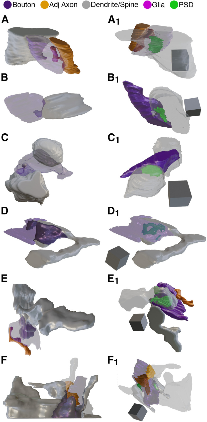
Figure 5.
3D Reconstructions of p21 and p46 SBBs. Focused ion beam serial electron microscopy 3D reconstructions of SBBs (purple) and their spinules from axon/boutons (yellow), and spines/dendrites (gray). Object color scheme shown at top. A–C, 3D reconstructions from a p21 ferret showing spinules from an adjacent axon (A, A1), postsynaptic dendrite (B, B1), and adjacent dendrite (C, C1) projecting into L4 SBBs. Note that B1 shows the spinule emerging from the edge (below and left) of the PSD. Reconstruction shown in A is the identical SBB shown in Figure 3A. D–F, 3D reconstructions from a p46 ferret showing hook-like spinule from a postsynaptic spine (D, D1), and spinules from adjacent axons and adjacent dendrites (E, E1, F, F1) projecting into L4 SBBs. Note the complex perforated PSD in D1, the identical SBB shown in Figure 3B. In F, F1, an SBB with synapses (green) onto two postsynaptic spines receives a large adjacent dendrite spinule (F) and a large adjacent axon spinule (F1). A1–F1, Identical SBBs as shown to the left in A–F, but with transparent postsynaptic neurites and/or SBBs to highlight the morphology and locations of the PSDs (green) or spinules. 3D scale cubes = 0.5 μm3.

