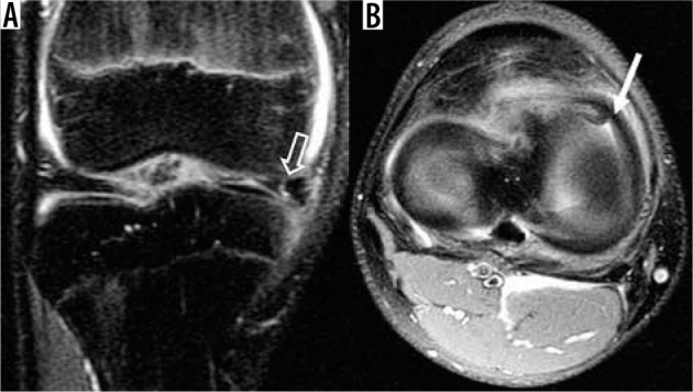
Figure 13.

Parrot beak tear. A) Coronal and B) axial proton density-weight- ed fat-saturated magnetic resonance images show a vertical tear that progresses from a radially oriented tear (open arrow in A) at the anterior horn of the medial meniscus. This tear has a curved appearance similar to a parrot’s beak on axial images (white arrow in B)
