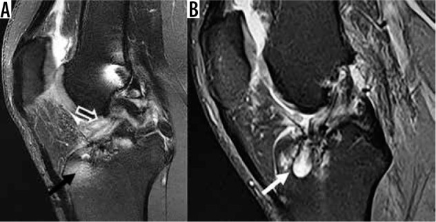
Figure 23.

Anterior cruciate ligament (ACL) reconstruction complications. A) Sagittal proton density-weighted (PDW) fat-saturated (FS) magnetic resonance (MR) image following ACL reconstruction, with an anteriorly positioned tibial tunnel (black arrow), associated with impingement of the ACL graft along the intercondylar roof (open arrow). B) Sagittal PDW FS MR image in a different patient following ACL reconstruction shows cystic de- generative changes along the tibial tunnel (white arrow)
