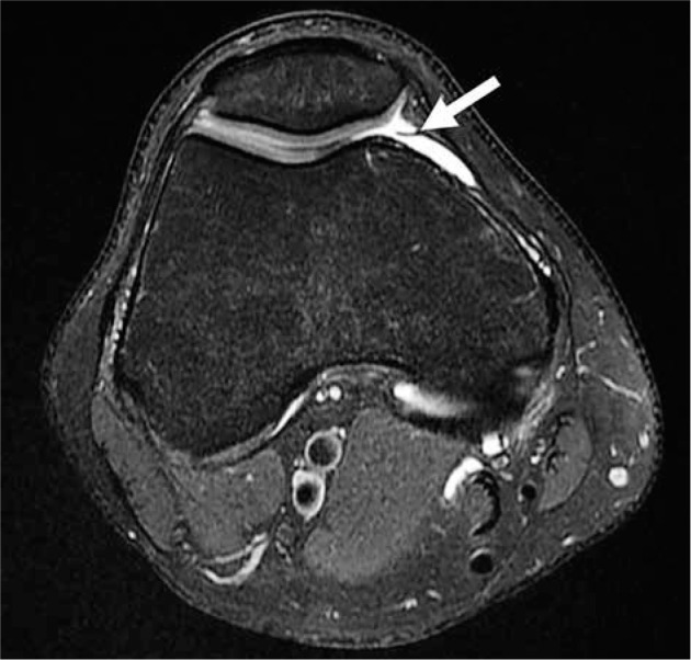
Figure 38.

Plica. Axial proton density-weighted fat-saturated magnetic resonance image in a 30-year-old male with linear low-signal intensity non-thickened tissue connected to the synovial lining and surrounded by joint fluid, compatible with a medial patellar plica (arrow)
