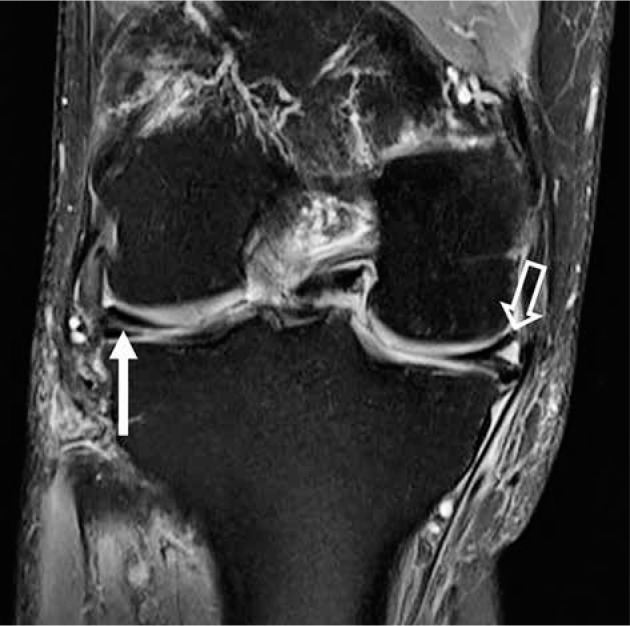
Figure 7.

Horizontal meniscal tear. Coronal proton density-weighted fat- saturated magnetic resonance image in a 63-year-old female with linear intrameniscal signal (closed arrow) extending to the undersurface of the body of the lateral meniscus, consistent with tear. High signal (open arrow) in the contralateral, medial meniscus does not extend to the meniscal arti- cular surface and is consistent with mucoid degeneration
