Abstract
The performance of electrocardiographic (ECG) voltage criteria to identify left and right ventricular hypertrophy (LVH and RVH) in young Asian female adults have not been clarified so far.
In a sample of 255 military young female adults, aged 25.2 years on average, echocardiographic LVH was respectively defined as the left ventricular mass (LVM) indexed by body surface area (BSA) (≥88 g/m2) and by height2.7 (≥41 g/m2.7), and RVH was defined as anterior right ventricular wall thickness >5.2 mm. The performance of ECG voltage criteria for the echocardiographic LVH and RVH were assessed by area under curve (AUC) of receiver operating characteristic (ROC) curve to estimate sensitivity and specificity.
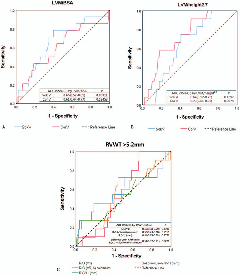
For the Sokolow-Lyon (the maximum of SV1 or SV2 + RV5 or RV6) and Cornell (RaVL + SV3) voltage criteria with the LVM/BSA ≥88 g/m2, the AUC of ROC curves were 0.66 (95% confidence intervals [CI]: 0.52–0.81, P = .039) and 0.61 (95% CI: 0.44–0.77, P = .18), respectively. For these 2 ECG voltage criteria with the LVM/height2.7 ≥41 g/m2.7, the AUC of ROC curves were 0.64 (95% CI: 0.52–0.75, P = 0.11) and 0.73 (95% CI: 0.61–0.85, P = 0.0074), respectively. The best cut-off points selected for the Sokolow-Lyon and Cornell voltage criteria with echocardiographic LVH in young Asian females were 26 mm and 6 mm, respectively. In contrast, all the AUC of ROC curves were less than 0.60 and not significant according to the Sokolow-Lyon (the maximum of RV1 + SV5 or V6) and Myers’ voltage criteria (eg, the voltage of R wave in V1 and the ratios of R/S in V1, V5 and V6) with echocardiographic RVH.
There was a suggestion that the ECG voltage criteria to screen the presence of LVH should be adjusted for the young Asian female adults, and with regard to RVH, the ECG voltage criteria were found ineffective.
Keywords: echocardiography, electrocardiographic criteria, left ventricular hypertrophy, right ventricular hypertrophy, young female adults
1. Introduction
The traditional electrocardiographic (ECG) voltage criteria in screening of the presence of left (LVH) and right ventricular hypertrophy (RVH) in the general population have been used for decades.[1–4] Most of the previous studies using Sokolow-Lyon and Cornell ECG voltage criteria to relate to LVH which was diagnosed by echocardiography or cardiac magnetic resonance imaging (MRI) showed consistent results that the sensitivity was very low, estimated merely 20%-30%, whereas the specificity was extremely high, estimated over 95%.[5,6] Similarly, using the ECG voltage criteria for the imaging-based RVH such as the Myers et al and Sokolow-Lyon revealed low sensitivity, commonly lower than 20% and high specificity which was up to 95% or more.[7,8]
It is notable that most studies examining the performance of the ECG voltage criteria to relate to the imaging-based LVH and RVH were carried out in White and Black, and middle to old aged persons of the Western world.[9–12] For the Asian individuals, the studies reporting the ECG criteria performance were relatively rare,[13–15] and in the same situation, most of these studies were aimed for the middle to old aged populations with several cardiovascular risk factors such like hypertension.[13,14] To our knowledge, since there have not established standard references with regard to the echocardiographic or other imaging based LVH or RVH for young Asian adults, only a few ECG studies were carried out to investigate the performance of the ECG voltage criteria in this population, particularly a lack of young Asian female adults so far.[8,15]
Military personnel are mainly composed of young adults, who have to receive regular exercise training. They are good samples to set standard references of imaging based LVH and RVH for the young adults. Therefore, the purpose of the study was firstly to clarify the standard references of echocardiographic LVH and RVH and then to examine the performance of ECG voltage criteria in a military young Asian female cohort in Taiwan.
2. Method
2.1. Study population
The ancillary cardiorespiratory fitness and hospitalization events in armed forces (CHIEF) Heart study included 1526 military young female adults, aged 18 to 42 years, in eastern Taiwan in 2014 to 2018.[16] All participants carried out a comprehensive health examination, and self-reported a questionnaire for their experience regarding toxic substances use and physical activity in the Hualien Armed Forces General Hospital of Taiwan. Of these, there were 265 subjects receiving a 12-lead ECG and a transthoracic echocardiography on the same day to ensure their cardiac health for the rank promotions and military awards, which were the inclusion criteria in this study, Those with hypertension (systolic/diastolic blood pressure ≥140/90 mm Hg, or on antihypertensive therapy) or an ECG finding of bundle branch block (n = 10) were excluded, leaving a sample of 255 females for the final analyses. The study design of CHIEF study has been described in detail previously.[17–21]
2.1.1. Measurements of 12-lead surface ECG
All 12-lead surface ECGs (Philips PageWriter Trim III) which were recorded at 25 mm/s paper speed and 1 mV/cm were prospectively performed by an experienced technician (Yu YS). The ECG variables including amplitudes of R and S waves in all limb and precordial leads and were retrospectively validated by 2 well-trained technicians (Yu YS and Lin F) and confirmed by a cardiologist (Lin GM) at the Hualien-Armed Forces General Hospital. For female adults, the Sokolow-Lyon voltage criterion for LVH was defined as the maximum of the amplitude (SV1 or SV2 + RV5 or RV6) ≥35 mm,[1] and the Cornell voltage criterion for LVH was defined as the amplitude (RaVL + SV3) ≥20 mm.[2] In addition, the Sokolow-Lyon voltage criterion for RVH was defined as the maximum of the amplitudes (RV1 + SV5 or SV6) > 10.5 mm,[3] and the Myers et al voltage criterion for RVH was defined as the R/S ratio of lead V1 > 1 or the R/S ratio of lead V5 or V6 < 1 or the R amplitude in lead V1 > 6 mm.[4]
2.1.2. Measurements of transthoracic echocardiography
All procedures of echocardiography using a 1 to 5 MHz transducer (iE33; Philips Medical Systems, Andover, MA) were performed by the same experienced technician (Yu YS) after the ECG and verified by a cardiologist (Lin GM) at the Hualien-Armed Forces General Hospital. All participants were examined using parasternal long-axis and short-axis approaches for the apical four and 2-chamber views in supine and left lateral positions. According to the suggestions of American Society of Echocardiography,[22] quantification of left ventricular wall thickness (interventricular septal and posterior walls) and chamber dimension were measured approximately at the onset of the QRS complex of end diastole and tips of the mitral valve by M-mode and 2-dimensional measurements in parasternal long axis view. Left ventricular mass (LVM) was thus calculated according to the corrected formula proposed by Devereux et al.[23] LVM = 0.8 × {1.04 × [(left ventricular end diastolic diameter (LVIDd) + end diastolic posterior wall thickness + end diastolic interventricular septal thickness]3 − left ventricular end diastolic diameter3} + 0.6. In addition, LVM was indexed for body surface area (LVM/BSA, g/m2), according to the Dubois formula,[24] and alternatively for height2.7 (LVM/height2.7, g/m2.7) suggested by de Simone et al.[25] The cut-off value for echocardiographic LVH was set as LVM/BSA ≥88 g/m2 and LVM/height2.7 ≥41 g/m2.7 which were the 95th percentile in the military young female adults in CHIEF study and according to another study finding for the young Asian female adults.[26,27] Measurements of anterior right ventricular wall thickness (RVWT) were by M-mode and 2-dimensional windows at the onset of the QRS complex of end diastole via the parasternal long-axis approaches.[28] Echocardiographic RVH was defined as RVWT > 5.2 mm, which was the 95th percentile in our young female cohort.[29]
2.2. Statistical analysis
Baseline characteristics of the CHIEF military female cohort were expressed as mean ± standard deviation (SD) for continuous variables and number (%) for categorical variables, respectively. Pearson correlation coefficient was used to determine the correlation of each ECG voltage criterion with the LVM indexes and RVWT, and was compared by the Fisher z test. Area under curves (AUC) of the receiver-operating characteristics (ROC) curves were used to evaluate and compare the performance of ECG voltage criteria for echocardiographic LVH and RVH. In addition, using the ROC curve to find the maximal sum of sensitivity and specificity was reclassified for each ECG criterion. A two-tailed value of P < .05 was considered significant. All analyses were performed using SAS version 9.4 (SAS Institute, Cary, NC).
2.2.1. Ethic statement
This study was reviewed and approved by the Institutional Review Board of the Mennonite Christian Hospital (No. 16-05-008) in Taiwan, and written informed consent was obtained from all participants.
3. Results
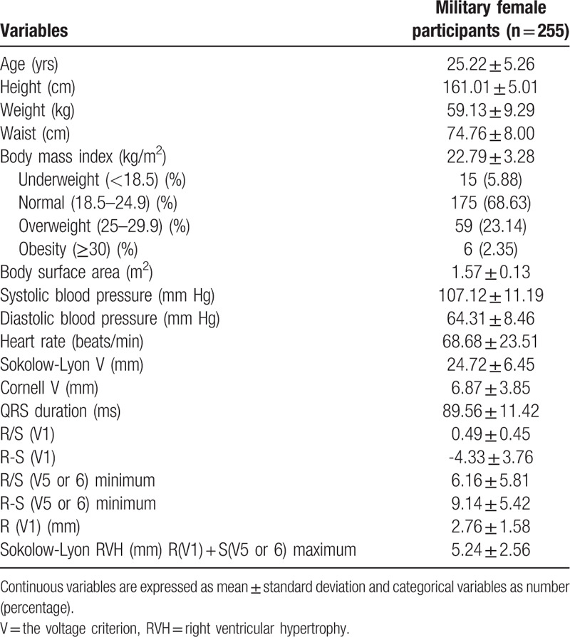
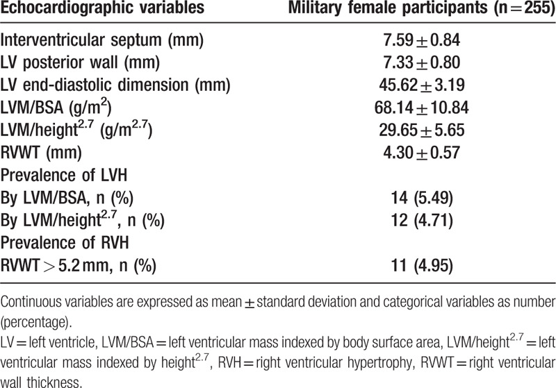
The baseline characteristics including ECG and echocardiographic data of the military female cohort are shown in Tables 1 and 2, respectively. Ages in the study subjects were between 18 and 42 years and averaged 25.2 years. The prevalence of echocardiographic LVH was 5.49% as LVM/BSA ≥88 g/m2 and 4.71% as LVM/height2.7 ≥41 g/m2.7, respectively. In addition, the prevalence of echocardiographic RVH was 4.95% as RVWT > 5.2 mm.
Table 1.
Baseline characteristics of demographic, anthropometric, and electrocardiographic measurements of the military female population.

Table 2.
Baseline echocardiographic parameters of the military population.

3.1. Correlation of each ECG criterion with the LVM indexes and RVWT
Table 3 reveals that there was a correlation of the Cornell criterion-based voltage (RaV + SV3) with the LVM indexes for BSA and LVM/height2.7 (r = 0.209 and 0.181, respectively), whereas there were no correlations between the Sokolow-Lyon criterion–based voltage (the maximal sum of SV1 or SV2 + RV5 or RV6) and the 2 LVM indexes (r = 0.045 and 0.025, respectively). In addition, there were no correlations between RVWT and each ECG voltage criterion for RVH. The greatest correlation coefficients with RVWT were observed in the Myers’ voltage criteria for the R/S ratio in lead 5 or 6 (r = –0.124), and in the Sokolow-Lyon criterion-based voltage (the maximum of RV1 + SV5 or SV6) (r = 0.017).
Table 3.
Pearson correlation coefficient (r) of electrocardiographic criteria with the left ventricular mass indexes and right ventricular wall thickness in the military female population.

3.2. Performance of the ECG voltage criteria for LVH and RVH using ROC curves
Figure 1A reveals the AUC of ROC curve using the Sokolow-Lyon voltage criterion for LVH ≥35 mm to detect the LVM/BSA index ≥88 g/m2 higher than that using the Cornell voltage criterion ≥20 mm for LVH (0.66 vs 0.61) in the young Asian females. On the contrary, Figure 1B shows the AUC of ROC curve using the Cornell voltage criterion ≥20 mm for LVH to identify the LVM/height2.7 index ≥41 g/m2.7 greater than that utilizing the Sokolow- Lyon voltage criterion ≥35 mm for LVH (0.73 vs 0.64) in the young Asian females. Figure 1C reveals the AUC of ROC curves using the Myers et al and Sokolow-Lyon voltage criteria for RVH to identify the RVWT > 5.2 mm (0.54–0.59) where all were nonsignificant in the young Asian females.
Figure 1.

The ROC curve with ECG criteria for identifying LVH and RVH in the military female population in Taiwan. (A) The ROC curve with 2 ECG criteria [the Sokolow-Lyon voltage (Sok V) and the Cornell voltage (Cor V)] for identifying LVH using left ventricular mass (LVM)/body surface area (BSA) ≥ 88 g/m2; (B) The ROC curve with 2 ECG criteria [the Sok V and the Cor V] for defining LVH using LVM/height2.7 ≥ 41 g/m2.7; (C) The ROC curve with the four ECG criteria [R/S (V1) and the minimum of R/S (V5 or V6), R(V1), and the maximum of R(V1) + S(V5 or V6) of Sokolow-Lyon RVH] for identifying RVH which was defined by the right ventricular wall thickness >5.2 mm. BSA = body surface area, ECG = electrocardiography, LVH = left ventricular hypertrophy, LVM = left ventricular mass, ROC = receiver operating characteristic, RVH = right ventricular hypertrophy.
3.3. Performance of the ECG voltage criteria for LVH and RVH using traditional and revised cut-off values
Table 4 demonstrates that the prevalence, sensitivity and positive predictive value of the ECG voltage criteria using traditional cut-off values to identify the presence of LVH and RVH in the young Asian females were extremely low, all estimated far less than 20%. In contrast, the specificity and negative predictive value of the ECG voltage criteria to identify the presence of LVH and RVH were extremely high, all estimated much greater than 92% in the young Asian females. When using the AUC of RUC curves to select the best cut-off points for echocardiographic LVH in the young Asian females, the Sokolow-Lyon voltage criterion was reclassified as ≥26 mm and the Cornell voltage criterion was ≥6 mm, respectively. In addition, the best cut-off point of Sokolow-Lyon criterion for echocardiographic RVH was reclassified as >5.2 mm.
Table 4.
The prevalence, sensitivity, specificity, predictive values and best cut-off point of each electrocardiographic criterion for echocardiographic left ventricular hypertrophy and right ventricular hypertrophy.

4. Discussion
Our principal findings were that firstly for the young Asian female adults, we established a standard of echocardiographic LVH as defied by the LVM/BSA index ≥88 g/m2 and by the LVM/height2.7 index ≥41 g/m2.7, respectively. In addition, we established a standard of echocardiographic RVH as defined by the RVWT > 5.2 mm. Second, using traditional ECG voltage criteria for LVH and RVH in the young Asian females consistently yielded low sensitivity and high specificity which were in line with the finding of previous studies. Finally, the best cut-off points for the ECG voltage criteria for LVH and RVH in the young Asian females should be lowered to obtain the maximal sum of sensitivity and specificity.
It is notable that among the premenopausal women in Asia, the prevalence of metabolic abnormalities such as obesity and hypertension, which are the risk factors of cardiac hypertrophy, is low.[20] In a previous study in Taiwan, the LVM and LVM index were lower in the female adults as compared with the male adults and the values decreased with younger ages.[30] The mean of LVM/BSA index was 63.6 g/m2 in women aged ≤30 years and 67.4 g/m2 in women aged 31 to 40 years. In addition, in another study for a multiethnic southeast Asian women cohort,[26] those aged <50 years had a mean LVM/BSA index with 64 (standard deviation = 14) g/m2 and the 95th percentile of the LVM/BSA index was 86 g/m2, close to our finding of 88 g/m2 as the cut-off point for echocardiographic LVH. Moreover, this is the first study clarifying the RVWT > 5.2 mm as echocardiographic RVH for the young Asian females.
As compared with the previous ECG studies for the middle-aged females,[14,28] Our findings revealed relevant results that there were low sensitivity and high specificity with regard to using the traditional ECG criteria to identify imaging-based LVH and RVH among the young Asian female adults. These findings were likely due to a very low prevalence of ECG-defined LVH, less than 5% in our subjects, reflecting a need to revise the cut-off values of ECG criteria-defined voltage for LVH. In addition, the correlation coefficients of the Cornell criterion-based voltage were found consistently better than that of the Sokolow-Lyon criterion-based voltage against the two LVM indexes in the young Asian female adults. Unlike the previous study findings for the middle-aged females, [14,31] the AUC of ROC curve was greater for the Sokolow-Lyon voltage criterion to detect the LVM/BSA index ≥88 g/m2 in the young Asian females, which might be due to a selection of different cut- off value for echocardiographic LVH. On the contrary, neither the Sokolow- Lyon nor the Myers et al criterion-based voltage to correlate with RVWT or to detect echocardiographic RVH was significant in the young Asian female adults. Although the ECG studies for the relationship with RVH in females were rare, the findings in our study were in line with that for males.
Our study had several strengths. First, both the ECG and echocardiographic examinations were performed in a strict manner and the procedures were standardized. Second, the military young females had to participate regular physical training that would modestly increase the level of 95th percentile to define the echocardiographic LVH and RVH compared with that of the age- matched general population of young females and avoid the selection bias. In contrast, this study had some limitations. First, this study was conducted on athletic military young Asian females and thus the results might not be appropriately applied to the general population of young females. Second, the female breast size might be a potential confounder for technicians to put the ECG precordial leads on the standard locations of chests wall, possibly leading to a bias. Third, there might be different results using other imaging diagnostic modalities such as cardiac magnetic resonance imaging for LVH and RVH.
5. Conclusion
There was a suggestion that the ECG voltage criteria to identify the presence of LVH as defined by echocardiographic LVM indexes should be adjusted for young Asian females, and for RVH as defined by echocardiographic RVWT, the ECG voltage criteria were found ineffective.
Author contributions
Conceptualization: Gen-Min Lin.
Data curation: Fang-Ying Su, Yen-Po Lin, Felicia Lin, Henry Horng-Shing Lu, Gen-Min Lin.
Formal analysis: Fang-Ying Su, Henry Horng-Shing Lu.
Funding acquisition: Gen-Min Lin.
Investigation: Yen-Po Lin, Felicia Lin, Yun-Shun Yu, Henry Horng-Shing Lu, Gen-Min Lin.
Methodology: Yen-Po Lin, Gen-Min Lin.
Project administration: Felicia Lin, Yun-Shun Yu, Gen-Min Lin.
Resources: Felicia Lin, Yun-Shun Yu, Gen-Min Lin.
Software: Henry Horng-Shing Lu.
Supervision: Yen-Po Lin, Younghoon Kwon, Henry Horng-Shing Lu, Gen-Min Lin.
Validation: Yen-Po Lin, Younghoon Kwon, Henry Horng-Shing Lu, Gen-Min Lin.
Visualization: Yen-Po Lin, Gen-Min Lin.
Writing – original draft: Fang-Ying Su, Yen-Po Lin.
Writing – review & editing: Yen-Po Lin, Gen-Min Lin.
Footnotes
Abbreviations: AUC = area under curve, BSA = body surface area, CHIEF = cardiorespiratory fitness and hospitalization events in armed forces, ECG = electrocardiography, LVH = left ventricular hypertrophy, LVM = left ventricular mass, ROC = receiver operating characteristic, RVH = right ventricular hypertrophy, RVWT = right ventricular wall thickness.
How to cite this article: Su FY, Lin YP, Lin F, Yu YS, Kwon Y, Lu HS, Lin GM. Comparisons of traditional electrocardiographic criteria for left and right ventricular hypertrophy in young Asian women: the CHIEF heart study. Medicine. 2020;99:42(e22836).
The study was supported by the grant from Hualien Armed Forces General Hospital (No. 805C-109-07).
The authors have no conflicts of interests to disclose.
The datasets generated during and/or analyzed during the current study are not publicly available, but are available from the corresponding author on reasonable request.
References
- [1].Sokolow M, Lyon TP. The ventricular complex in left ventricular hypertrophy as obtained by unipolar precordial and limb leads. Am Heart J 1949;37:161–86. [DOI] [PubMed] [Google Scholar]
- [2].Okin PM, Roman MJ, Devereux RB, et al. Electrocardiographic identification of increased left ventricular mass by simple voltage-duration products. J Am Coll Cardiol 1995;25:417–23. [DOI] [PubMed] [Google Scholar]
- [3].Sokolow M, Lyon TP. The ventricular complex in right ventricular hypertrophy as obtained by unipolar precordial and limb leads. Am Heart J 1949;38:273–94. [DOI] [PubMed] [Google Scholar]
- [4].Myers GB, Klein HA, Stofer BE. The electrocardiographic diagnosis of right ventricular hypertrophy. Am Heart J 1948;35:1–40. [DOI] [PubMed] [Google Scholar]
- [5].Jain A, Tandri H, Dalal D, et al. Diagnostic and prognostic utility of electrocardiography for left ventricular hypertrophy defined by magnetic resonance imaging in relationship to ethnicity: the Multi-Ethnic Study of Atherosclerosis (MESA). Am Heart J 2010;159:652–8. [DOI] [PMC free article] [PubMed] [Google Scholar]
- [6].Grossman A, Prokupetz A, Koren-Morag N, et al. Comparison of usefulness of Sokolow and Cornell criteria for left ventricular hypertrophy in subjects aged <20 years versus >30 years. Am J Cardiol 2012;110:440–4. [DOI] [PubMed] [Google Scholar]
- [7].Whitman IR, Patel VV, Soliman EZ, et al. Validity of the surface electrocardiogram criteria for right ventricular hypertrophy: the MESA-RV Study (Multi-Ethnic Study of Atherosclerosis-Right Ventricle). J Am Coll Cardiol 2014;63:672–81. [DOI] [PMC free article] [PubMed] [Google Scholar]
- [8].Meng FC, Lin YP, Su FY, et al. Association between electrocardiographic and echocardiographic right ventricular hypertrophy in a military cohort in Taiwan: The CHIEF study: ECG criteria for RVH. Indian Heart J 2017;69:331–3. [DOI] [PMC free article] [PubMed] [Google Scholar]
- [9].Truong QA, Ptaszek LM, Charipar EM, et al. Performance of electrocardiographic criteria for left ventricular hypertrophy as compared with cardiac computed tomography: from the Rule Out Myocardial Infarction Using Computer Assisted Tomography Trial. J Hypertens 2010;28:1959–67. [DOI] [PMC free article] [PubMed] [Google Scholar]
- [10].Okin PM, Devereux RB, Jern S, et al. Regression of electrocardiographic left ventricular hypertrophy by losartan versus atenolol: The Losartan Intervention for Endpoint reduction in Hypertension (LIFE) Study. Circulation 2003;108:684–90. [DOI] [PubMed] [Google Scholar]
- [11].Okin PM, Wright JT, Nieminen MS, et al. Ethnic differences in electrocardiographic criteria for left ventricular hypertrophy: the LIFE study. Losartan Intervention for Endpoint. Am J Hypertens 2002;15:663–71. [DOI] [PubMed] [Google Scholar]
- [12].Chapman JN, Mayet J, Chang CL, et al. Ethnic differences in the identification of left ventricular hypertrophy in the hypertensive patient. Am J Hypertens 1999;12:437–42. [DOI] [PubMed] [Google Scholar]
- [13].Xie L, Wang Z. Correlation between echocardiographic left ventricular mass index and electrocardiographic variables used in left ventricular hypertrophy criteria in Chinese hypertensive patients. Hellenic J Cardiol 2010;51:391–401. [PubMed] [Google Scholar]
- [14].Park JK, Shin JH, Kim SH, et al. A comparison of cornell and sokolow-lyon electrocardiographic criteria for left ventricular hypertrophy in korean patients. Korean Circ J 2012;42:606–13. [DOI] [PMC free article] [PubMed] [Google Scholar]
- [15].Su FY, Li YH, Lin YP, et al. A comparison of Cornell and Sokolow-Lyon electrocardiographic criteria for left ventricular hypertrophy in a military male population in Taiwan: the Cardiorespiratory fitness and HospItalization Events in armed Forces study. Cardiovasc Diagn Ther 2017;7:244–51. [DOI] [PMC free article] [PubMed] [Google Scholar]
- [16].Lin GM, Li YH, Lee CJ, et al. Rationale and design of the cardiorespiratory fitness and hospitalization events in armed forces study in Eastern Taiwan. World J Cardiol 2016;8:464–71. [DOI] [PMC free article] [PubMed] [Google Scholar]
- [17].Chao WH, Su FY, Lin F, et al. Association of electrocardiographic left and right ventricular hypertrophy with physical fitness of military males: the CHIEF study. Eur J Sport Sci 2019;19:1214–20. [DOI] [PubMed] [Google Scholar]
- [18].Tsai KZ, Lai SW, Hsieh CJ, et al. Association between mild anemia and physical fitness in a military male cohort: The CHIEF study. Sci Rep 2019;9:11165. [DOI] [PMC free article] [PubMed] [Google Scholar]
- [19].Lin JW, Tsai KZ, Chen KW, et al. Sex-specific association between serum uric acid and elevated alanine aminotransferase in a military cohort: The CHIEF Study. Endocr Metab Immune Disord Drug Targets 2019;19:333–40. [DOI] [PubMed] [Google Scholar]
- [20].Chen KW, Meng FC, Shih YL, et al. Sex-specific association between metabolic abnormalities and elevated alanine aminotransferase levels in a military cohort: The CHIEF Study. Int J Environ Res Public Health 2018;15:E545. [DOI] [PMC free article] [PubMed] [Google Scholar]
- [21].Lin GM, Nagamine M, Yang SN, et al. Machine learning based suicide ideation prediction for military personnel. IEEE J Biomed Health Inform 2020;24:1907–16. [DOI] [PubMed] [Google Scholar]
- [22].Sahn DJ, DeMaria A, Kisslo J, et al. Recommendations regarding quantitation in M-mode echocardiography: results of a survey of echocardiographic measurements. Circulation 1978;58:1072–83. [DOI] [PubMed] [Google Scholar]
- [23].Devereux RB, Alonso DR, Lutas EM, et al. Echocardiographic assessment of left ventricular hypertrophy: comparison to necropsy findings. Am J Cardiol 1986;57:450–8. [DOI] [PubMed] [Google Scholar]
- [24].Du Bois D, Du Bois EF. A formula to estimate the approximate surface area if height and weight be known. Nutrition 1989;5:303–11. [PubMed] [Google Scholar]
- [25].de Simone G, Kizer JR, Chinali M, et al. Normalization for body size and population-attributable risk of left ventricular hypertrophy: the Strong Heart Study. Am J Hypertens 2005;18:191–6. [DOI] [PubMed] [Google Scholar]
- [26].Wong RC, Yip JW, Gupta A, et al. Echocardiographic left ventricular mass in a multiethnic Southeast Asian population: proposed new gender and age-specific norms. Echocardiography 2008;25:805–11. [DOI] [PubMed] [Google Scholar]
- [27].Lin GM, Liu K. An electrocardiographic system with anthropometrics via machine learning to screen left ventricular hypertrophy among young adults. IEEE J Transl Eng Health Med 2020;8:1800111. [DOI] [PMC free article] [PubMed] [Google Scholar]
- [28].Rudski LG, Lai WW, Afilalo J, et al. Guidelines for the echocardiographic assessment of the right heart in adults: a report from the American Society of Echocardiography endorsed by the European Association of Echocardiography, a registered branch of the European Society of Cardiology, and the Canadian Society of Echocardiography. J Am Soc Echocardiogr 2010;23:685–713. [DOI] [PubMed] [Google Scholar]
- [29].Lin GM, Lu HH. A 12-lead ECG-based system with physiological parameters and machine learning to identify right ventricular hypertrophy in young adults. IEEE J Transl Eng Health Med 2020;8:1900510. [DOI] [PMC free article] [PubMed] [Google Scholar]
- [30].Chen C, Sung KT, Shih SC, et al. Age, gender and load-related influences on left ventricular geometric remodeling, systolic mid-wall function, and NT-ProBNP in asymptomatic Asian population. PLoS One 2016;11:e0156467. [DOI] [PMC free article] [PubMed] [Google Scholar]
- [31].Ishikawa J, Yamanaka Y, Toba A, et al. Gender-adjustment and cutoff values of Cornell product in hypertensive Japanese patients. Int Heart J 2017;58:933–8. [DOI] [PubMed] [Google Scholar]


