
Fig. 3. Dara induces CD38 protein reduction in NK cells and Ca2+ mobilization.
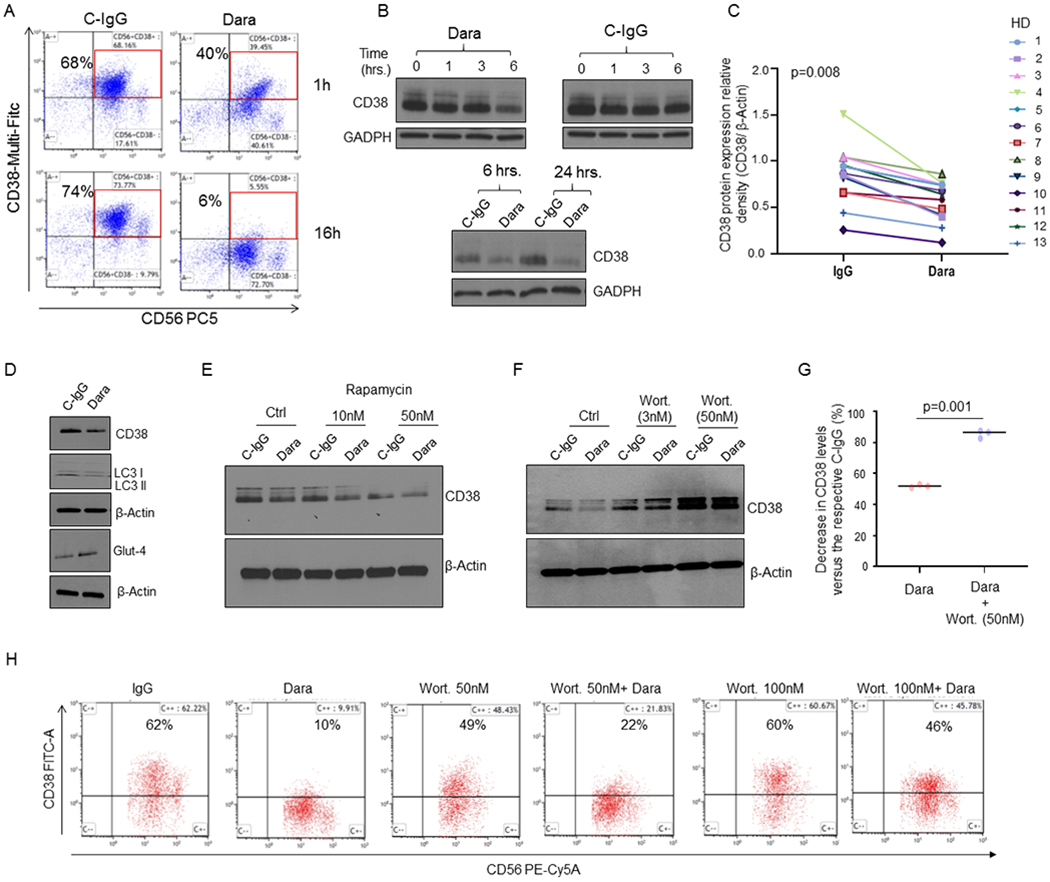
A) CD38 surface expression on NK cells at 1hr and 16hrs as detected by flow cytometry, showing down-regulation upon Dara treatment at 1hr and 16hrs compared to C-IgG; B) Western Blot analysis of CD38 protein level expression in NK cells treated over time with Dara (10 μg/mL) or IgG (10 μg/mL). C) Thirteen tumor-free donors (HD) were analyzed, and CD38 levels were assessed by densitometry analysis using β-actin as internal housekeeping. The Shapiro-Wilk test suggests that the samples are generated from a Gaussian distribution (α=0.05). Welch t-test statistic = 2.5963 with p=0.008; D) Western Blot analysis showing CD38 levels in primary NK cells treated for 6 hrs with Dara (10 μg/ml) or C-IgG, showing LC3II protein downregulation and GLUT4 upregulation; The experiment was performed in at least n=3 independent donors; E) Western Blot analysis showing CD38 levels in primary NK cells treated for 16 hours with rapamycin (10-50nM) as indicated in the presence of either C-IgG or Dara; F,G) Western Blot analysis showing CD38 levels in primary NK cells treated for 6 hours with wortmannin (Wort.) as indicated in the presence of either C-IgG or Dara. The experiment was repeated in biological triplicate, and CD38 relative density levels (CD38/β-actin) were normalized to C-IgG–treated NK cells for each HD donor, the differences reported (G); H) Flow cytometry analysis showing that Wort. treatment can partially rescue CD38 surface expression on CD56+CD3(−) NK cells after 6 hrs of Dara treatment.