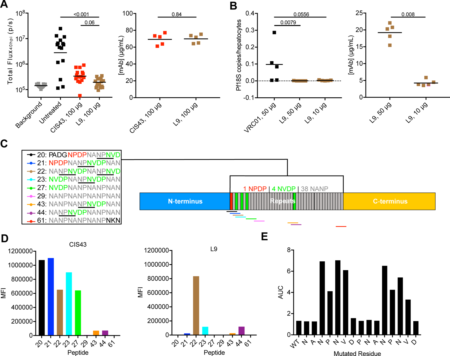
Figure 1. L9 is a neutralizing human mAb that binds the NPNV motif of PfCSP.

(A) Left: liver burden reduction (bioluminescence; total flux, photons/sec) in B6-albino mice 40 hrs post-infection (hpi; n=15/group; data pooled from three independent experiments) mediated by 100 µg CIS43 or L9 administered 2 hours before IV challenge with 2,000 Pb-PfCSP-GFP/Luc-SPZ. P-values were determined by comparing L9 to CIS43 and untreated control using the Kruskal-Wallis test with Dunn’s post-hoc correction. Right: serum mAb titers 2 hours after administration of 100 µg CIS43 or L9 in separate mice (n=5/group) determined through ELISA. Differences between CIS43 and L9 were determined using the two-tailed Mann–Whitney test. (B) Left: liver burden reduction (Pf 18S rRNA normalized to number of human hepatocytes) in FRG-huHep mice (n=5/group) administered 50 or 10 µg L9 and challenged IV with 100,000 PfSPZ. Right: serum mAb titers in challenged FRG-huHep mice. Differences between VRC01 (anti-HIV-1 isotype control IgG) and L9 (50 vs. 10 µg) were determined using the two-tailed Mann–Whitney test. (C) Schematic of PfCSP_3D7 depicting the N-terminus, repeat region (with color-coded overlapping 15mer peptides 20–61), and C-terminus. Every NPNV motif in each peptide is underlined. (D) ELISA (MFI, median fluorescence intensity) of CIS43 and L9 binding to peptides 20–61. (E) Competition ELISA (AUC, area under the curve) of L9 binding to rPfCSP_FL in the presence of varying concentrations of peptide 22 (WT, leftmost bar) or variant peptides (subsequent bars) where the indicated residue was mutated to alanine or serine. A-B: lines represent geometric mean. See also Figure S1.
