Abstract
Myositis ossificans (MO) is the abnormal formation of benign heterotopic bone tissue in soft tissues or muscles, mostly in sites of trauma. Though it has been described in most parts of the body, less than a dozen cases involving the chest wall have been reported. It is known to resolve spontaneously and various medical treatments have been suggested to hasten its resolution. Large tumors, suspicion of malignancy, and presence of symptoms are indications for surgical intervention. The differential diagnoses include sarcomas, infections, callous, calcified hematomas, and cysts. We present the clinical, radiological, and pathological images of a post traumatic MO of the chest wall, arising from under the medial third of the clavicle and growing into the deeper surface of the pectoralis major muscle. The patient is doing well eight months after the excision of the same.
Keywords: Myositis ossificans, Chest wall, Tumor, Calcification, Muscle injury, Soft tissues
Myositis ossificans (MO) is a benign tumor caused by heterotopic bone formation in soft tissues or muscles, often in an area of trauma [1]. It has been described in most regions of the body; but chest wall involvement is rare with less than a dozen cases reported so far [1]. We present the clinical, radiological, and pathological images of this rare chest wall lesion.
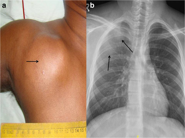
A 16-year-old boy presented with a progressively growing painful swelling over the right anterior chest wall which started immediately after a trauma 10 weeks earlier. On examination, there was a tender and hard 10 cm diameter tumor in the infraclavicular region, restricting shoulder movements (Fig. 1a). Clinically, it was arising from the chest wall, deep to the pectoralis major muscle.
Fig. 1.
(a) Clinical photograph showing the large right pectoral tumor. (b) Chest radiograph showing an ill-defined increased density in the right upper hemithorax (arrows) with no fractures or lytic lesions in adjacent bones
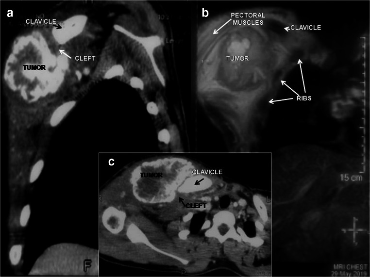
Chest x-ray showed increased density over the right hemithorax with no lytic lesions or fractures in the underlying bones (Fig. 1b). Computerized tomogram (CT) and magnetic resonance imaging were suggestive of a MO, characterized by a well-defined, irregular, heterogeneously enhancing, peripherally calcified mass. It was arising from the chest wall from under the medial third of the clavicle and was growing into the overlying pectoralis major muscle. There were no features of malignancy (Fig. 2a–c). The diagnosis was confirmed by a needle biopsy and the tumor was excised completely. The patient had an uneventful recovery and is doing well 8 months after surgery.
Fig. 2.
Reconstructed sagittal CT image (a), magnetic resonance image (b), and axial CT image (c) of the chest, showing the irregular peripherally calcified mass, closely related to the medial end of the right clavicle, but separated by a cleft of lucency
MO is commonest in the 2nd and 3rd decades of life, with a male preponderance. It is considered to be due to an aberrant repair response of tissues to trauma, a history of which may be present in only 40–75% of the patients [1, 2].
Small lesions are known to resolve spontaneously, and medical managements have been suggested [3]. Malignant transformation is considered unlikely [4, 5], but at times, it is difficult to distinguish a MO from a sarcoma [1, 3–5]. Besides this indication, the presence of pain, restriction of movements, and a large size may warrant its excision. If surgery is contemplated, a simple excision of the mass is considered sufficient, with a little or no chance of it recurring [3, 4].
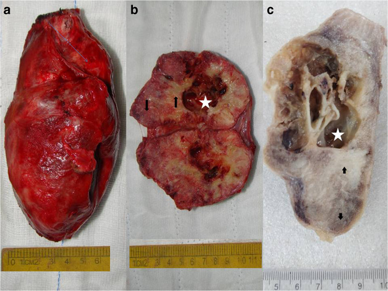
In the early stages (< 4 weeks), CT shows a lesion iso- to hypo-dense to muscle without calcification. In the intermediate period (4–8 weeks), amorphous patchy calcifications appear, which later becomes a dense peripheral shell of calcification. With further maturation (> 8 weeks), the lesion becomes smaller and may exhibit more diffuse calcification and ossification [5, 6]. It is typically separated from the adjacent bone by lucency or a cleft with no evidence of local invasion [1, 2]. Histologically, pluripotent cells and periosteal osteoblasts in the injured tissue are thought to initiate this abnormal repair process centripetally, resulting in a MO [1, 5]. Thus, in a mature lesion, three zones are seen, a central undifferentiated zone, surrounded by areas of osteoid and mature bone formation with calcification (Figs. 3a–c and 4a, b). This phenomenon named zonation is not seen in malignancies [1, 5]. Thus, a needle biopsy from the central area may show atypical cells suspicious of malignancy [5].
Fig. 3.
(a) Excised bosselated myositis ossificans tumor. (b) Bisected tumor showing zonation, a central multicystic area (star) with thin walls, filled with hemorrhagic fluid, surrounded by dense white areas (up arrow), which in turn are surrounded by hard bony granular foci (down arrow) at the periphery. (c) Formalin-fixed bisected tumor with markings as in (b)
Fig. 4.
(a) Histopathology showing an outer zone of mature cancellous bony spicules (left facing arrow), a central zone of proliferating fibroblastic spindle cells (star) and a transition zone of islands of immature woven bone (down arrows), rimmed by osteoblasts (hematoxylin and eosin, × 40). (b) The center of the lesion with fibrous components (up arrows) and pseudocystic spaces (triangle) filled with blood and lined by loose spindle cell stroma and scattered osteoclast-like giant cells (hematoxylin and eosin, × 40)
We present this interesting case to focus on its rarity, benign nature, and the good result achieved by surgery. Common differentials would include sarcomas, aneurysmal bone cysts, calcifying hematomas, infections, and fracture callus [2, 3, 5, 6].
Funding
There is no funding involved in this study.
Compliance with ethical standards
Ethical approval
This study article was in accordance and compliance with the ethical standards of the institution.
Informed consent
Informed consent was taken from the patient whose images are being published.
Conflict of interest
The authors declare that there is no conflict of interest.
Statement of human and animal rights
This is an article on images, so there is no research involving human or animal participant.
Footnotes
Publisher’s note
Springer Nature remains neutral with regard to jurisdictional claims in published maps and institutional affiliations.
References
- 1.Nisolle JF, Delaunois L, Trigaux JP. Myositis ossificans of the chest wall. Eur Respir J. 1996;9:178–9. [DOI] [PubMed]
- 2.Tyler P, Saifuddin A. The imaging of myositis ossificans. Semin Musculoskelet Radiol. 2010;14:201–16. [DOI] [PubMed]
- 3.Kim JT, Yoon YH, Baek WK, Han JY, Chu YC, Kim H-J. Myositis ossificans of the chest wall simulating malignant neoplasm. Ann Thorac Surg. 2000;70:1718–20. [DOI] [PubMed]
- 4.Sumiyoshi K, Tsuneyoshi M, Enjoji M. Myositis ossificans. A clinicopathologic study of 21 cases. Acta Pathol Jpn. 1985;35:1109–1122. [PubMed] [Google Scholar]
- 5.Harmon D. Case 38, 1994. N Engl J Med. 1994;331:1079–84.
- 6.Zhang L, Hwang S, Benayed R, et al. Myositis ossificans-like soft tissue aneurysmal bone cyst: a clinical, radiological, and pathological study of seven cases with COL1A1-USP6 fusion and a novel ANGPTL2-USP6 fusion. Mod Pathol. 2020. 10.1038/s41379-020-0513-4. [DOI] [PMC free article] [PubMed]


