Correction to: J Exp Clin Cancer Res 33, 99 (2014)
https://doi.org/10.1186/s13046-014-0099-0
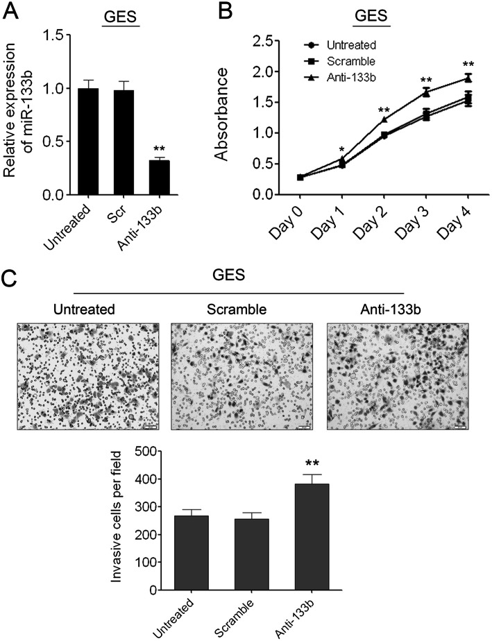
Following publication of the article [1], the authors identified errors in Figs. 3, 4 and 6; specifically panels Fig. 3c (HGC-27 ‘untreated’), Fig. 4c (GES ‘untreated’), Fig. 6c and d. The corrections do not change the results or the conclusions of this paper.
Fig. 3.
Enforced expression of miR-133b can inhibit GC cell migration and invasion. (A) The pictures of wound healing and the percentages of open wound of HGC-27 cells at 0, 24, 48 hours after scratching. Data are shown as mean + s.d. (n = 3); ** indicates P-value <0.01. (B) The pictures of wound healing and the percentages of open wound of MGC-803 cells at 0, 24, 48 hours after scratching. Data are shown as mean + s.d. (n = 3); ** indicates P-value <0.01. (C) The invaded HGC-27 cells in the Matrigel transwell invasion assay. Data are shown as mean + s.d. (n = 3); ** indicates P-value <0.01. (D) The invaded MGC-803 cells in the Matrigel transwell invasion assay. Data are shown as mean + s.d. (n = 3); ** indicates P-value <0.01
Fig. 4.
Knockdown of miR-133b in GES cells can promote cell proliferation and migration. (A) Inhibition of miR-133b in GES cells was confirmed by qRT-PCR. (B) The cell growth of GES cells at day 0, 1, 2, 3, 4 post transfection which was detected by CCK-8 assay. Data are shown as mean ± s.d. (n = 3); * indicates P-value <0.05;** indicates P-value <0.01. (C) The invaded GES cells in the Matrigel transwell invasion assay. Data are shown as mean + s.d. (n = 3); ** indicates P-value <0.01
Fig. 6.
Knock down of FSCN1 can inhibit GC cell growth and invasion. (A) Western blot analysis of FSCN1 expression in HGC-27 and MGC-803 cells transfected with negative control or FSCN1 siRNAs. (B) The cell growth of HGC-27 and MGC-803 cells at day 0, 1, 2, 3, 4 post transfection which was detected by CCK-8 assay. Data are shown as mean + s.d. (n = 3); * indicates P-value <0.05. ** indicates P-value <0.01. *** indicates P-value <0.001. (C) The invaded HGC-27 cells in the Matrigel transwell invasion assay. Data are shown as mean + s.d. (n = 3); ** indicates P-value <0.01. (D) The invaded MGC-803 cells in the Matrigel transwell invasion assay. Data are shown as mean + s.d. (n = 3); ** indicates P-value <0.01. (E) Western blot analysis of FSCN1 expression in 6 pairs of GC tissues (C) and the adjacent non-neoplastic tissues (N)
Footnotes
Lihua Guo, Hua Bai and Dongling Zou contributed equally to this work.
Contributor Information
Qi Zhou, Email: qizhou9128@163.com.
Jinsheng He, Email: jshhe@bjtu.edu.cn.
Reference
- 1.Guo L, Bai H, Zou D, et al. The role of microRNA-133b and its target gene FSCN1 in gastric cancer. J Exp Clin Cancer Res. 2014;33:99. doi: 10.1186/s13046-014-0099-0. [DOI] [PMC free article] [PubMed] [Google Scholar]


