Abstract
Rice foot rot caused by Dickeya zeae is an important bacterial disease of rice worldwide. In this study, we identified a new strain EC2 from rice in Guangdong province, China. This strain differed from the previously identified strain from rice in its biochemical characteristics, pathogenicity, and genomic constituents. To explore genomic discrepancies between EC2 and previously identified strains from rice, a complete genome sequence of EC2 was obtained and used for comparative genomic analyses. The complete genome sequence of EC2 is 4,575,125 bp in length. EC2 was phylogenetically closest to previously identified Dickeya strains from rice, but not within their subgroup. In terms of secretion systems, genomic comparisons revealed that EC2 harbored only type I (T1SS), typeⅡ (T2SS), and type VI (T6SS) secretion systems. The flagella cluster of this strain possessed specific genomic characteristics like other D. zeae strains from Guangdong and from rice; within this locus, the genetic diversity among strains from rice was much lower than that of within strains from non-rice hosts. Unlike other strains from rice, EC2 lost the zeamine cluster, but retained the clustered regularly interspaced short palindromic repeats-1 (CRISPR-1) array. Compared to the other D. zeae strains containing both exopolysaccharide (EPS) and capsular polysaccharide (CPS) clusters, EC2 harbored only the CPS cluster, while the other strains from rice carried only the EPS cluster. Furthermore, we found strain MS1 from banana, carrying both EPS and CPS clusters, produced significantly more EPS than the strains from rice, and exhibited different biofilm-associated phenotypes. Comparative genomics analyses suggest EC2 likely evolved through a pathway different from the other D. zeae strains from rice, producing a new type of rice foot rot pathogen. These findings emphasize the emergence of a new type of D. zeae strain causing rice foot rot, an essential step in the early prevention of this rice bacterial disease.
Introduction
Rice foot rot is an important bacterial disease affecting rice (Oryza sativa L.), and first reported in Japan [1], and spread to the other important rice-growing countries, including China, South Korea, the Philippines, India, Indonesia, and Bangladesh [2–4]. It has become one of the most important bacterial diseases of rice in India [5]. In China, the disease has caused significant losses in Zhejiang, Shanghai, Jiangsu, Hubei, and Fujian provinces before 1983 [6]. Currently, it is omnipresent in most of the major rice-growing areas in China, and has become an important threat to rice production [4]. Therefore, the epidemiologic mechanisms of this disease, and the pathogenesis of the causal pathogen have raised extensive concerns.
The causal agent of rice foot rot disease is Dickeya zeae (previously Erwinia chrysanthemi pv. zeae), family Pectobacteriaceae. The genus includes: D. zeae, D. chrysanthemi, D. dianthicola, D. paradisiaca, D. dadantii subsp. dadantii, D. dadantii subsp. dieffenbachiae, D. solani, D. aquatica, D. fangzhongdai, D. undicola, and D. lacustris [7–14]. D. zeae contains a group of pathogens infecting important plants in the Poaceae like rice and maize, which indicate distinct genetic variants in D. zeae [15, 16]. This was consistent with the genetic evolution analysis of Dickeya species based on recA gene sequences; indicated that there were different sequence variants (sequevars) isolated from different host plants existed among D. zeae strains [17]. Moreover, no common antigens among D. zeae strains from rice or maize were found in serological tests. The strains from rice, however, exhibited higher pathogenicity and broader host range than strains from maize [18]. These findings indicate the differentiation and genetic evolution of D. zeae strains might be an outcome of long-term interactions between pathogens and hosts.
Until now, there were 11 complete or draft genomes of D. zeae deposited at the National Center for Biotechnology Information (NCBI) GenBank Genome database. Among them, three strains were from rice, including EC1 (complete, Guangdong, China), ZJU1202 (draft, Guangdong, China) and DZ2Q (draft, Italy); in a phylogenetic analyses, all three strains from rice were grouped in a clade separated from the other D. zeae strains [15]. The D. zeae strains from rice may fall in a distinct group, as they showed some unique genomic features, like the zeamine phytotoxins biosynthesis gene cluster [15], and incomplete subtype I-F CRISPR array. In this study, however, we isolated a new D. zeae strain from rice in Guangdong, China, with obvious differences in cultural and biochemical characteristics, and genomic determinants from the typical strain EC1 from rice. Previous studies on the virulence factors of D. zeae strains from rice focused on the acyl homoserine-lactone regulated quorum-sensing system [3], zeamine biosynthesis gene cluster [19–21], zeamine biosynthesis-related transcription factors SlyA [22] and Fis [23], as well as the twin-arginine translocation system [24]. These genomic traits might not reflect the comprehensive strain differentiation and genetic evolution of the identified strains from rice. In this study, therefore, we characterized the complete genome sequence of a newly identified strain D. zeae EC2 isolated from diseased rice plant and compared with previously identified strains from rice, EC1, ZJU1202, and DZ2Q, and other D. zeae strains. We also analyzed the differences in some cultural and pathogenicity characteristics among the newly identified strain from rice, D. zeae EC2, the previously identified strain, D. zeae EC1, and the strain from banana, D. zeae MS1. The genome-wide comparison and culture and pathogenicity would help to clarify the differentiation among D. zeae strains from rice and their genetic adaptation to the host. This information might raise more attention to this newly emerging rice pathogen and provide better insight into the early prevention of rice foot rot disease.
Materials and methods
Bacterial strains and culture media
Three rice plants with typical symptoms of rice foot rot were collected from the fields of Qingyuan city, Guangdong province, China. The margins between diseased and healthy areas at the base of stems were cut into pieces, surface-sterilized in 75% ethanol for 30 s and 1% NaOCl for 1 min, and then rinsed three times with sterile water. The pieces were macerated in 100 μL of sterile water, and the mixture were streaked onto nutrient agar (NA) plates (3 g/L beef extract, 0.5 g/L yeast extract paste, 5 g/L peptone, and 10 g/L agar, pH 7.0) and incubated for 48 h at 30°C. Single colonies were picked and subcultured three times; one typical strain, EC2, was confirmed through Koch's postulates and identity was confirmed using 16S rDNA primers [25] and recA [26]. South China Agricultural University provided D. zeae EC1 strain, while our group isolated MS1 and CE1 (Canna edulis strain) strains. These strains were stored in glycerol at -80°C at the Key Laboratory of New Technique for Plant Protection in Guangdong province, China. For subculture, bacteria were grown to a concentration of 108 CFU/mL at 30°C in Luria-Bertani (LB) broth (10 g/L bacto tryptone, 5 g/L yeast extract, and 10 g/L NaCl, pH 7.0) in an incubator shaker at 100 rpm.
Biolog analysis
Dickeya zeae strains, EC1, EC2, MS1 and CE1, were characterized using GEN III MicroStation (Biolog, Hayward, CA, USA). A 150-μL bacterial suspension of 108 CFU/mL was added to each well of a GEN III micro-plate (Biolog) containing 95 sugars, alcohols, acids, amines, and other substrates. Culture suspensions were incubated and read, and then compared to the databases using the automated microbial analysis system, Biolog.
Pathogenicity test
Bacteria were grown to a concentration of 108 CFU/mL in LB broth, and diluted fivefold. A 200-μL bacterial dilution of each strain EC1 and EC2 was injected into the basal stem of each rice seedling at the tillering stage. Rice seedlings inoculated with sterile water served as a negative control. The rice seeds were provided by the College of Agriculture, South China Agricultural University, China, and were grown in the greenhouse of our lab. The injection sites were wrapped in cotton balls moisturized with sterile water. The inoculated plants were placed in the greenhouse at 30°C and 90% relative humidity. Seedlings with soft rot symptoms were used for bacterial isolation, and re-isolated strains were confirmed by 16S rDNA amplification and sequencing.
Bacteria were grown to a concentration of 109 CFU/mL in LB broth. Half of the culture was kept as culture crude, and the remaining half was centrifuged for 10 min at 12,000 rpm; the supernatant pipetted as culture extract. The non-inoculated LB broth was used as a negative control. Each of the 20 rice seeds was immersed in 20 mL of prepared culture crude or culture extract for 6 h at room temperature. The treated seeds were washed three times in sterilized water and transferred onto moistened filter papers in petri dishes. The seeds were incubated for one week at 28°C and 14-h-light and 10-h-dark conditions.
Genomic DNA extraction and genome sequencing
Total DNA was extracted from 2-mL bacterial suspensions (108 CFU/mL) using the TIANamp Bacterial DNA Kit (Tiangen Biotech, Beijing, China) according to manufacturer's directions. The fragmented genomic DNAs were treated with G-tubes (Covaris) and end-reparation. The treated DNAs were used to construct a SMRTbell DNA library with fragment sizes of >10 Kb. Next, SMRT sequencing was conducted using the Pacific Biosciences RSII sequencer (Pacific Biosciences, Menlo Park, United States) at Genedenovo (Guangzhou, China), according to standard protocols using P4-C2 chemistry. Reads (≥500 bp) with a quality value of over 0.75 were processed for next step. A Hierarchical genome-assembly process pipeline [27] was used to correct random errors in long seed reads (≥ 6 Kb) by aligning shorter reads against them. A total of 115,312 reads with an average length of 9,047 bp and totaling 1,043,332,658 bp, were obtained, and de novo assembled using the Celera assembler with overlap-layout-consensus strategy [28]. The Quiver consensus algorithm [27] was used to validate the quality of the assembled genome sequence. A circularized genome of 4,574,986 bp with 53.34% GC content was generated after trimming the ends of the assembled sequence. To correct this assembly, another DNA library was constructed for next-generation sequencing. The paired-end library was sequenced on an Illumina HiSeq 4000 (Illumina, San Diego, CA, USA) at Genedenovo (Guangzhou, China). A total of 14,457,752 output reads were aligned to the assembly of PacBio sequencing using a Burrows-Wheeler alignment tool with default parameters [29], and 14,255,069 of them were matched to the circularized genome. Pilon was also used to improve the genome sequence by searching for inconsistencies between the genome sequence and the reads [30]. In terms of insertions, deletions, and substitutions, 170, 31, and 1 correction were made, respectively, to the circularized genome. The closed genome was corrected to 4,575,125 bp in length with a GC content of 53.34%.
Annotation of the D. zeae EC2 genome
RepeatMasker [31], rRNAmmer [32], and tNRAscan [33] were applied to search for repetitive elements, noncoding RNAs, and tRNAs, respectively, and gene finding was performed with the GeneMarkS program [34]. Next, we annotated the functions of the predicted genes through similarity searches against several databases: NCBI Non-redundant Protein Database (ftp://ftp.ncbi.nih.gov/blast/db/FASTA/nr.gz), UniProt/Swiss-Prot (http://www.uniprot.org/downloads), Kyoto Encyclopedia of Genes and Genomes (http://www.genome.jp/kegg/), Gene Ontology, Cluster of orthologous groups of proteins (COG), and Protein Families (ftp://ftp.ebi.ac.uk/pub/databases/Pfam). GC contents, GC-skew value (GC skew = [G-C]/[G+C]), tRNA, rRNA, predicted genes in positive and negative strands, and COG annotation, were presented using Circos [35]. We also used CRISPRFinder [36] and antiSMASH 4.0 [37] to predict CRISPRs and gene clusters of polyketides (PKs) and nonribosomal peptides (NRPs), respectively.
Phylogenetic analysis of Dickeya strains
Twenty-three Dickeya strains with complete or draft genome sequences were used to extract the gene sequences of dnaX, recA, dnaN, fusA, gapA, purA, rplB, rpoS, and gyrA [11], plus an out-group control, Pectobacterium carotovorum subsp. carotovorum PC1. The concatenated sequences of these nine genes were processed on MEGA 5.1 using a neighbor-joining algorithm with 1,000 bootstrapped replications. To obtain an estimate of overall genomic similarities, the genome sequences of 13 D. zeae strains were processed to calculate average nucleotide identity (ANI) and alignment percentage (AP) using CLC Genomics Workbench 12.0.3 (Qiagen, USA), and in silico DNA-DNA hybridization (isDDH) [38] by adapting formula 2.
Genomic comparison of available genomes of D. zeae strains or strains from rice
Genomic annotation of 13 whole-genome sequences of D. zeae strains and one well-sequenced strain, D. dadantii 3937, were retrieved from NCBI and used for genomic comparison. Synteny analysis was performed on Mummer (http://mummer.sourceforge.net/) by conducting nucleotide-nucleotide sequence comparisons. The alignments of pairs of nucleic acid sequences were marked at their coordinate positions in the size-reduced synteny graphs. To determine the orthologous relationships of coding proteins for D. zeae strains, orthoMCL [39] was used to identify the set of common genes or specific genes through a BLAST search with the following parameters: p-value cut-off = 1 × 10⁻⁵; identity cut-off = 80%.
Assays of exopolysaccharide (EPS) production
Single colonies of strains EC2, EC1, and MS1, were transferred to 15 mL nutrient broth (NB; 3 g/L beef extract, 0.5 g/L yeast extract paste, and 5 g/L peptone, pH 7.0) and incubated at 28°C until OD600 1.0. For colony morphology assay, one-μL bacteria culture was spotted onto NA plates supplemented with 2% glucose, and sub-cultured for 4 d at 28°C [40]. EPS production was measured as follow: three-mL bacteria culture was transferred into 300 mL NB medium, and grown for 5 d at 200 rpm and 28°C. The culture was centrifuged for 30 min at 10,000 rpm, and 250 mL of supernatant collected; 20% KC1 (w/v) was added to make a final KC1 concentration of 1%. After this, a double volume of ethanol was added to this solution; the mixture was placed at 4°C for 24 h followed by centrifugation for 30 min at 10,000 rpm. The pellet was dried at 55°C and measured the weight [41]. The experiment was repeated in triplicate, and two-tailed t-tests were performed to evaluate the differences.
Results
The D. zeae pathogen was found to cause rice foot rot in Qingyuan city, China
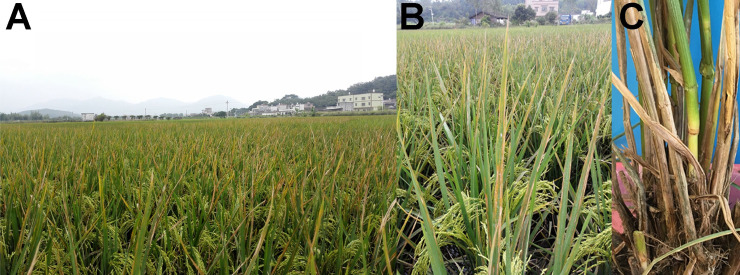
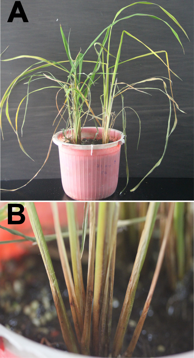
In October 2016, rice plants from the fields of Qingyuan city showed symptoms of leaf yellowing and necrosis (Fig 1A), and rice spikes failed to pollinate and form grains (Fig 1B). The rice stems turned dark with soft rot near the soil line, and were easily broken (Fig 1C). Typical strain EC2 was isolated from the diseased rice plant. Pathogenicity tests showed that the new strain isolated from rice can cause typical foot rot symptoms as previously verified for strain EC1 that was also isolated from rice in Guangdong. These symptoms included shrinking and yellowing of newly emerging and older leaves, respectively (Fig 2A); soft rot at the foot of stems (Fig 2B). Biolog analysis confirmed EC2 as the strain from genus Dickeya, as well as other D. zeae strains from Guangdong Province (EC1 isolated from rice, MS1 isolated from banana, and CE1 isolated from C. edulis ker.). Principal coordinates analysis revealed that this new strain EC2 was much different from previously identified strain from rice, EC1, in the use of different substrates as well as other D. zeae Guangdong strains (S1 Fig).
Fig 1. Symptoms of rice foot rot observed in the fields of Qingyuan, Guangdong province, China.
(A) Yellowing and necrosis of rice leaves. (B) Rice spikes fail to pollinate and form grains and leaves are yellow and necrotic. (C) The crowns of diseased rice stems turn dark and rot.
Fig 2. Typical rice foot rot symptoms observed after inoculation with new strain, EC2, isolated from rice.
(A) New leaves are wilting and older leaves are yellowing 11 d after inoculation with strain EC2. (B) The base of rice stems are dark and rotten 11 d after inoculation with strain EC2.
Genome assembly and annotation
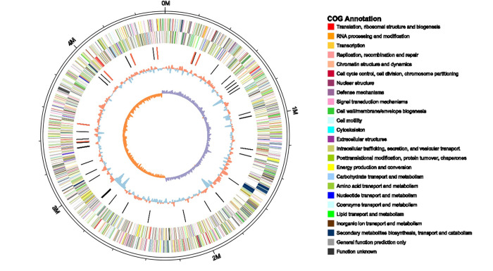
The complete genome of strain EC2 was 4,575,125 bp in length with a 53.34% GC content, and predicted 3,999 coding sequences. Information on predicted gene distribution, the COG annotation, and GC content was represented on the circular chromosome beginning at the dnaA start codon (Fig 3). In the genome, we found 75 tRNA and 7 rRNA regions. Within the predicted rRNA regions, two and four common organization types (16S-23S-5S) were predicted on the positive and negative strands, respectively, while an unusual organization type (16S-23S-5S-5S) was predicted on the negative strand. The complete genome project of EC2 was deposited under NCBI GenBank accession no. CP031515.
Fig 3. Circular visualization of the complete genome of D. zeae EC2.
From outside to inside, the circles indicate predicted genes on the positive strand, predicted genes on the negative strand, ncRNAs (black indicates tRNAs, red indicates rRNAs), G+C contents and GC skew values (GC skew = (G-C)/(G+C); purple indicates >0, orange indicates <0), respectively.
Phylogenetic analysis of Dickeya strains
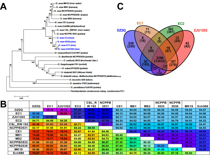
Phylogenetic analysis of concatenated sequences of nine genes distinctly separated D. zeae strains from the other nine species (Fig 4A). Among the D. zeae strains, strains EC1, ZJU1202, DZ2Q, and EC2 from rice were grouped into a clade differentiated from other D. zeae strains. However, EC2 was placed at a different branch from the previously identified strains from rice (Fig 4A). This was also demonstrated by the phylogenetic analysis based on whole-genome sequences (S2 Fig). ANI analysis indicated that the ANI values between EC2 and previously identified strains from rice ranged from 97.24% to 97.27% and reached the suggested cutoff (96.00%) for species delineation [42], but they were lower than the values between any two previously identified strains from rice (Fig 4B); AP values between EC2 and previously identified strains from rice were also based on lower alignment coverages ranging from 84.26% to 84.71%. Alternatively, EC2 also showed a close relationship with the other two D. zeae strains, NCPPB 3531 and CSL_RW192 (Fig 4A and 4B).These suggested that EC2 experienced a genetic divergence from previously identified D. zeae strains from rice.
Fig 4. Phylogenetic placement of D. zeae strain EC2 and its common or specific genes with other strains isolated from rice.
(A) Phylogenetic analysis of Dickeya strains based on concatenated sequences of the genes, dnaX, recA, dnaN, fusA, gapA, purA, rplB, rpoS and gyrA; twenty-three Dickeya strains were included in a phylogenetic analysis with a neighbor joining algorithm and bootstrapped at 1,000 replications. (B) Calculation of ANI and AP between pairs of genome sequences of D. zeae strains. Upper numbers indicate the values of ANI, and lower numbers indicate the values of AP. (C) Numbers of common or specific genes among D. zeae rice strains in an orthoMCL analysis. Upper numbers indicate the number of groups of genes, and lower numbers in parentheses indicate the number of genes.
Genome dissimilarities between EC2 and other strains isolated from rice
Synteny analysis between EC2 and other complete D. zeae genomes indicated that EC2 was highly co-linear with D. zeae genomes, except for a number of genomic dissimilarities like inversion and rearrangement events (S3 Fig). Comparing the genomes of four strains from rice including EC2, EC1, ZJU1202, and DZ2Q, orthoMCL cluster analysis revealed that 3,298 orthologous gene groups containing 13,432 genes were conserved in all of the four strains (Fig 4C). There were 365 groups and 391 genes specific to EC2, and 236 groups and 717 genes conserved within the previously identified strains from rice, but not in EC2. The specific genes in EC2 included those in a CPS biosynthesis cluster, T6SS gene clusters, a subtype I-F CRISPR cluster, and an NRPs- PKs cluster similar to a known viscosin cluster (S1 Table). Genes absent in EC2 included those in an EPS biosynthesis cluster, a zeamine cluster, and hrp-type T3SS, T4SS, and T5SS clusters (S2 Table). The zeamine cluster in EC1 encodes the phytotoxins which have a toxic effect against rice seed germination [22]. Accordingly, both culture crude and culture extract of EC1 significantly inhibited rice seed germination, but those of EC2 and MS1 did not (S4 Fig).
Secretion systems
T1SS cluster composed of prtG–prtA (DWV07_10200–DWV07_10235) and out-type T2SS consisted of outS, outB–outO operon (DWV07_13930, DWV07_13925–DWV07_13865) were both conserved in EC2, but not hrp-type T3SS, T4SS, and T5SS. This was further confirmed by PCR, the genes of hrpL, hrpY, and hrpS of hrp-type T3SS, virB4 and virBll of T4SS, and hecA2 and hecB of T5SS of D. zeae, were not able to be amplified in EC2; we successfully amplified these gene products in EC1 and MS1 (Fig 5). This was consistent with orthoMCL cluster analysis. Compared with EC2, the previously identified strains from rice and other two close stains, NCPPB 3531 and CSL_RW192, had T3SS–T5SS, except that T4SS was not found in the genome of NCPPB 3531.
Fig 5. PCR detection of genes of T3SS, T4SS, and T5SS from D. zeae strains.
hrpL, hrpY, and hrpS are genes of T3SS; virB4 and virB11 are genes of T4SS; hecA2 and hecB are genes of T5SS. Lane M indicates molecular weight markers from top to bottom (2,000, 1,000, 750, 500, 250, 100 bp); lane EC1 indicates D. zeae EC1 from rice; lane MS1 indicates D. zeae MS1 from banana; lane EC2 indicates D. zeae EC2 from rice; and lane N indicates ddH2O template.
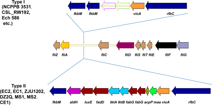
The flagellar cluster considered a subtype of T3SS was found in EC2, including the flagellin gene fliC, 38 flagellar biosynthesis genes, 2 flagellar motor genes, and 7 chemotaxis-associated genes (EC2: DWV07_12460–DWV07_12760, spanning 60.6 Kb). This was conserved in EC1, ZJU1202, DZ2Q, NCPPB 3531, and CSL_RW192, plus other D. zeae strains like MS1 and Ech586 (Fig 6). Like EC2, all of the other Guangdong strains, including strains EC1 and ZJU1202 from rice, strains MS1 and MS2 from banana, and strain CE1 from C. edulis, harbored a gene cluster (fkbM-aldH-luxE-fadD-tktA-tktB-fabG-fabG-acpP-maa-vioA-rfbC) upstream of flic, as did the Italy strain DZ2Q isolated from rice (Fig 6). This gene cluster encoded glycosyltransferase RfbC; aminotransferase VioA; maltose O-acetyltransferase Maa; the sugar biosynthetic proteins TktA and TktB; the fatty acid biosynthesis proteins AcpP, FabG, FadD, LuxE, and AldH/LuxC; and methyltransferase FkbM. Unlike EC2, other strains in the genus Dickeya including NCPPB 3531, CSL_RW192, and Ech586, lacked maltose O-acetyltransferase, sugar biosynthetic proteins, or fatty acid biosynthesis proteins and contained only an fkbM-fkbM–vioA-rfbC gene cluster, where a carbamoyl-phosphate synthase gene and a dehydrogenase gene were inserted between fkbM and vioA. GC contents of the former type of larger gene clusters (37.02%–39.22%) were much lower than those of the latter types of shorter gene clusters (47.96%–48.30%).
Fig 6. Two types of genomic organization found in the flagellar-type T3SS of D. zeae strains.
Compared to other D. zeae srains, D. zeae strains from rice have a longer gene cluster between the genes of fliA and fliC, as do the other Guangdong strains from banana and C. edulis. Gene fkbM encodes a methyltransferase; aldH/luxC, luxE, fadD, fabG, acpP encode fatty acid biosynthesis proteins; tktA and tktB encode sugar biosynthetic proteins; maa encodes a maltose O-acetyltransferase; vioA encodes an aminotransferase; and rfbC encodes a glycosyltransferase.
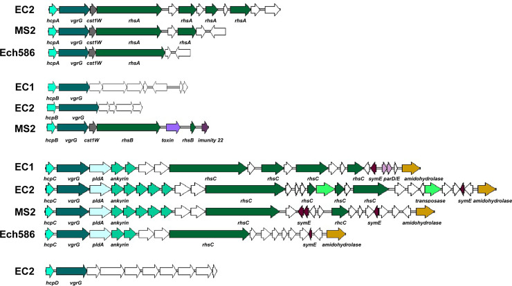
A gene cluster encoding T6SS was also present in EC2 (DWV07_06350–DWV07_06570). The complete set of genes included: the hemolysin-coregulated gene (hcp) and valine-glycine repeat G gene (vgrG); rearrangement hotspot elements (rhs); and the core T6SS genes impB/tssB, impC/tssC, gp25 (lysozyme)/ tssE, impG/tssF, impH/tssG, filamentous hemagglutinin (FHA)/tagH, vasD/tssJ, impJ/tssK, impK, clpB/tssH, sfa (sigma-54-dependent Fis family), vasI/tagO, impA /tssA, icmF/tssM, vasL, and tetratricopeptide repeats (TPRs). This T6SS cluster was also found in the other strains from rice, EC1, ZJU1202, and DZ2Q, as well as other D. zeae strains like MS2 and Ech586 (Fig 7). Among these T6SS genes, hcp and vgrG not only existed within this gene cluster, but they also had homologous genes at other genomic positions. The number of pairs of hcp and vgrG varied among EC2 and other D. zeae strains. For example, EC2 had four, while MS2 had three and EC1 and Ech586 each contained two pairs. Compared to EC2 and MS2, EC1 did not have an hcpA-vgrG locus and Ech586 did not have an hcpB-vgrG locus; besides, the fourth hcp-vgrG locus (hcpD-vgrG) was found in the genome of EC2 rather than EC1 or MS2 or Ech586 (Fig 7). Furthermore, locus hcpD-vgrG was only found within EC2 among all of the available D. zeae genomes.
Fig 7. Genomic organization of the T6SS in D. zeae strains.
The four hcp genes in D. zeae EC2 are at loci DWV07_07765, DWV07_03820, DWV07_06350, and DWV07_18735. Genes hcp and vgrG are present in pairs in T6SS and encode an extracellular substrate. The rhs genes encode nuclease effector proteins of T6SS. Homologous genes are presented in the same color.
Among these hcp-vgrG loci, only hcpC-vgrG contained a complete set of T6SS core genes, and only some of them were linked to the rhs gene or gene cluster (Fig 7). As with EC1, EC2 did not contain the rhs gene or gene cluster at the locus of hcpB-vgrG, nor at hcpD-vgrG. EC2 contained the rhs genes at the loci of hcpA-vgrG and hcpC-vgrG. MS2 have the rhs genes at all of the loci of hcpA-vgrG–hcpC-vgrG. In another case, these strains varied in the number of additional orphan rhs toxin-immunity pairs downstream of the main rhs genes. EC2 harbored three more rhs genes in tandem arrays downstream of both rhsA and rhsC, EC1 also shown three additional rhs genes downstream of rhsC, and MS2 contained one additional rhs gene downstream of all three rhs loci, while Ech586 possessed only the main rhs genes at the loci of both rhsA and rhsC (Fig 7). However, the main rhs genes, rhsAs, rhsBs, rhsCs, in the different D. zeae strains significantly differed in their C-terminal toxin domains (Rhs-CTs). These genes contained extensive polymorphisms at the C-terminal toxin moiety and encoded conserved large N-domain YD-peptide repeats.
EPS and CPS biosynthesis clusters
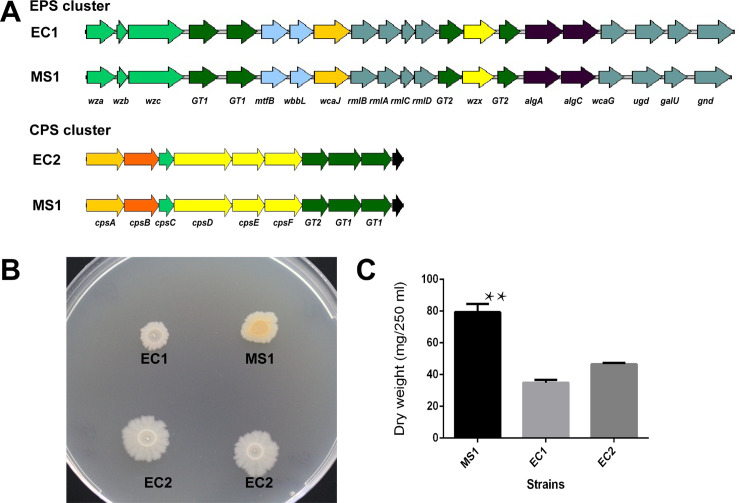
An EPS and CPS biosynthesis clusters were found in the genomes of D. zeae strains, but strains from rice did not have both of them. Taking D. zeae MS1 as an example, the EPS cluster in MS1 consisted of a large operon of 21 genes (J417_RS0113995 –J417_RS0114100), while the operon of the CPS cluster had 10 genes (J417_RS0103105–J417_RS0103150) (Fig 8A). Three D. zeae strains from rice, EC1, ZJU1202, and DZ2Q, lacked the CPS cluster but contained the EPS cluster (EC1: W909_RS06075– W909_RS06175; ZJU1202: WYU_RS0108445–WYU_RS0108340; DZ2Q: J134_RS12880–J134_RS12780) (Fig 8A), as did D. zeae CSL RW192. In a different instance, the new strain from rice, EC2, was the only D. zeae strain that lacked the EPS cluster, but harbored the CPS cluster (DWV07_02695–DWV07_02740). EPS cluster in D. zeae strains included genes encoding the polysaccharide export protein Wza, glycosyl transferase family proteins GT1 and GT2, undecaprenyl-phosphate glucose phosphotransferase WcaJ, polysaccharide biosynthesis protein Wzx, and others. Within CPS cluster, the predicted functions of some genes were similar to the EPS genes, such as cpsA encoding undecaprenyl-phosphate glucose phosphotransferase like wcaJ, cpsC encoding polysaccharide export protein like wza, cpsE encoding multidrug and toxic compound extrusion family and similar proteins in polysaccharide biosynthesis like wzx, as well as genes encoding glycosyl transferase family proteins GT1 and GT2. Further, the CPS cluster encoded another two capsular polysaccharide biosynthesis proteins, CpsD and CpsF, and an OM_channel superfamily protein CpsB. On the 2% glucose-supplemented NA plates, MS1 showed different colony morphology in comparison to EC1 and EC2. The colonies of MS1 were more prominent than the other two strains. EC1 showed the smallest colonies and EC2 exhibited colonies with a distinct, serrated border, indicating different EPS-associated phenotypes (Fig 8B). In turn, significantly more EPS was produced by MS1 than by those two strains from rice (Fig 8C).
Fig 8. EPS and CPS biosynthesis clusters in D. zeae strains and their EPS production.
(A) Genomic organization of EPS and CPS biosynthesis clusters in D. zeae strains EC2, EC1, and MS1. Strain from banana, MS1, has both EPS and CPS clusters. Previously identified strains from rice, EC1, ZJU1202, and DZ2Q, only have the EPS cluster, and the newly identified strain from rice, EC2, only has the CPS cluster. Homologous genes are presented in the same color. (B) Colony morphology of D. zeae strains, EC2, EC1, and MS1, on NA plates supplemented with 2% glucose. Colonies of strain from banana, MS1, were more prominent than the flat colonies of strains from rice, EC1 and EC2. Colonies of EC1 grew more slowly and EC2 had obvious serrated borders. (C) Dry weight of EPS produced by D. zeae strains EC2, EC1, and MS1. ** indicates significant difference observed between MS1 and either of strains from rice, EC1 and EC2, by two-tailed t-test, and the p values of difference statistics were 0.0002 and 0.0004, respectively.
Discussion
D. zeae strain EC2 was closest to previously identified strains from rice, but did not belong to same subgroup
Among the strains from rice, the previously identified strains from rice in China and Italy were much more conserved, except for EC2. Pairwise isDDH analysis (S5 Fig) found that these previously identified strains from rice reached the cut-off (90.00%) for a subspecies [43], and they were also suggested to be a subspecies within D. zeae [15].On other hand, ANI and AP analyses suggested EC2 was also close to the other two strains from non-rice hosts, NCPPB 3531 and CSL_RW192 (S5 Fig). In accordance with this, strains EC1, ZJU1202, DZ2Q, NCPPB 3531, and CSL RW192, were classified into a genomospecies named D. oryzae [44]. However, most of the genome sequences used in ANI and AP analyses were draft genomes, and ANI and AP analyses were based on pairwise comparisons. Thus, the comprehensive phylogenetic analyses using concatenated sequences of multiple genes of multiple stains might be better for explaining their genetic relationships and classification. Moreover, ANI and AP analyses showed EC2 and other strains from rice had higher similarities than strains from non-rice hosts. Thus, EC2 was a distant strain from the conserved subgroup of previous identified strains from rice, but still a member of the group of strains isolated from rice.
Fatty acid and sugar clusters inserted within fli/che locus might be associated with flagellin glycosylation and its modification
Flagellin FliC contained a motif similar to Flg22 that is implicated in plant recognition. Similar to DspE, this flagellin protein is considered a cause of plant cell death in Pectobacterium spp. [45]. Further, flagellin glycosylation is ubiquitous in most phytopathogenic bacteria, including D. dadantii strains, and is important for the virulence of some phytopathogens [46]. Glycosyltransferase plays key roles in the formation of glycosylation protein by catalyzing the transfer of a saccharide onto the acceptors [47] and is used in the glycosylation of flagellin [48]. For D. dadantii 3937, mutation of glycosyltransferase RfbC (Dda3937_03424) affected the glycosylation of Flic and further diminished its pathogenicity on chicory plants. The genes rfbC–fkbM (located between fliA and fliC) were supposedly involved in flagellin glycosylation [49].
Among the Guangdong strains (strains from rice, EC1, EC2, and ZJU1202, strain from banana, MS1, and strain from C. edulis, CE1), and the Italy strain DZ2Q from rice, their rfbC–fkbM clusters were inserted with genes encoding maltose O-acetyltransferase, sugar biosynthetic proteins, and fatty acid biosynthesis proteins. This is consistent in that the glycosyltransferase and sugar biosynthetic genes responsible for flagellin glycosylation are usually adjacent to the flagellin gene [50]. Moreover, the flagellin glycan chains of some bacteria can be methyl-, formyl-, acetyl-, or amino-modified [51]. Their essential role in flagellin glycosylation, the modification of flagellin glycan, is important for other virulence factors like motility, exopolysaccharide production, and biofilm formation [52]. Compared to the previously explored D. dadantii gene cluster, the additional sugar biosynthetic and fatty acid biosynthesis genes might play essential roles in flagellin glycosylation. Of note, several fatty acid genes were involved in this cluster, suggesting that the flagellin glycan of these D. zeae strains may incorporate the liposugar component [51]. The versatility and complexity of the flagellin glycan structure in these pathogens might help them adapt to monocotyledonous hosts like rice, banana, and C. edulis. Interestingly, these strains from rice, banana, and Canna were mostly from Guangdong province, China, except for DZ2Q from Italy. Using the sequences of this inserted gene cluster among strains from rice, banana, and Canna, phylogenetic analysis revealed that EC2 was closer to other strains from rice. Those four strains from rice had 320 polymorphic sites and 0.020 of nucleotide diversity (Pi), whereas the other three strains from non-rice hosts showed 1,010 polymorphic sites and 0.085 of nucleotide diversity (Pi). The strains from non-rice had much higher genetic diversity, suggesting they were older than the strains from rice. Additionally, the observation of lower GC contents indicating the inserted gene cluster might be acquired by horizontal gene transfer (HGT), and these strains with this inserted gene cluster might differentiate from a same evolutionary event and were closely related. Thus, the D. zeae strains from rice might differentiate from D. zeae strains infecting other hosts in adjacent geographical areas, and retained this genomic characteristic either in the previously identified strains from rice in China and Italy or the newly identified strain from rice. This also indicated that the strains from rice was related to those other D. zeae strains carrying the similar inserted gene cluster and it might not be appropriate to simply classify the strains from rice into the genomospecies D. oryzae and to keep them apart in this study.
T6SS was a potential important virulent factor of the newly identified strain from rice
T3SS–T5SS were not found in the newly identified strain from rice, EC2. These three secretion systems could be vital for the virulence of soft-rot Pectobacteriaceae, including Dickeya pathogens [53–55]. Pectobacterium strains naturally lacking T3SS have been isolated, suggesting that T3SS is not required for their survival [56, 57]. This is also common in D. paradisiaca strains. The functions of T3SS in Dickeya strains are different from the “stealth” mechanisms of T3SS used by Pseudomonas syringae or other plant-pathogenic bacteria to attack plants [58]. However, the method that soft-rot strains in the Pectobacteriaceae use to compensate for these lost secretion systems in the plant infection process remains unknown [45].
T1SS, T2SS, and T6SS were conserved in all analyzed D. zeae genomes. T1SS-secreting metalloproteases can affect plant cell wall proteins or the activities of pathogen-secreting enzymes [59]. T2SS-secreting plant cell wall-degrading enzymes (PCWDEs) are a group of enzymes active in plant cell-wall digestion [60]. Mutants of T1SS showed delayed symptom expression [59] and mutations in out-type T2SS inhibited soft rot symptoms [61], indicating important roles of these two secretion systems in pathogenesis. Moreover, T2SS was conserved among different Dickeya species, including D. paradisiaca, as PCWDEs were considered “brute force” in creating soft rot symptoms [62].
Genetic elements besides T1SS and T2SS, however, are required for the efficient colonization of host plants and adaption to new growth conditions encountered in the hosts [58]. T6SS is the most recently discovered secretion system in gram-negative bacteria [63] and is well conserved among them [64]. T6SS has been shown to mediate inter-bacterial antagonism through competition with other bacteria for space and resources [65] and facilitates pathogen invasion through the killing of commensal species [66], and could also affect the production of EPS and reduce virulence [67]. For Dickeya pathogens, there are very few reports on the functional analyses of T6SS components. Rhs effector proteins in D. dadantii 3937 carry nuclease domains that degrade target cell DNA and play roles in contact-dependent growth inhibition [68], as is consistent with the important function of T6SS in inter-bacterial antagonism [65]. In this study, a diverse repertoire was observed in the T6SS Hcp-VgrG component and Rhs effectors among D. zeae strains. The Hcp-VgrG component is an essential part of T6SS, which is the extracellular substrate activating T6SS [69]; it is also always present in the same operon with Rhs effectors and interacts with those effector proteins [65]. For example, VgrG is required for RhsB-mediated inhibition in D. dadantii 3937 [68]. Thus, those differentiations within T6SS loci might contribute to their virulence by enabling them to compete more effectively with other host-associated bacteria and exploit a specific host niche [65, 70].
The term effective competition applies especially to the newly identified strain from rice, EC2, which did not have the zeamine or T3SS clusters. Besides the inter-bacterial antagonism of T6SS, T6SS is closely related to T3SS in bacterial pathogens. The transition between T3SS and T6SS could coordinate bacterial lifecycles, like planktonic and biofilm formation in Pseudomonas aeruginosa, and production of these secretion systems could be switched by RetS/GacS sensors and the second messenger c-di-GMP [71]. Moreover, mutation of the T6SS core gene, tssM, also affects the expression of T3SS genes in Ralstonia solanacearum [72]. Collectively, T6SS might play important roles in the pathogenesis of EC2, such as effective competition with host-associated bacteria and adaption to the host environment. Its mechanism of pathogenesis might be different than in previously isolated strains from rice and require further exploration in the future. EC2 could be a potential type strain for the investigation of T6SS functions independent of the effects of other virulence determinants, like T3SS.
Loss of EPS or CPS biosynthesis cluster might be associated with EPS production and biofilm phenotypes
EPS is a major virulence factor, increasing the disease severity of bacterial pathogens in their hosts [73]. In this study, EPS cluster found in previously identified strains from rice was homologous with the EPS biosynthesis cluster (wza-wzb-wzc-wzx) encoding the group 1 and 4 capsules in Escherichia coli [74]. This locus was also considered in relation to the EPS biosynthesis of Erwinia pyrifoliae [75] and Pectobacterium brasiliense [76]. EPSs have two types of polysaccharides: one released as an exopolysaccharide (EPS), or slime polysaccharide, and the other as a capsular polysaccharide (CPS) that can form a discrete capsule around the cell and is intimately associated with the cell surface [77]. A CPS cluster was found in EC2 rather than EC1 and other strains from rice. Among gram-negative bacteria, EPS and CPS are often essential virulence determinants in plant pathogens, and the processes of their biosynthesis and export are indistinguishable [77], some of CPS genes were also predicted to play the functions similar with EPS genes in this study. Thus, loss of one of them might cause EC1 and EC2 to produce significantly less EPS than MS1 (Fig 8), as was consistent with the Hu’s observation that appreciably less EPS was produced by EC1 than other D. zeae strains [16].
EPS is a major component of the bacterial biofilm matrix, forming the scaffold for the biofilm architecture. It is also responsible for adhesion to surfaces and the cohesiveness of biofilm [78]. In this study, we found that only MS1 could form intact, regular biofilm on biofilm-inducing SOBG medium. EC1 biofilm appeared clumps, and EC2 biofilm was thinner (S6 Fig). These results indicated that the loss of either the EPS or CPS cluster could limit biofilm formation by affecting the EPS-associated phenotype. The effect on biofilm formation was further demonstrated by the different colony morphology on Congo red (CR; indicative of curli fibers and cellulose production) plates or Calcofluor (CF; indicative of cellulose production) plates. Curli fibers and cellulose synthesis have been considered a primary cause of biofilm formation [79]. We found that MS1 and EC2 formed wrinkled, red colonies on CR plates while EC1 produced smooth blue colonies (S6 Fig). These three strains all fluoresced on CF plates under UV conditions (S6 Fig). Thus, we speculated that loss of the CPS cluster in EC1 might cause defective curli production, limiting the functional amyloids that form the extracellular matrix of biofilms [80].
An evolutionary pathway for the new strain EC2 isolated from rice was different from previously identified strains
CRISPRs protect against bacteriophages and plasmids, and are widely distributed among bacteria [81]. Two types were found in most D. zeae strains. CRISPR-1 (subtype I-F) consisted of CRISPR-associated (Cas) core proteins (Cas1, Cas3) and Csy proteins (Csy1–Csy4), and CRISPR-2 (subtype I-E) contained the Cas core proteins (Cas1, Cas2, Cas3, Cas5) and Cse proteins (Cse1–Cse4). In a previous study, we found the CRISPR-1 array was conserved in D. zeae species, but the strains from rice contained either one simple direct repeat sequence or lacked the whole array [82]. Strain EC2, however, made this differentiation more complicated as it had the complete CRISPR-1 array (position: 3,690,316–3,709,212 bp) at the allele locus.
Six PKs/NRPs gene clusters were predicted in EC1, including gene clusters related to the biosynthesis of zeamine, indigoidine, bicornutin, siderophore, O-antigen saccharide, and turnerbactin. These PKs/NRPs clusters were also found in EC2, except for the zeamine cluster (zmsO–zmsN). It was only found in the D. zeae strains from rice, ZJU1202 and DZ2Q, D. solani strains, and D. fangzhongdai strains [15, 82]. The non-acquisition of the zeamine cluster encoding the phytotoxins which affected the rice seed germination, however, did not affect the pathogenicity of EC2 on rice plants, indicating the zeamine cluster might not be a necessary virulence factor for this new strain from rice.
CRISPR-1 is universal in all Dickeya species and might have been acquired before the differentiation of the genus Dickeya [82]. Lower genomic GC contents revealed that the zeamine clusters were likely derived from HGT [15]. Thus, strains from rice in China and Italy, EC1, ZJU1202, and DZ2Q, were much conserved, and all of them lost CRISPR-1 and acquired the zeamine cluster late in their evolutional process. In another case, missing of the T3SS–T5SS and EPS cluster, retention of the CRISPR-1 array, and failure of the zeamine cluster in EC2 suggests that EC2 was likely on an evolutionary pathway different to previously identified strains infecting rice. However, EC2 was still phylogenetically closest to that subgroup of strains previously isolated from rice. This was also indicated by the closer relationship between EC2 and other strains from rice in the genomic trait of fli/che loci incorporated with potential flagellin glycosylation and modification, in comparison with the other strains from non-rice hosts.
Conclusions
We isolated a new strain of D. zeae, EC2, from rice plant with typical rice foot rot symptoms. It was different from previously identified causal agents in biochemical characteristics and seed germination inhibition capability. Compared to the genomes of other strains from rice, this new strain lacked the hrp-type T3SS, T4SS, T5SS, and zeamine clusters, contained only a CPS cluster rather than the customary EPS cluster, and retained a subtype I-F CRISPR cluster. These differences indicated EC2 was likely on an evolutionary pathway different to the conserved subgroup consisted of previously identified strains from rice in China and Italy. However, EC2 was still phylogenetically closest to other Dickeya strains from rice, indicating all strains from rice have evolved toward a same direction for the adaption to rice plants. They retained common virulence traits on rice, like the characteristic fli/che loci and the varied T6SSs. Thus, we report a new type of foot rot pathogen in the rice fields of China that requires our future attention.
Supporting information
The analyzed strains include strains from rice, EC1 and EC2, strain from banana, MS1, and strain from C. edulis, CE1. Biochemical characteristics were based on their use of different substrates and obtained from Biolog analysis.
(TIF)
Dickeya dadantii 3937 was used as the out-group control.
(TIF)
MS2 is a strain from banana in Guangdong, China. Ech586 is a strain from Philodendron Schott in Florida, USA. Red indicates the homologous regions present in the same orientation; blue indicates the homologous regions present in an inverted orientation.
(TIF)
The analyzed strains include strains from rice, D. zeae EC1 and EC2, strain from banana, D. zeae MS1. (A) Inhibitory activity of culture crude from different strains. (B) Inhibitory activity of culture extract from different strains. CK indicates negative control.
(TIF)
The single genome-to-genome distance value was calculated at the GGDC web service (http://ggdc.dsmz.de/ggdc.php) using formula 2.
(TIF)
(A) Phenotypes of CR-binding strains from rice, EC1 and EC2, and strain from banana, MS1. (B) Phenotypes of CF-binding strains from rice, EC1 and EC2, and strain from banana, MS1. CR and CF plates were incubated at 25°C for 4 d. (C) Biofilm formation after growth in SOBG medium for 24 h. (D) Biofilm formation after growth in SOBG medium for 48 h. The static SOBG cultures were incubated at 30°C for 24 to 48 h.
(TIF)
(PDF)
The previously identified strains from rice include D. zeae EC1, ZJU1202, and DZ2Q. The 391 specific genes were grouped into 365 orthoMCL groups.
(XLSX)
The previously identified strains from rice include D. zeae EC1, ZJU1202, and DZ2Q. The 717 specific genes were grouped into 236 orthoMCL groups.
(XLSX)
Acknowledgments
We appreciate Dr. Jianuan Zhou from South China Agricultural University for providing the Dickeya zeae rice strain EC1.
Data Availability
The sequencing read data from the PacBio RS II sequencing platform and Illumina HiSeq 4000 platform were deposited in NCBI SRA under accession nos. SRR7630398 and SRR8181654, respectively. These SRA data are under the BioProject accession no. PRJNA483372, Sample accession no. SAMN09742555. Genome assembly and annotation were deposited in GenBank under accession no. CP031515.
Funding Statement
This research was supported by grants from the Natural Science Foundation of Guangdong province (2015A030312002, 2016A030311017) awarded to BL and QY, National Natural Science Foundation of China (31300118) awarded to JZ, Science and Technology Project of Guangdong province (2016B020202003) awarded to HS, the Science and Technology Project of Guangzhou city (201704030120) awarded to DS, and the Special fund for scientific innovation strategy-construction of high level Academy of Agriculture Science (R2017PY-QY004, R2018QD-056) awarded to JZ. The funders had no role in study design, data collection and analysis, decision to publish, or preparation of the manuscript.
References
- 1.Goto M. Bacterial foot rot of rice caused by a strain of Erwinia chrysanthemi. Phytopathology. 1979; 69:213–6. [Google Scholar]
- 2.Liu QG, Wang ZZ. Infection characteristics of Erwinia chrysanthemi pv. zeae on rice. Journal of South China Agricultural University. 2004; 25:55–7. (in Chinese) [Google Scholar]
- 3.Hussain MB, Zhang HB, Xu JL, Liu Q, Jiang Z, Zhang LH. The acyl-homoserine lactone-type quorum-sensing system modulates cell motility and virulence of Erwinia chrysanthemi pv. zeae. Journal of Bacteriology. 2008; 190(3):1045–53. 10.1128/JB.01472-07 [DOI] [PMC free article] [PubMed] [Google Scholar]
- 4.Liu QG, Zhang Q, Wei CD. Advances in Research of Rice Bacterial Foot Rot. Scientia Agricultura Sinica. 2013; 46(14):2923–31. (in Chinese) [Google Scholar]
- 5.Amarjit S, Brar JS, Kang IS, Daljit S. Plant disease scenario in Punjab. Plant Disease Research. 2009; 24(2):135–41. [Google Scholar]
- 6.Hong JM, Di GX, Xie LT, Guan MP. The identification of the pathogen of rice bacterial foot rot. Journal of Zhejiang Agricultural University. 1983; 9(4):339–42. (in Chinese) [Google Scholar]
- 7.Dye DW, Bradbury JF, Goto M, Hayward AC, Lelliott RA, Schroth MN. International standards for naming pathovars of phytopathogenic bacteria and a list of pathovar names and pathotype strains. Review of Plant Pathology. 1980; 59:153–68. [Google Scholar]
- 8.Samson R, Legendre JB, Christen R, Achouak W, Gardan L. Transfer of Pectobecterium chrysanthemi (Burkholder et al., 1953) Brenner et al. 1973 and Brenneria paradisiaca to the genus Dickeya gen. nov. as Dickeya chrysanthemi comb. nov. and Dickeya paradisiaca comb. nov. and delineation of four novel species, Dickeya dadantii sp. nov., Dickeya dianthicola sp. nov., Dickeya dieffenbachiae sp. nov. and Dickeya zeae sp. nov. International Journal of Systematic and Evolutionary Microbiology. 2005; 55:1415–27. 10.1099/ijs.0.02791-0 [DOI] [PubMed] [Google Scholar]
- 9.Brady C, Cleenwerck I, Denman S, Venter S, Rodríguez-Palenzuela P, Coutinho TA, et al. Proposal to reclassify Brenneria quercina (Hildebrand & Schroth 1967) Hauben et al. 1999 into a novel genus, Lonsdalea gen. nov., as Lonsdalea quercina comb. nov., descriptions of Lonsdalea quercina subsp. quercina comb. nov., Lonsdalea quercina subsp. iberica subsp. nov. and Lonsdalea quercina subsp. britannica subsp. nov., emendation of the description of the genus Brenneria, reclassification of Dickeya dieffenbachiae as Dickeya dadantii subsp. dieffenbachiae comb. nov., and emendation of the description of Dickeya dadantii. International Journal of Systematic and Evolutionary Microbiology. 2012; 62:1592–602. 10.1099/ijs.0.035055-0 [DOI] [PubMed] [Google Scholar]
- 10.Parkinson N, De Vos P, Pirhonen M, Elphinstone J. Dickeya aquatica sp. nov., isolated from waterways. International Journal of Systematic and Evolutionary Microbiology. 2014; 64(7):2264–6. [DOI] [PubMed] [Google Scholar]
- 11.van der Wolf JM, Nijhuis EH, Kowalewska MJ, Saddler GS, Parkinson N, Elphinstone JG, et al. Dickeya solani sp. nov. a pectinolytic plant-pathogenic bacterium isolated from potato (Solanum tuberosum). International Journal of Systematic and Evolutionary Microbiology. 2014; 64(3):768–74. [DOI] [PubMed] [Google Scholar]
- 12.Tian Y, Zhao Y, Yuan X, Yi J, Fan J, Xu Z, et al. Dickeya fangzhongdai sp. nov., a plant-pathogenic bacterium isolated from pear trees (Pyrus pyrifolia). International Journal of Systematic and Evolutionary Microbiology. 2016; 66(8):2831–5. 10.1099/ijsem.0.001060 [DOI] [PubMed] [Google Scholar]
- 13.Oulghazi S, Pédron J, Cigna J, Lau Y, Moumni M, Van Gijsegem F, et al. Dickeya undicola sp. nov., a novel species for pectinolytic isolates from surface waters in Europe and Asia. International Journal of Systematic and Evolutionary Microbiology. 2019; 69(8). 10.1099/ijsem.0.003497 [DOI] [PubMed] [Google Scholar]
- 14.Hugouvieux-Cotte-Pattat N, Jacot-des-Combes C, Briolay J. Dickeya lacustris sp. nov., a water-living pectinolytic bacterium isolated from lakes in France. International Journal of Systematic and Evolutionary Microbiology. 2019; 69(3). 10.1099/ijsem.0.003208 [DOI] [PubMed] [Google Scholar]
- 15.Zhou J, Cheng Y, Lv M, Liao L, Chen Y, Gu Y, et al. The complete genome sequence of Dickeya zeae EC1 reveals substantial divergence from other Dickeya strains and species. BMC Genomics. 2015; 16:571 10.1186/s12864-015-1545-x [DOI] [PMC free article] [PubMed] [Google Scholar]
- 16.Hu M, Li J, Chen R, Li W, Feng L, Shi L, et al. Dickeya zeae strains isolated from rice, banana and clivia rot plants show great virulence differentials. BMC Microbiology. 2018; 18(1):136 10.1186/s12866-018-1300-y [DOI] [PMC free article] [PubMed] [Google Scholar]
- 17.Parkinson N, Stead D, Bew J, Heeney J, Tsror L, Elphinstone J. Dickeya species relatedness and clade structure determined by comparison of recA sequences. International Journal of Systematic and Evolutionary Microbiology. 2009; 59:2388–93. 10.1099/ijs.0.009258-0 [DOI] [PubMed] [Google Scholar]
- 18.Wang JS, Yang XY. Comparative study on the causal bacteria of rice foot rot and corn stalk rot. Acta Phytopathologica Sinica. 1991; 21(3):181–4. (in Chinese) [Google Scholar]
- 19.Wu J, Zhang HB, Xu JL, Cox RJ, Simpson TJ, Zhang LH. (13)C labeling reveals multiple amination reactions in the biosynthesis of a novel polyketidepolyamine antibiotic zeamine from Dickeya zeae. Chemical Communications. 2010; 46:333–5. 10.1039/b916307g [DOI] [PubMed] [Google Scholar]
- 20.Zhou J, Zhang H, Wu J, Liu Q, Xi P, Lee J, et al. A novel multidomain polyketide synthase is essential for zeamine production and the virulence of Dickeya zeae. Molecular Plant-Microbe Interactions. 2011; 24(10):1156–64. 10.1094/MPMI-04-11-0087 [DOI] [PubMed] [Google Scholar]
- 21.Cheng Y, Liu X, An S, Chang C, Zou Y, Huang L, et al. A nonribosomal peptide synthase containing a stand-alone condensation domain is essential for phytotoxin zeamine biosynthesis. Molecular Plant-Microbe Interactions. 2013; 26(11):1294–301. 10.1094/MPMI-04-13-0098-R [DOI] [PubMed] [Google Scholar]
- 22.Zhou JN, Zhang HB, Lv MF, Chen YF, Liao LS, Cheng YY, et al. SlyA regulates phytotoxin production and virulence in Dickeya zeae EC1. Molecular Plant Pathology. 2016; 17(9):1398–408. 10.1111/mpp.12376 [DOI] [PMC free article] [PubMed] [Google Scholar]
- 23.Lv MF, Chen YF, Liao LS, Liang ZB, Shi ZR, Tang YX, et al. Fis is a global regulator critical for modulation of virulence factor production and pathogenicity of Dickeya zeae. Scientific Reports. 2018; 8(1):341 10.1038/s41598-017-18578-2 [DOI] [PMC free article] [PubMed] [Google Scholar]
- 24.Zhang Q, Yu C, Wen L, Liu Q. Tat system is required for the virulence of Dickeya zeae on rice plants. Journal of Plant Pathology. 2018; 100(3):409–18. [Google Scholar]
- 25.Weisberg WG. 16S ribosomal DNA amplification for phylogenetic study. Journal of Bacteriology. 1991; 173:697–703. 10.1128/jb.173.2.697-703.1991 [DOI] [PMC free article] [PubMed] [Google Scholar]
- 26.Waleron M, Waleron K, Podhajska A, Lojkowka E. Genotyping of bacteria belonging to the former Erwinia genus by PCR-RFLP analysis of a recA gene fragment. Microbiology. 2002; 48:583–95. [DOI] [PubMed] [Google Scholar]
- 27.Chin CS, Alexander DH, Marks P, Klammer AA, Drake J, Heiner C, et al. Nonhybrid, finished microbial genome assemblies from long-read SMRT sequencing data. Nature Methods. 2013; 10(6):563–9. 10.1038/nmeth.2474 [DOI] [PubMed] [Google Scholar]
- 28.Myers EW, Sutton GG, Delcher AL, Dew IM, Fasulo DP, Flanigan MJ, et al. A whole-genome assembly of Drosophila. Science. 2000; 287(5461):2196–204. 10.1126/science.287.5461.2196 [DOI] [PubMed] [Google Scholar]
- 29.Li H, Durbin R. Fast and accurate short read alignment with Burrows-Wheeler transform. Bioinformatics. 2009; 25: 1754–60. 10.1093/bioinformatics/btp324 [DOI] [PMC free article] [PubMed] [Google Scholar]
- 30.Walker BJ, Abeel T, Shea T, Priest M, Abouelliel A, Sakthikumar S, et al. Pilon: An Integrated Tool for Comprehensive Microbial Variant Detection and Genome Assembly Improvement. PLoS ONE. 2014; 9(11):e112963 10.1371/journal.pone.0112963 [DOI] [PMC free article] [PubMed] [Google Scholar]
- 31.Chen N. Using RepeatMasker to identify repetitive elements in genomic sequences. Current Protocols in Bioinformatics. 2004; 5(1):4.10.1–14. [DOI] [PubMed] [Google Scholar]
- 32.Lagesen K, Hallin P, Rødland EA, Staerfeldt HH, Rognes T, Ussery DW. RNAmmer: consistent and rapid annotation of ribosomal RNA genes. Nucleic Acids Research. 2007; 35(9):3100–8. [DOI] [PMC free article] [PubMed] [Google Scholar]
- 33.Lowe TM, Eddy SR. tRNAscan-SE: a program for improved detection of transfer RNA genes in genomic sequence. Nucleic Acids Research. 1997; 25(5): 955–64. [DOI] [PMC free article] [PubMed] [Google Scholar]
- 34.Besemer J, Lomsadze A, Borodovsky M. GeneMarkS: a self-training method for prediction of gene starts in microbial genomes. Implications for finding sequence motifs in regulatory regions. Nucleic Acids Research. 2001; 29(12):2607–18. [DOI] [PMC free article] [PubMed] [Google Scholar]
- 35.Krzywinski M, Schein J, Birol I, Connors J, Gascoyne R, Horsman D, et al. Circos: an information aesthetic for comparative genomics. Genome Research. 2009; 19:1639–45. 10.1101/gr.092759.109 [DOI] [PMC free article] [PubMed] [Google Scholar]
- 36.Grissa I, Vergnaud G, Pourcel C. CRISPRFinder: a web tool to identify clustered regularly interspaced short palindromic repeats. Nucleic Acids Research. 2007; 35 (Suppl 2):52–7. [DOI] [PMC free article] [PubMed] [Google Scholar]
- 37.Blin K, Wolf T, Chevrette M, Lu X, Schwalen C, Kautsar S, et al. AntiSMASH 4.0—improvements in chemistry prediction and gene cluster boundary identification. Nucleic Acids Research. 2017; 45(W1):W36–41. 10.1093/nar/gkx319 [DOI] [PMC free article] [PubMed] [Google Scholar]
- 38.Meier-Kolthoff JP, Auch AF, Klenk HP, Göker M. Genome sequence-based species delimitation with confidence intervals and improved distance functions. BMC Bioinformatics. 2013; 14:60 10.1186/1471-2105-14-60 [DOI] [PMC free article] [PubMed] [Google Scholar]
- 39.Li L, Stoeckert CJ, Roos DS. OrthoMCL: identification of ortholog groups for eukaryotic genomes. Genome Research. 2003; 13:2178–89. [DOI] [PMC free article] [PubMed] [Google Scholar]
- 40.Tang JL, Liu YN, Barber CE, Dow JM, Wootton JC, Daniels MJ. Genetic and molecular analysis of a cluster of rpf genes involved in positive regulation of synthesis of extracellular enzymes and polysaccharide in Xanthomonas campestris pathovar campestris. Molecular and General Genetics. 1991; 226:409–17. [DOI] [PubMed] [Google Scholar]
- 41.Barrére GC, Barber CE, Daniels MJ. Molecular cloning of genes involved in the biosynthesis of the extracellular polysaccharide xanthan by Xanthomonas campestris pv. campestris. International Journal of Biological Macromolecules. 1986; 8:372–4. [Google Scholar]
- 42.Richter M, Rosselló-Mora R. Shifting the genomic gold standard for the prokaryotic species definition. Proceedings of the National Academy of Science of the United States of America. 2009; 106 (45):19126–31. [DOI] [PMC free article] [PubMed] [Google Scholar]
- 43.Meier-Kolthoff JP, Hahnke RL, Petersen J, Scheuner C, Michael V, Fiebig A, et al. Complete genome sequence of DSM 30083T, the type strain (U5/41T) of Escherichia coli, and a proposal for delineating subspecies in microbial taxonomy. Standards in Genomic Sciences. 2014; 9:2 10.1186/1944-3277-9-2 [DOI] [PMC free article] [PubMed] [Google Scholar]
- 44.Wang X, He SW, Guo HB, Han JG, Thin K, Gao JS, et al. Dickeya oryzae sp. nov., isolated from the roots of rice. International Journal of Systematic and Evolutionary Microbiology. 2020; 70(7). 10.1099/ijsem.0.004265 [DOI] [PubMed] [Google Scholar]
- 45.Charkowski A, Blanco C, Condemine G, Expert D, Franza T, Hayes C, et al. The role of secretion systems and small molecules in soft-rot Enterobacteriaceae pathogenicity. Annual Review of Phytopathology. 2012; 50:425–49. [DOI] [PubMed] [Google Scholar]
- 46.Ichinose Y, Taguchi F, Yamamoto M, Ohnishikameyama M, Atsumi T, Iwaki M, et al. Flagellin glycosylation is ubiquitous in a broad range of phytopathogenic bacteria. Journal of General Plant Pathology. 2013; 79: 359–65. [Google Scholar]
- 47.Coutinho PM, Deleury E, Davies GJ, Henrissat B. An evolving hierarchical family classification for glycosyltransferases. Journal of Molecular Biology. 2003; 328:307–17. [DOI] [PubMed] [Google Scholar]
- 48.Iwashkiw JA, Vozza NF, Kinsella RL, Feldman MF. Pour some sugar on it: the expanding world of bacterial protein O-linked glycosylation. Molecular microbiology. 2013; 89:14–28. [DOI] [PubMed] [Google Scholar]
- 49.Royet K, Parisot N, Rodrigue A, Gueguen E, Condemine G. Identification by Tn-seq of Dickeya dadantii genes required for survival in chicory plants. Molecular plant pathology. 2019; 20:287–306. [DOI] [PMC free article] [PubMed] [Google Scholar]
- 50.Power PM, Jennings MP. The genetics of glycosylation in Gram-negative bacteria. FEMS microbiology letters. 2003; 218:211–22. [DOI] [PubMed] [Google Scholar]
- 51.De Maayer P, Cowan DA. Flashy flagella: flagellin modification is relatively common and highly versatile among the Enterobacteriaceae. BMC Genomics. 2016; 17:377 10.1186/s12864-016-2735-x [DOI] [PMC free article] [PubMed] [Google Scholar]
- 52.Li H, Yu C, Chen H, Tian F, He C. PXO_00987, a putative acetyltransferase, is required for flagellin glycosylation, and regulates flagellar motility, exopolysaccharide production, and biofilm formation in Xanthomonas oryzae pv. oryzae. Microbial pathogenesis. 2015; 85:50–7. [DOI] [PubMed] [Google Scholar]
- 53.Rojas CM, Ham JH, Deng WL, Doyle JJ, Collmer A. HecA is a member of a class of adhesins produced by diverse pathogenic bacteria and contributes to the attachment, aggregation, epidermal cell killing, and virulence phenotypes of Erwinia chrysanthemi EC16 on Nicotiana clevelandii seedlings. Proceedings of the National Academy of Sciences of the United States of America. 2002; 99(20):13142–7. [DOI] [PMC free article] [PubMed] [Google Scholar]
- 54.Bell KS, Sebaihia M, Pritchard L, Holden MTG, Hyman LJ, Holeva MC, et al. Genome sequence of the enterobacterial phytopathogen Erwinia carotovora subsp. atroseptica and characterization of virulence factors. Proceedings of the National Academy of Sciences of the United States of America. 2004; 101(30):11105–10. [DOI] [PMC free article] [PubMed] [Google Scholar]
- 55.Yang S, Peng Q, San Francisco M, Wang Y, Zeng Q, Yang CH. Type III Secretion System Genes of Dickeya dadantii 3937 Are Induced by Plant Phenolic Acids. PLoS ONE. 2008; 3(8):e2973 10.1371/journal.pone.0002973 [DOI] [PMC free article] [PubMed] [Google Scholar]
- 56.Kim HS, Ma B, Perna NT, Charkowski AO. Prevalence and virulence of natural type III secretion system deficient Pectobacterium strains. Applied and Environmental Microbiology. 2009; 75: 4539–49. [DOI] [PMC free article] [PubMed] [Google Scholar]
- 57.Pitman AR, Harrow SA, Visnovsky SB. Genetic characterisation of Pectobacterium wasabiae causing soft rot disease of potato in New Zealand. European Journal of Plant Pathology. 2010; 126:423–35. [Google Scholar]
- 58.Reverchon S, Muskhelisvili G, Nasser W. Virulence Program of a Bacterial Plant Pathogen: The Dickeya Model. Progress in Molecular Biology and Translational Science. 2016; 142:51–92. [DOI] [PubMed] [Google Scholar]
- 59.Hommais F, Oger-Desfeux C, Van Gijsegem F, Castang S, Ligori S, Expert D, et al. PecS is a global regulator of the symptomatic phase in the phytopathogenic bacterium Erwinia chrysanthemi 3937. Journal of Bacteriology. 2008; 190(22):7508–22. [DOI] [PMC free article] [PubMed] [Google Scholar]
- 60.Kazemi-Pour N, Condemine G, Hugouvieux-Cotte-Pattat N. The secretome of the plant pathogenic bacterium Erwinia chrysanthemi. Proteomics. 2004; 4:3177–86. [DOI] [PubMed] [Google Scholar]
- 61.Andro T, Chambost JP, Kotoujansky A, Cattaneo J, Bertheau Y, Barras F, et al. Mutants of Erwiniachrysanthemi defective in secretion of pectinase and cellulase. Journal of Bacteriology. 1984; 160(3):1199–203. [DOI] [PMC free article] [PubMed] [Google Scholar]
- 62.Toth IK, Birch PRJ. Rotting softly and stealthily. Current Opinion in Plant Biology 2005; 8:424–429. [DOI] [PubMed] [Google Scholar]
- 63.Mougous JD, Cuff ME, Raunser S, Shen A, Zhou M, Gifford CA, et al. A virulence locus of Pseudomonas aeruginosa encodes a protein secretion apparatus. Science. 2006; 312:1526–30. [DOI] [PMC free article] [PubMed] [Google Scholar]
- 64.Russell AB, Hood RD, Bui NK, LeRoux M, Vollmer W, Mougous JD. Type VI secretion delivers bacteriolytic effectors to target cells. Nature. 2011; 475(7356):343–7. [DOI] [PMC free article] [PubMed] [Google Scholar]
- 65.Russell AB, Peterson SB, Mougous JD. Type VI secretion system effectors: poisons with a purpose. Nature Reviews Microbiology. 2014; 12(2):37–148. [DOI] [PMC free article] [PubMed] [Google Scholar]
- 66.Smith WPJ, Vettiger A, Winter J, Ryser T, Comstock LE, Basler M, et al. The evolution of the type VI secretion system as a disintegration weapon. PLoS Biology. 2020; 18(5): e3000720 10.1371/journal.pbio.3000720 [DOI] [PMC free article] [PubMed] [Google Scholar]
- 67.Tian Y, Zhao Y, Shi L, Cui Z, Hu B, Zhao Y. Type VI Secretion Systems of Erwinia amylovora, Contribute to Bacterial Competition, Virulence, and Exopolysaccharide Production. Phytopathology. 2017; 107(6):654–61. [DOI] [PubMed] [Google Scholar]
- 68.Koskiniemi S, Lamoureux JG, Nikolakakis KC, t'Kint de Roodenbeke C, Kaplan MD, Low DA, et al. Rhs proteins from diverse bacteria mediate intercellular competition. Proceedings of the National Academy of Sciences of the United States of America. 2013; 110:7032–7. [DOI] [PMC free article] [PubMed] [Google Scholar]
- 69.Silverman JM, Brunet YR, Cascales E, Mougous JD. Structure and regulation of the type VI secretion system. Annual Review of Microbiology. 2012; 66:453–72. 10.1146/annurev-micro-121809-151619 [DOI] [PMC free article] [PubMed] [Google Scholar]
- 70.Green E, Mecsas J. Bacterial secretion systems: an overview. Microbiology Spectrum. 2016; 4(1). 10.1128/microbiolspec.VMBF-0012-2015 [DOI] [PMC free article] [PubMed] [Google Scholar]
- 71.Moscoso JA, Mikkelsen H, Heeb S, Williams P, Filloux A. The Pseudomonas aeruginosa sensor RetS switches Type III and Type VI secretion via c-di-GMP signalling. Environmental Microbiology. 2011; 13(12):3128–38. [DOI] [PubMed] [Google Scholar]
- 72.Zhang L, Xu JS, Xu J, Chen K, He L, Feng J. TssM is essential for virulence and required for type VI secretion in Ralstonia solanacearum. Journal of Plant Disease and Protection. 2012; 119(4):125–34. [Google Scholar]
- 73.Prakasha A, Darren Grice I, Vinay Kumar KS, Sadashivac MP, Shankard HN, Umesha S. Extracellular polysaccharide from, Ralstonia solanacearum; a strong inducer of eggplant defense against bacterial wilt. Biological Control. 2017; 110: 107–16. [Google Scholar]
- 74.Whitfield C. Biosynthesis and Assembly of Capsular Polysaccharides in Escherichia coli. Annual Review of Biochemistry. 2006; 75:39–68. [DOI] [PubMed] [Google Scholar]
- 75.Smits TH, Jaenicke S, Rezzonico F, Kamber T, Goesmann A, Frey JE, et al. Complete genome sequence of the fire blight pathogen Erwinia pyrifoliae DSM 12163T and comparative genomic insights into plant pathogenicity. BMC Genomics. 2010; 11:2 10.1186/1471-2164-11-2 [DOI] [PMC free article] [PubMed] [Google Scholar]
- 76.Bellieny-Rabelo D, Tanui CK, Miguel N, Kwenda S, Shyntum DY, Moleleki LN. Transcriptome and Comparative Genomics Analyses Reveal New Functional Insights on Key Determinants of Pathogenesis and Interbacterial Competition in Pectobacterium and Dickeya spp. Applied and Environmental Microbiology. 2019; 85(2):e02050–18. 10.1128/aem.02050-18 [DOI] [PMC free article] [PubMed] [Google Scholar]
- 77.Roberts IS. The biochemistry and genetics of capsular polysaccharide production in bacteria. Annual review of microbiology. 1996; 50:285–315. [DOI] [PubMed] [Google Scholar]
- 78.Flemming HC, Wingender J. The biofilm matrix. Nature Reviews Microbiology. 2010; 8(9):623–33. [DOI] [PubMed] [Google Scholar]
- 79.Da Re S, Ghigo JM. A CsgD-Independent Pathway for Cellulose Production and Biofilm Formation in Escherichia coli. Journal of Bacteriology. 2006; 188(8):3073–87. [DOI] [PMC free article] [PubMed] [Google Scholar]
- 80.Sleutel M, Van den Broeck I, Van Gerven N, Feuillie C, Jonckheere W, Valotteau C, et al. Nucleation and growth of a bacterial functional amyloid at single-fiber resolution. Nature Chemical Biology. 2017; 13, 902–8. [DOI] [PMC free article] [PubMed] [Google Scholar]
- 81.Mojica FJM, Díez-Villase˜nor C, García-Martínez J, Soria E. Intervening sequences of regularly spaced prokaryotic repeats derive from foreign genetic elements. Journal of Molecular Evolution. 2005; 60:174–82. 10.1007/s00239-004-0046-3 [DOI] [PubMed] [Google Scholar]
- 82.Zhang J, Hu J, Shen H, Zhang Y, Sun D, Pu X, et al. Genomic analysis of the Phalaenopsis pathogen Dickeya sp. PA1, representing the emerging species Dickeya fangzhongdai. BMC Genomics. 2018; 19(1):782 10.1186/s12864-018-5154-3 [DOI] [PMC free article] [PubMed] [Google Scholar]
Associated Data
This section collects any data citations, data availability statements, or supplementary materials included in this article.
Supplementary Materials
The analyzed strains include strains from rice, EC1 and EC2, strain from banana, MS1, and strain from C. edulis, CE1. Biochemical characteristics were based on their use of different substrates and obtained from Biolog analysis.
(TIF)
Dickeya dadantii 3937 was used as the out-group control.
(TIF)
MS2 is a strain from banana in Guangdong, China. Ech586 is a strain from Philodendron Schott in Florida, USA. Red indicates the homologous regions present in the same orientation; blue indicates the homologous regions present in an inverted orientation.
(TIF)
The analyzed strains include strains from rice, D. zeae EC1 and EC2, strain from banana, D. zeae MS1. (A) Inhibitory activity of culture crude from different strains. (B) Inhibitory activity of culture extract from different strains. CK indicates negative control.
(TIF)
The single genome-to-genome distance value was calculated at the GGDC web service (http://ggdc.dsmz.de/ggdc.php) using formula 2.
(TIF)
(A) Phenotypes of CR-binding strains from rice, EC1 and EC2, and strain from banana, MS1. (B) Phenotypes of CF-binding strains from rice, EC1 and EC2, and strain from banana, MS1. CR and CF plates were incubated at 25°C for 4 d. (C) Biofilm formation after growth in SOBG medium for 24 h. (D) Biofilm formation after growth in SOBG medium for 48 h. The static SOBG cultures were incubated at 30°C for 24 to 48 h.
(TIF)
(PDF)
The previously identified strains from rice include D. zeae EC1, ZJU1202, and DZ2Q. The 391 specific genes were grouped into 365 orthoMCL groups.
(XLSX)
The previously identified strains from rice include D. zeae EC1, ZJU1202, and DZ2Q. The 717 specific genes were grouped into 236 orthoMCL groups.
(XLSX)
Data Availability Statement
The sequencing read data from the PacBio RS II sequencing platform and Illumina HiSeq 4000 platform were deposited in NCBI SRA under accession nos. SRR7630398 and SRR8181654, respectively. These SRA data are under the BioProject accession no. PRJNA483372, Sample accession no. SAMN09742555. Genome assembly and annotation were deposited in GenBank under accession no. CP031515.


