Summary:
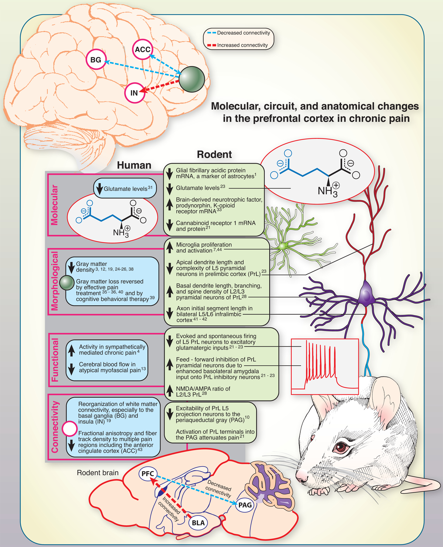
The prefrontal cortex undergoes functional and structural reorganization in chronic pain conditions in both rodents and humans. We provide an illustrated overview of the molecular, functional, and connectivity pathology occurring in the prefrontal cortex in chronic pain states.
The brain integrates information about the intensity, quality and location of noxious inputs with other states such as attention, anxiety, fear and expectation. A key brain region where this integration occurs is the prefrontal cortex (PFC). Importantly, the PFC is also responsible for higher executive functioning [11]. While acute pain is critical for survival, chronic pain is a detrimental, pathological state that drives changes in the PFC, culminating in pain amplification and cognitive problems. Here, we summarize how chronic pain affects the PFC in patients and in preclinical rodent models and why this is an important area of research in pain neuroscience.
Decades of lesion studies in rodents have demonstrated that the medial portion of the PFC (mPFC) controls higher executive functioning [8; 14; 27; 32]. The mPFC’s subregions, the prelimbic (PrL; or dorsolateral PFC in humans) and the infralimbic (IL; ventromedial PFC in humans) cortices are highly interconnected with one another as well as with other regions such as the amygdala, hippocampus, nucleus accumbens, and striatum [20; 29; 46]. The PrL and IL are responsible for modulating goal directed behaviors by integrating thought, motivation and action to achieve a goal. As such, a cardinal sign of PFC dysfunction in chronic pain patients presents as cognitive impairment which occurs in a variety of chronic pain conditions [2; 5; 6; 9; 15–18; 30; 34; 37]. Interestingly, pain relief using currently prescribed analgesics is insufficient to reverse cognitive impairments as the deficits persist and even worsen after analgesic treatment [16; 17; 34; 37], indicating that PFC dysfunction is resistant to transient analgesia.
Another sign of PFC dysfunction in chronic pain patients is gray matter loss. Shrinkage of the frontal cortical gray matter has been identified consistently across a variety of pain conditions [3; 12; 19; 24–26; 38]. This anatomical abnormality is severe and has been equated to the cortical loss seen over 10–20 years of normal aging in a healthy individual [3]. As none of these neuroimaging studies excluded patients who were taking analgesics, it is unlikely that cortical thinning is reversed by currently prescribed pain therapeutics. In fact, to date, the only two methods shown to reverse cortical thinning are cognitive behavioral therapy [39] and effective interventional pain management [35; 36; 40]. Cortical gray matter restoration has been observed in patients with hip osteoarthritic pain which dissipated after undergoing total hip replacement surgery [35; 36] and in chronic low back pain after spinal surgery or facet joint injections [40]. There is a clear need to investigate treatment options that can target both pain and its PFC-driven comorbidities.
Importantly, restoration of frontal cortical gray matter in chronic pain indicates the disease is not neurodegenerative, suggesting that structural reorganization of resident neurons and/or glia in the PFC account for the abnormality. Indeed, PFC morphological plasticity has been identified in rodents with neuropathic pain. Layer 2/3 pyramidal neurons display increased spine density and basal dendritic branching in the PrL contralateral (right) to nerve injury [28], while the apical dendrites of layer 5 pyramidal neurons are shrunken and less complex [23]. Axon initial segments are shrunken in the bilateral IL in mice with neuropathic pain [41; 42]. Microglia also proliferate [7], activate [7], and appear to take on an M1 phenotype as proinflammatory cytokines such as interleukin-6 and interleukin-1β are markedly increased in the PFC of rodents with neuropathic pain [44].
Neuroinflammation or structural pruning are either the cause or consequence of physiological dysfunction in PFC neurons. Reductions in PFC glutamate levels have been detected in rats [23] and humans [31] with chronic pain. Correspondingly, layer 5 pyramidal neurons display a loss in spontaneous and evoked firing arising from enhanced peri-somatic inhibition by local GABAergic interneurons in the PrL [21–23]. This disruption in excitation-inhibition balance is driven by augmented monosynaptic connections from basolateral amygdala (BLA) projection neurons onto layer 5 PrL inhibitory interneurons, resulting in feed-forward inhibition [21; 22]. Interestingly, strengthened glutamatergic inputs onto PrL inhibitory interneurons is due to a loss of the Gi-coupled cannabinoid receptor 1, resulting in disinhibition of glutamatergic afferents into the PrL [21]. While detection of PFC activity changes in humans has been inconsistent [4; 13], the emerging picture is that the PFC is deactivated in chronic pain.
Additionally, rodents with chronic pain show a loss in activity of PrL L5 pyramidal neurons that signal to the periaqueductal gray (PAG) [10; 21], a region responsible for mediating endogenous analgesia. Restoration of PrL cortical activity or activation of PrL afferents to the PAG can attenuate nociceptive behaviors in rodents with neuropathic pain [21; 45]. The chronification of pain may result in part from disruption of this descending analgesic circuitry that stems from PFC deactivation. Human data shows there is a loss of fiber track density and reorganization of white matter connectivity from the PFC to the insula, basal ganglia and other regions involved in pain processing such as the anterior cingulate cortex [19; 43], indicating that PFC output may be disrupted in human chronic pain patients as well.
Transcriptome analysis of the mPFC using quantitative real‐time PCR or sequencing has also identified specific mRNA transcripts that are dysregulated in chronic pain. Rodents with neuropathic pain display an increase in brain-derived neurotrophic factor, prodynorphin, and κ-opioid receptor [33]. Interestingly, mRNA for glial fibrillary acidic protein (GFAP), a commonly-used marker of astrocytes, was shown to be down-regulated in the mPFC of mice that had neuropathic pain for 6 months, suggesting that astrocyte populations may be diminishing at these later time points [1].
The work summarized here shows that complementary human and rodent studies have led to important insight into how the PFC changes in chronic pain states. A reverse translational approach has clearly been embraced wherein human symptomology and neuroimaging is directing preclinical investigations. Although there is still much unknown, the current picture is that the PFC modulates the pain experience in critical ways and that many comorbidities of painful disease are driven by PFC changes. Continuously growing insight into this pathology has great promise for improving pain care.

Funding:
NIH grants NS065926 and NS102161 to TJP
Footnotes
The authors declare no conflicts of interest.
References Cited
- [1].Alvarado S, Tajerian M, Millecamps M, Suderman M, Stone LS, Szyf M. Peripheral nerve injury is accompanied by chronic transcriptome-wide changes in the mouse prefrontal cortex. Mol Pain 2013;9:21. [DOI] [PMC free article] [PubMed] [Google Scholar]
- [2].Apkarian AV, Sosa Y, Krauss BR, Thomas PS, Fredrickson BE, Levy RE, Harden RN, Chialvo DR. Chronic pain patients are impaired on an emotional decision-making task. Pain 2004;108(1–2):129–136. [DOI] [PubMed] [Google Scholar]
- [3].Apkarian AV, Sosa Y, Sonty S, Levy RM, Harden RN, Parrish TB, Gitelman DR. Chronic back pain is associated with decreased prefrontal and thalamic gray matter density. J Neurosci 2004;24(46):10410–10415. [DOI] [PMC free article] [PubMed] [Google Scholar]
- [4].Apkarian AV, Thomas PS, Krauss BR, Szeverenyi NM. Prefrontal cortical hyperactivity in patients with sympathetically mediated chronic pain. Neurosci Lett 2001;311(3):193–197. [DOI] [PubMed] [Google Scholar]
- [5].Attal N, Masselin-Dubois A, Martinez V, Jayr C, Albi A, Fermanian J, Bouhassira D, Baudic S. Does cognitive functioning predict chronic pain? Results from a prospective surgical cohort. Brain 2014;137(Pt 3):904–917. [DOI] [PubMed] [Google Scholar]
- [6].Baker KS, Gibson SJ, Georgiou-Karistianis N, Giummarra MJ. Relationship between self-reported cognitive difficulties, objective neuropsychological test performance and psychological distress in chronic pain. Eur J Pain 2018;22(3):601–613. [DOI] [PubMed] [Google Scholar]
- [7].Barcelon EE, Cho WH, Jun SB, Lee SJ. Brain Microglial Activation in Chronic Pain-Associated Affective Disorder. Front Neurosci 2019;13:213. [DOI] [PMC free article] [PubMed] [Google Scholar]
- [8].Birrell JM, Brown VJ. Medial frontal cortex mediates perceptual attentional set shifting in the rat. J Neurosci 2000;20(11):4320–4324. [DOI] [PMC free article] [PubMed] [Google Scholar]
- [9].Bosma FK, Kessels RP. Cognitive impairments, psychological dysfunction, and coping styles in patients with chronic whiplash syndrome. Neuropsychiatry Neuropsychol Behav Neurol 2002;15(1):56–65. [PubMed] [Google Scholar]
- [10].Cheriyan J, Sheets PL. Altered Excitability and Local Connectivity of mPFC-PAG Neurons in a Mouse Model of Neuropathic Pain. J Neurosci 2018;38(20):4829–4839. [DOI] [PMC free article] [PubMed] [Google Scholar]
- [11].Chudasama Y Animal models of prefrontal-executive function. Behav Neurosci 2011;125(3):327–343. [DOI] [PubMed] [Google Scholar]
- [12].Davis KD, Pope G, Chen J, Kwan CL, Crawley AP, Diamant NE. Cortical thinning in IBS: implications for homeostatic, attention, and pain processing. Neurology 2008;70(2):153–154. [DOI] [PubMed] [Google Scholar]
- [13].Derbyshire SW, Jones AK, Devani P, Friston KJ, Feinmann C, Harris M, Pearce S, Watson JD, Frackowiak RS. Cerebral responses to pain in patients with atypical facial pain measured by positron emission tomography. J Neurol Neurosurg Psychiatry 1994;57(10):1166–1172. [DOI] [PMC free article] [PubMed] [Google Scholar]
- [14].Dias R, Robbins TW, Roberts AC. Primate analogue of the Wisconsin Card Sorting Test: effects of excitotoxic lesions of the prefrontal cortex in the marmoset. Behav Neurosci 1996;110(5):872–886. [DOI] [PubMed] [Google Scholar]
- [15].Dick B, Eccleston C, Crombez G. Attentional functioning in fibromyalgia, rheumatoid arthritis, and musculoskeletal pain patients. Arthritis Rheum 2002;47(6):639–644. [DOI] [PubMed] [Google Scholar]
- [16].Dick BD, Rashiq S. Disruption of attention and working memory traces in individuals with chronic pain. Anesth Analg 2007;104(5):1223–1229, tables of contents. [DOI] [PubMed] [Google Scholar]
- [17].Eccleston C Chronic pain and distraction: an experimental investigation into the role of sustained and shifting attention in the processing of chronic persistent pain. Behav Res Ther 1995;33(4):391–405. [DOI] [PubMed] [Google Scholar]
- [18].Galvez R, Marsal C, Vidal J, Ruiz M, Rejas J. Cross-sectional evaluation of patient functioning and health-related quality of life in patients with neuropathic pain under standard care conditions. Eur J Pain 2007;11(3):244–255. [DOI] [PubMed] [Google Scholar]
- [19].Geha PY, Baliki MN, Harden RN, Bauer WR, Parrish TB, Apkarian AV. The brain in chronic CRPS pain: abnormal gray-white matter interactions in emotional and autonomic regions. Neuron 2008;60(4):570–581. [DOI] [PMC free article] [PubMed] [Google Scholar]
- [20].Hoover WB, Vertes RP. Anatomical analysis of afferent projections to the medial prefrontal cortex in the rat. Brain Struct Funct 2007;212(2):149–179. [DOI] [PubMed] [Google Scholar]
- [21].Huang J, Gadotti VM, Chen L, Souza IA, Huang S, Wang D, Ramakrishnan C, Deisseroth K, Zhang Z, Zamponi GW. A neuronal circuit for activating descending modulation of neuropathic pain. Nat Neurosci 2019;22(10):1659–1668. [DOI] [PubMed] [Google Scholar]
- [22].Ji G, Sun H, Fu Y, Li Z, Pais-Vieira M, Galhardo V, Neugebauer V. Cognitive impairment in pain through amygdala-driven prefrontal cortical deactivation. J Neurosci 2010;30(15):5451–5464. [DOI] [PMC free article] [PubMed] [Google Scholar]
- [23].Kelly CJ, Huang M, Meltzer H, Martina M. Reduced Glutamatergic Currents and Dendritic Branching of Layer 5 Pyramidal Cells Contribute to Medial Prefrontal Cortex Deactivation in a Rat Model of Neuropathic Pain. Front Cell Neurosci 2016;10:133. [DOI] [PMC free article] [PubMed] [Google Scholar]
- [24].Kuchinad A, Schweinhardt P, Seminowicz DA, Wood PB, Chizh BA, Bushnell MC. Accelerated brain gray matter loss in fibromyalgia patients: premature aging of the brain? J Neurosci 2007;27(15):4004–4007. [DOI] [PMC free article] [PubMed] [Google Scholar]
- [25].Liao X, Mao C, Wang Y, Zhang Q, Cao D, Seminowicz DA, Zhang M, Yang X. Brain gray matter alterations in Chinese patients with chronic knee osteoarthritis pain based on voxel-based morphometry. Medicine (Baltimore) 2018;97(12):e0145. [DOI] [PMC free article] [PubMed] [Google Scholar]
- [26].Lin C, Lee SH, Weng HH. Gray Matter Atrophy within the Default Mode Network of Fibromyalgia: A Meta-Analysis of Voxel-Based Morphometry Studies. Biomed Res Int 2016;2016:7296125. [DOI] [PMC free article] [PubMed] [Google Scholar]
- [27].Livingston-Thomas JM, Jeffers MS, Nguemeni C, Shoichet MS, Morshead CM, Corbett D. Assessing cognitive function following medial prefrontal stroke in the rat. Behav Brain Res 2015;294:102–110. [DOI] [PubMed] [Google Scholar]
- [28].Metz AE, Yau HJ, Centeno MV, Apkarian AV, Martina M. Morphological and functional reorganization of rat medial prefrontal cortex in neuropathic pain. Proc Natl Acad Sci U S A 2009;106(7):2423–2428. [DOI] [PMC free article] [PubMed] [Google Scholar]
- [29].Mukherjee A, Caroni P. Infralimbic cortex is required for learning alternatives to prelimbic promoted associations through reciprocal connectivity. Nat Commun 2018;9(1):2727. [DOI] [PMC free article] [PubMed] [Google Scholar]
- [30].Nadar MS, Jasem Z, Manee FS. The Cognitive Functions in Adults with Chronic Pain: A Comparative Study. Pain Res Manag 2016;2016:5719380. [DOI] [PMC free article] [PubMed] [Google Scholar]
- [31].Naylor B, Hesam-Shariati N, McAuley JH, Boag S, Newton-John T, Rae CD, Gustin SM. Reduced Glutamate in the Medial Prefrontal Cortex Is Associated With Emotional and Cognitive Dysregulation in People With Chronic Pain. Front Neurol 2019;10:1110. [DOI] [PMC free article] [PubMed] [Google Scholar]
- [32].Paine TA, Asinof SK, Diehl GW, Frackman A, Leffler J. Medial prefrontal cortex lesions impair decision-making on a rodent gambling task: reversal by D1 receptor antagonist administration. Behav Brain Res 2013;243:247–254. [DOI] [PMC free article] [PubMed] [Google Scholar]
- [33].Palmisano M, Caputi FF, Mercatelli D, Romualdi P, Candeletti S. Dynorphinergic system alterations in the corticostriatal circuitry of neuropathic mice support its role in the negative affective component of pain. Genes Brain Behav 2019;18(6):e12467. [DOI] [PMC free article] [PubMed] [Google Scholar]
- [34].Povedano M, Gascon J, Galvez R, Ruiz M, Rejas J. Cognitive function impairment in patients with neuropathic pain under standard conditions of care. J Pain Symptom Manage 2007;33(1):78–89. [DOI] [PubMed] [Google Scholar]
- [35].Rodriguez-Raecke R, Niemeier A, Ihle K, Ruether W, May A. Brain gray matter decrease in chronic pain is the consequence and not the cause of pain. J Neurosci 2009;29(44):13746–13750. [DOI] [PMC free article] [PubMed] [Google Scholar]
- [36].Rodriguez-Raecke R, Niemeier A, Ihle K, Ruether W, May A. Structural brain changes in chronic pain reflect probably neither damage nor atrophy. PLoS One 2013;8(2):e54475. [DOI] [PMC free article] [PubMed] [Google Scholar]
- [37].Schiltenwolf M, Akbar M, Hug A, Pfuller U, Gantz S, Neubauer E, Flor H, Wang H. Evidence of specific cognitive deficits in patients with chronic low back pain under long-term substitution treatment of opioids. Pain Physician 2014;17(1):9–20. [PubMed] [Google Scholar]
- [38].Schmidt-Wilcke T, Leinisch E, Straube A, Kampfe N, Draganski B, Diener HC, Bogdahn U, May A. Gray matter decrease in patients with chronic tension type headache. Neurology 2005;65(9):1483–1486. [DOI] [PubMed] [Google Scholar]
- [39].Seminowicz DA, Shpaner M, Keaser ML, Krauthamer GM, Mantegna J, Dumas JA, Newhouse PA, Filippi CG, Keefe FJ, Naylor MR. Cognitive-behavioral therapy increases prefrontal cortex gray matter in patients with chronic pain. J Pain 2013;14(12):1573–1584. [DOI] [PMC free article] [PubMed] [Google Scholar]
- [40].Seminowicz DA, Wideman TH, Naso L, Hatami-Khoroushahi Z, Fallatah S, Ware MA, Jarzem P, Bushnell MC, Shir Y, Ouellet JA, Stone LS. Effective treatment of chronic low back pain in humans reverses abnormal brain anatomy and function. J Neurosci 2011;31(20):7540–7550. [DOI] [PMC free article] [PubMed] [Google Scholar]
- [41].Shiers S, Mwirigi J, Pradhan G, Kume M, Black B, Barragan-Iglesias P, Moy JK, Dussor G, Pancrazio JJ, Kroener S, Price TJ. Reversal of peripheral nerve injury-induced neuropathic pain and cognitive dysfunction via genetic and tomivosertib targeting of MNK. Neuropsychopharmacology 2019. [DOI] [PMC free article] [PubMed]
- [42].Shiers S, Pradhan G, Mwirigi J, Mejia G, Ahmad A, Kroener S, Price T. Neuropathic Pain Creates an Enduring Prefrontal Cortex Dysfunction Corrected by the Type II Diabetic Drug Metformin But Not by Gabapentin. J Neurosci 2018;38(33):7337–7350. [DOI] [PMC free article] [PubMed] [Google Scholar]
- [43].Woodworth D, Mayer E, Leu K, Ashe-McNalley C, Naliboff BD, Labus JS, Tillisch K, Kutch JJ, Farmer MA, Apkarian AV, Johnson KA, Mackey SC, Ness TJ, Landis JR, Deutsch G, Harris RE, Clauw DJ, Mullins C, Ellingson BM, Network MR. Unique Microstructural Changes in the Brain Associated with Urological Chronic Pelvic Pain Syndrome (UCPPS) Revealed by Diffusion Tensor MRI, Super-Resolution Track Density Imaging, and Statistical Parameter Mapping: A MAPP Network Neuroimaging Study. PLoS One 2015;10(10):e0140250. [DOI] [PMC free article] [PubMed] [Google Scholar]
- [44].Xu N, Tang XH, Pan W, Xie ZM, Zhang GF, Ji MH, Yang JJ, Zhou MT, Zhou ZQ. Spared Nerve Injury Increases the Expression of Microglia M1 Markers in the Prefrontal Cortex of Rats and Provokes Depression-Like Behaviors. Front Neurosci 2017;11:209. [DOI] [PMC free article] [PubMed] [Google Scholar]
- [45].Zhang Z, Gadotti VM, Chen L, Souza IA, Stemkowski PL, Zamponi GW. Role of Prelimbic GABAergic Circuits in Sensory and Emotional Aspects of Neuropathic Pain. Cell Rep 2015;12(5):752–759. [DOI] [PubMed] [Google Scholar]
- [46].Zhou H, Martinez E, Lin HH, Yang R, Dale JA, Liu K, Huang D, Wang J. Inhibition of the Prefrontal Projection to the Nucleus Accumbens Enhances Pain Sensitivity and Affect. Front Cell Neurosci 2018;12:240. [DOI] [PMC free article] [PubMed] [Google Scholar]


