

To the Editor,
Clinical assessments of an overall pigment disorder or that of an individual skin lesion may greatly differ from that perceived by a patient. Being able to objectively quantify the melanin content of each skin lesion would greatly reduce these discrepancies, help gain patient trust and establish pre- and post-treatment baselines. Clinically, this would be difficult without objective tools that quantify epidermal melanin concentration, such as in vivo microscopy or pathological analysis (Verschoore et al. 2012).
Electronic devices, called melanometers, are useful tools in measuring in vivo epidermal melanin concentration and personal sensitivity to ultraviolet light (Kanellis 2019). To standardize epidermal melanin concentration measurements, the average proportion of melanin to total pigment concentration is typically described in terms of a “pigment index” or “melanin index” (Arisi et al. 2019; Liakoni et al. 2019). Conversely, to determine an individual’s skin sensitivity to ultraviolet light or phototherapy, these devices calculate parameters, such as the minimal erythema dose, erythema index, and individual maximum safe radiant exposure (Asawanonda et al. 2000; Dolotov et al. 2004). The significant inter-individual and intra-individual variability of these parameters, which partly arise from variations in the epidermal melanin concentration, could easily be quantified by melanometers (Kanellis 2019). These devices could also be used to accurately map melasma dermal melanin deposition in persons of colour in a way not possible using methods, such as the Wood’s lamp.
Clinically, the inherently subjective and time-intensive Melasma Area and Severity Index score is typically used to quantify the severity of melasma before, during and after therapy (Kimbrough-Green et al. 1994). Assessing changes in epidermal melanin concentration in melasma is useful in predicting treatment duration, dose tapering and even early recognition and prognostication of treatment-resistant skin lesions. Regarding melasma diagnosis and management, it is striking to note the abundance of promising topical, phototherapy and oral melasma therapies that required further validation (Rodrigues and Pandya 2015; Kwon et al. 2019). Encouragingly, quantifying melasma severity before, during and after therapy with commercially available melanometers is simpler, easier and faster than other objective methods, such as microscopy. Therefore, it is proposed that melanometers could expedite the validation of promising melasma therapies for integration into mainstream practise through rapid and reproducible comparative studies of different treatment modalities.
Besides comparative studies, melanometers could be used in other aspects of patient care. Recently, Sarkar et al. (2019) emphasized the importance of photoprotection through thrice daily sunscreen use as a treatment for melasma as opposed to cosmetic or skin-lightening agents. They used the Melasma Area and Severity Index score to quantify improvements in melasma due to regular sunscreen use and demonstrated significantly improved quality of life using the Melasma Quality of Life Index. Melanometers could be used to describe the relationship between the sun protection factor rating of broad-spectrum sunscreen and the efficacy of sunscreen in the treatment or prevention of melasma. Melanometers could be used to emphasize the importance of photoprotection and reassure patients suffering from melasma and other conditions by quantifying the beneficial effects of sun-safety behaviours (Fig. 1) and avoidance of other known exacerbating factors (Rodrigues and Pandya 2015). Finally, these devices could also reassure the treating physician that sunscreen is not just as an adjunct to other treatment modalities of melasma.
Fig. 1.
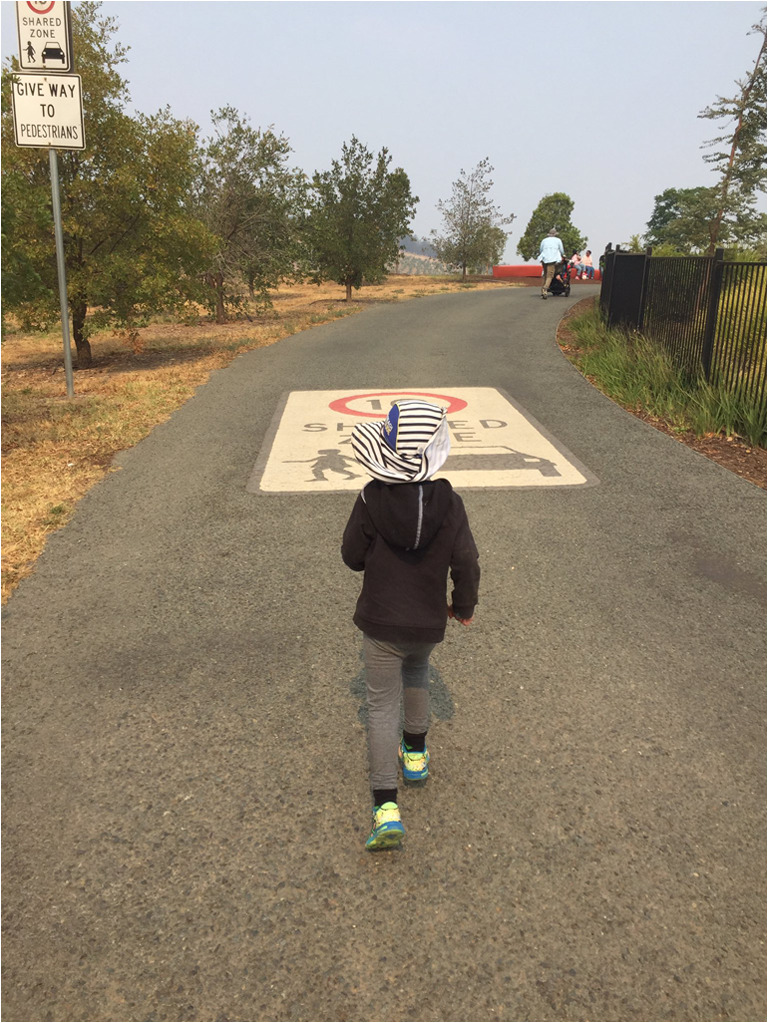
Emphasizing photoprotection and other sun safety practices plays an integral part in preventing skin damage from excessive ultraviolet light exposure
Footnotes
Publisher’s note
Springer Nature remains neutral with regard to jurisdictional claims in published maps and institutional affiliations.
References
- Arisi M, Rossi MT, Fusano M, Gualini A, Tomasi C, Moggio E, et al. Clinical and spectrophotometric evaluation of skin photoadaptation in vitiligo patients after a short cycle of NB-UVB phototherapy. Dermatology. 2019;235(6):509–515. doi: 10.1159/000502853. [DOI] [PubMed] [Google Scholar]
- Asawanonda P, Anderson RR, Chang Y, Taylor CR. 308-nm Excimer laser for the treatment of psoriasis: a dose-response study. JAMA Dermatol. 2000;136(5):619–624. doi: 10.1001/archderm.136.5.619. [DOI] [PubMed] [Google Scholar]
- Dolotov LE, Sinichkin YP, Tuchin VV, Utz SR, Altshuler GB, Yaroslavsky IV. Design and evaluation of a novel portable erythema-melanin-meter. Lasers Surg Med. 2004;34(2):127–135. doi: 10.1002/lsm.10233. [DOI] [PubMed] [Google Scholar]
- Kanellis VG. A review of melanin sensor devices. Biophys Rev. 2019;11:843–849. doi: 10.1007/s12551-019-00581-8. [DOI] [PMC free article] [PubMed] [Google Scholar]
- Kimbrough-Green CK, Griffiths CEM, Finkel LJ, Hamilton TA, Bulengo-Ransby SM, Ellis CN, Voorhees JJ (1994) Topical retinoic acid (tretinoin) for melasma in black patients. A vehicle-controlled clinical trial. Arch Dermatol 130(727). 10.1001/archderm.1994.01690060057005 [PubMed]
- Kwon SH, Na JI, Choi JY, Park KC. Melasma: updates and perspectives. Exp Dermatol. 2019;28:704–708. doi: 10.1111/exd.13844. [DOI] [PubMed] [Google Scholar]
- Liakoni E, St. Helen G, Dempsey DA, Jacob P, Tyndale RF, Benowitz NL (2019) Relationship between skin melanin index and nicotine pharmacokinetics in African American smokers. Drug Alcohol Depend 204(107474). 10.1016/j.drugalcdep.2019.04.039 [DOI] [PMC free article] [PubMed]
- Rodrigues M, Pandya AG. Melasma: clinical diagnosis and management options. Australas J Dermatol. 2015;56:151–163. doi: 10.1111/ajd.12290. [DOI] [PubMed] [Google Scholar]
- Sarkar R, Ghunawat S, Narang I, Verma S, Garg VK, Dua R. Role of broad-spectrum sunscreen alone in the improvement of melasma area severity index (MASI) and melasma quality of life index in melasma. J Cosmet Dermatol. 2019;18:1066–1073. doi: 10.1111/jocd.12911. [DOI] [PubMed] [Google Scholar]
- Verschoore M, Gupta S, Sharma VK, Ortonne JP. Determination of melanin and haemoglobin in the skin of idiopathic cutaneous hyperchromia of the orbital region (ICHOR): a study of Indian patients. J Cutan Aesthet Surg. 2012;5:176–182. doi: 10.4103/0974-2077.101371. [DOI] [PMC free article] [PubMed] [Google Scholar]