Figure 10.

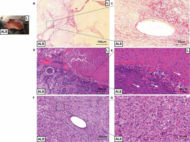
ALS could be recellularized after partial orthotopic transplantation. Macroscopic view of tissue connection area between recipient liver (represented by “L”) and acellular liver scaffold (represented by “ALS”) 3-d post-partial orthotopic transplantation (a). Microscopic view of liver sections obtained post 30 d of transplantation (B, C, D, E, F and G). Sirius red staining show delimitation between transplanted ALS area and recipient liver area. The white dotted line represents acellular liver scaffold and recipient liver tissue connection area (injured/fibrotic area) (b). Magnification of blood vessel-like structure formed into transplanted ALS (c). Recipient liver area (represented by “L”) appears a normal liver parenchyma 30-d post-transplantation (C). Infiltration of inflammatory cells (white circle) were detected into acellular scaffold area (represented by “ALS” (d). Red blood cells (white arrows) were detected into ALS vessel and recipient liver sinusoids (e). The acellular liver scaffold was completely recellularized post-orthotopic transplantation (f and g). Scale bars: 100 µm (B, D and F), 50 µm (C, E and G), 10x and 20x, respectively.
