Abstract
Honey bees are critical pollinators in ecosystems and agriculture, but their numbers have significantly declined. Declines in pollinator populations are thought to be due to multiple factors including habitat loss, climate change, increased vulnerability to disease and parasites, and pesticide use. Neonicotinoid pesticides are agonists of insect nicotinic cholinergic receptors, and sub-lethal exposures are linked to reduced honey bee hive survival. Honey bees are highly dependent on circadian clocks to regulate critical behaviors, such as foraging orientation and navigation, time-memory for food sources, sleep, and learning/memory processes. Because circadian clock neurons in insects receive light input through cholinergic signaling we tested for effects of neonicotinoids on honey bee circadian rhythms and sleep. Neonicotinoid ingestion by feeding over several days results in neonicotinoid accumulation in the bee brain, disrupts circadian rhythmicity in many individual bees, shifts the timing of behavioral circadian rhythms in bees that remain rhythmic, and impairs sleep. Neonicotinoids and light input act synergistically to disrupt bee circadian behavior, and neonicotinoids directly stimulate wake-promoting clock neurons in the fruit fly brain. Neonicotinoids disrupt honey bee circadian rhythms and sleep, likely by aberrant stimulation of clock neurons, to potentially impair honey bee navigation, time-memory, and social communication.
Subject terms: Neuroscience, Physiology
Introduction
Honey bees are critical pollinators in ecosystems and agriculture, but their numbers have significantly declined over the past three decades1. The observed declines in pollinator populations are thought to be due to multiple factors including habitat loss, climate change, increased vulnerability to disease and parasites, and pesticide use2. Neonicotinoid pesticides are agonists of insect nicotinic cholinergic receptors, which mediate excitatory neurotransmission within the insect brain, and sub-lethal exposures to neonicotinoids are linked to reduced honey bee hive survival3,4.
Honey bees are highly dependent on circadian clocks to regulate critical behaviors, such as foraging orientation and navigation, time-memory for food sources, sleep, and learning/memory processes5–8. Circadian clock neurons in Drosophila receive light input through cholinergic signaling9, and honey bees possess homologous clusters of putative clock neurons10,11, thus honey bee clock neurons are potential targets for neonicotinoid effects. Sub-lethal doses of neonicotinoid pesticides have been shown to disrupt navigation by forager honey bees12,13. In addition, neonicotinoid pesticides disturb honey bee learning and memory14,15 The mechanisms by which neonicotinoids disrupt honey bee orientation and learning are unknown, however both of these complex behaviors are highly regulated by endogenous honey bee circadian clocks8,16. Sleep is an additional critical clock-regulated neural process, and sleep in honey bees supports navigational memory consolidation17 and social communication through the waggle dance by which foragers communicate the location of food sources18.
Here we tested for effects of ingestion of neonicotinoids through feeding on honey bee circadian rhythms and sleep. We find that neonicotinoid ingestion over several days results in neonicotinoid accumulation in the bee brain, disrupts circadian rhythmicity in many individual bees, shifts the timing of behavioral circadian rhythms in bees that remain rhythmic, and impairs sleep. Neonicotinoids, thus, potentially impair key clock-based aspects of honey bee navigation, time-memory, and social communication.
Results
Honey bee (Apis mellifera ligustica) hives were maintained on the Vanderbilt University campus. Vanderbilt does not use neonicotinoid pesticides, and naïve forager bees from our hives did not show detectable levels of thiamethoxam or clothianidin, the two neonicotinoids used in this study (Supplemental Fig. 1).
To study effects of neonicotinoid pesticides on individual honey bee circadian rhythms and sleep, forager honey bees were captured at hive entrances and maintained in individual infrared activity monitors in the laboratory for up to 8 days with ad libitum access to bee candy (honey and powdered sugar, with or without the addition of thiamethoxam or clothianidin)19. Neonicotinoids in the food ranged from 0 to 140 ng/g (or parts per billion, ppb), which is within the concentration range reported in flower nectar and pollen encountered by foraging bees20–23. To determine the amounts of neonicotinoids present in dosed bees, we isolated whole brains from control bees that consumed bee candy with no added neonicotinoid, and from bees that consumed bee candy dosed with thiamethoxam at two concentrations (25 ppb and 70 ppb). We collected brains from each group at 24-h intervals over 4 days and then tested for thiamethoxan and its metabolite clothianidin by mass spectrometry (see “Materials and methods”). Thiamethoxam and clothianidin were undetectable in control bee brains (Supplemental Fig. 1). Thiamethoxam was undetectable, or detected at low picogram levels, in bee brains at both doses. The active metabolite of thiamethoxam, clothianidin24,25, was readily detected at higher levels (0–220 pg/brain, Supplemental Fig. 1), consistent with the previously described rapid conversion of thiamethoxam to clothianidin in insects24,25. Forager honey bees exposed to thiamethoxam-dosed food exhibited sustained levels of clothianidon of ca. 50–70 pg/brain through day 4 at the 70 ppb dose, with lower levels detected at the 25 ppb dose (Supplemental Fig. 1).
To obtain sufficient circadian cycles for analysis, only data from control and dosed bees that survived a minimum of 4 days in light:dark (LD) experiments, or a minimum of 5 days in constant dark (DD) and constant light (LL) experiments, were included in datasets (see “Materials and methods”). For those bees meeting these criteria for inclusion, survivorship analysis indicated that there was no additional mortality of neonicotinoid-exposed bees compared to control unexposed bees at any of the neonicotinoid concentrations (25–140 ppb) used in the LD, DD, and LL datasets (N = 944, Supplemental Fig. 2). Survivorship analysis of all bees that began LD, DD and LL experiments (including those that did not survive sufficient days to contribute to the dataset) showed that there was an initial significant loss of dosed bees compared to controls primarily limited to the first day of exposure to both 70 ppb and 140 ppb levels of thiamethoxam or clothianidin in the food, followed by comparable survivorship to controls on subsequent days (Supplemental Fig. 3). Thus, our study reports results from bees for which consumption of food with these levels of neonicotinoids for 4–5 days was not lethal and did not reduce survivorship compared to controls, while consumption of food with the 70 ppb and 140 ppb levels of thiamethoxam and clothianidin was lethal to some bees in the first 24 h.
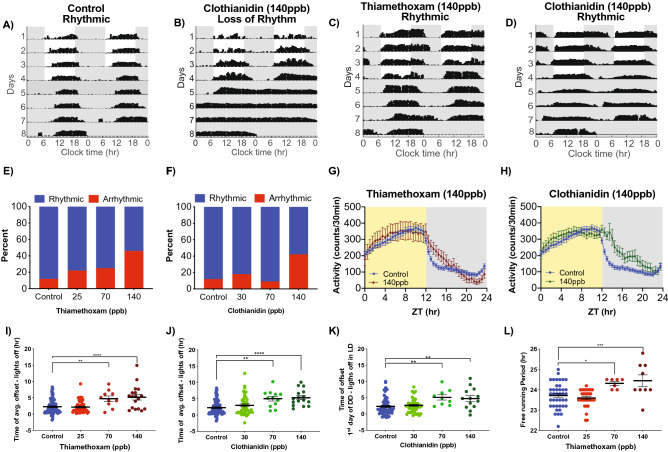
We first measured entrained and free-running circadian rhythms of captured foragers for 4 days of 12:12 LD followed by 4 days of DD in the presence and absence of neonicotinoids in the food. Ingestion of neonicotinoids completely disrupted locomotor circadian rhythms in a significant portion of bees, inducing arrhythmic behavior in an increasing proportion of bees in a dose-dependent manner (Fig. 1A,B). While only a small fraction (12%) of unexposed control bees exhibited arrhythmic locomotor behavior, nearly half of thiamethoxam-dosed (46%) and clothianidin-dosed (42%) bees lost circadian behavioral rhythms following consumption of these neonicotinoids (140 ppb, Fig. 1E,F; Pearson Chi-Square test, p = 0.0043 Thiamethoxam; p = 0.0142 Clothianidin).
Figure 1.
Neonicotinoid ingestion alters circadian locomotor rhythms of honey bee foragers in LD and DD. Representative actograms of forager bees showing (A) control rhythmic activity, (B) loss of rhythms following ingestion of clothianidin (140 ppb) and altered locomotor rhythm patterns after ingestion of (C) thiamethoxam (140 ppb) and (D) clothianidin (140 ppb). Contingency plots reporting the percent of arrhythmic individuals for bees exposed to (E) thiamethoxam (Pearson X2 = 13.14; p = 0.0043, **) or (F) clothianidin (Pearson X2 = 10.59; p = 0.0142, *). 30 min binned activity profiles of control bees vs. bees exposed to either 140 ppb of (G) thiamethoxam (Two-way RM ANOVA, Time p < 0.0001, ****; Dose p = 0.7150; Interaction p < 0.0001, ****) or (H) clothianidin (Two-way RM ANOVA, Time p < 0.0001, ****; Dose p = 0.1014; Interaction p < 0.0001, ****). Average time of activity offset following lights off for (I) thiamethoxam-exposed bees (One-way ANOVA F = 11.23; p < 0.0001, ****; Dunnett’s multiple comparison 70 ppb p = 0.0054, **; 140 ppb p < 0.0001, ****) and (J) clothianidin-exposed bees (One-way ANOVA F = 9.664; p < 0.0001, ****; Dunnett’s multiple comparison 70 ppb p = 0.0012, **; 140 ppb p < 0.0001, ****). (K) Offset of activity during the first day of constant darkness relative to time of lights off in previous LD for clothianidin-exposed bees (One-way ANOVA F = 5.805; p < 0.0009, ***; Dunnett’s multiple comparison 70 ppb p = 0.0077, **; 140 ppb p = 0.0063, **). (L) Free-running period of bees under constant darkness after 4 days of LD and ingestion of different doses of thiamethoxam (One-way ANOVA F = 7.323; p < 0.0002, ***; Dunnett’s multiple comparison 70 ppb p = 0.0374, *; 140 ppb p = 0.0028, **). Dunnett’s multiple comparisons were performed, only significant values are shown for panels (I–L).
In addition to disrupting honey bee locomotor circadian rhythms in many individuals, neonicotinoid ingestion also dramatically altered the rhythmic behavior of the dosed bees that remained rhythmic. Individual and average actogram activity profiles show that honey bees that ingested neonicotinoids delayed termination of their active period well into the dark phase following lights off, and increased their activity at night (Fig. 1C,D,G,H). There was a more than 3-h delay in the time of activity offset in bees that consumed either thiamethoxam or clothianidin compared to undosed bees (Fig. 1I,J; One-Way ANOVA p < 0.0001). This marked delay in activity offset persisted into the first day of DD in bees that consumed clothianidin (Fig. 1K, p = 0.0007), indicating a shift in the alignment of circadian rhythms relative to the light cycle. While honey bees that consumed neonicotinoids had statistically significant increases in the proportion of locomotor activity at night, and in the overall duration of activity each day, there was no significant increase in the total activity counts, indicating that neonicotinoids induced changes in the overall temporal structure of activity, but not the total amount of daily activity (Supplemental Fig. 4). In addition, ingestion of thiamethoxam, but not clothianidin, lengthened the free-running period of honey bee locomotor rhythms measured in DD following LD (Fig. 1L; One-Way ANOVA, p = 0.0002), demonstrating an effect on a core property of the underlying circadian clock.
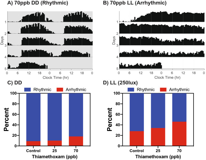
Surprisingly, when honey bees were exposed to neonicotinoids solely in DD (without first being exposed during LD), the treatment effects were blunted. There was no significant increase in behavioral arrhythmicity (Fig. 2A,C) and only a trend toward lengthening of the period (Supplemental Fig. 5). This result, and the central role of cholinergic signaling in clock light input in Drosophila, suggested to us the possibility of a key interaction between light input and neonicotinoid ingestion. To examine this potential interaction further, we combined continuous background illumination (LL, 250 lx) with exposure to thiamethoxam-dosed bee candy for a period of 5 days (Fig. 2B,D). Constant illumination is an abnormal signal to the circadian clock which renders some bees arrhythmic26. We found that that LL itself disrupted rhythms in 28% of undosed bees compared to just 9% arrhythmicity in DD. When thiamethoxam was combined with LL, a dose-dependent pattern in circadian disruption emerged, such that 34% of bees exposed to 25 ppb and 46% of bees exposed to 70 ppb became arrhythmic (Fig. 2B,D), but no significant effects occurred in response to exposure to these doses of thiamethoxam in DD (Fig. 2A). The differential effects between neonicotinoid exposure in LL vs DD suggests synergy between continuous light input and neonicotinoids in altering honey bee circadian behaviors.
Figure 2.
Constant light and thiamethoxam ingestion act synergistically to disrupt circadian rhythmicity of honey bees. (A) Representative actogram of foragers showing rhythmic activity of a bee exposed to 70 ppb thiamethoxam in constant darkness (DD). (B) Representative actogram of arrhythmic behavior after exposure to 70 ppb thiamethoxam in constant light (LL). Contingency plots showing the percentage of honey bees that were arrhythmic after exposure to thiamethoxam at various concentrations (C) under constant darkness (DD) (X2 = 2.101 p = 0.3498, ns), or (D) constant light (LL) (250 lx; X2 = 12.16 p = 0.0023, **).
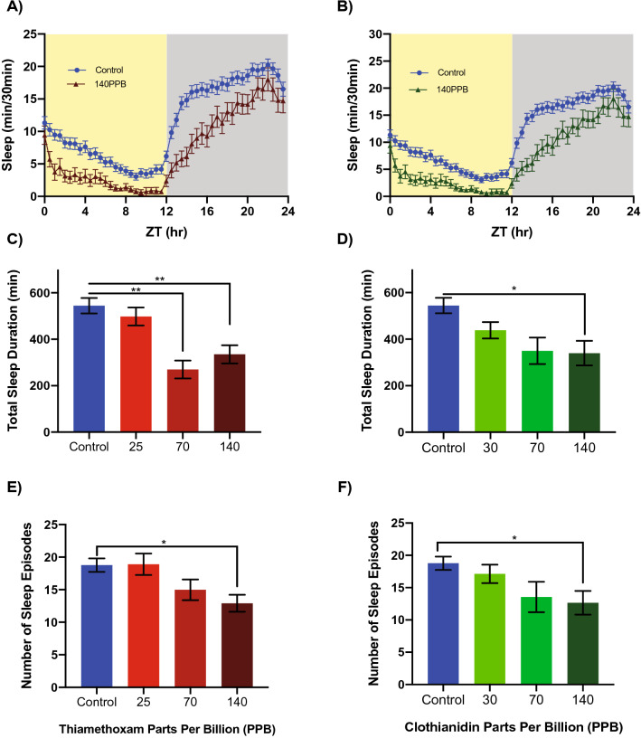
Light input mediated by nicotinic cholinergic neurotransmission targets the large ventral lateral neurons (l-LNvs), a specific subset of PDF-expressing clock neurons that antagonize sleep in fruit flies27,28. We therefore tested for neonicotinoid effects on bee sleep using previously established methods for sleep analysis in flies and bees that define sleep as episodes of inactivity lasting 5 min or more29–31. Ingestion of neonicotinoids significantly impaired sleep in honey bees (Fig. 3), with average daily sleep profiles showing that neonicotinoid-dosed bees in LD exhibited reduced sleep across both night and day (Fig. 3A,B). Overall sleep duration was reduced by up to 50% (Fig. 3C,D, One-way ANOVA p = 0.0004 Thiamethoxam; p = 0.0067 Clothianidin), while the number of sleep bouts were reduced as much as 24% (Fig. 3E,F, One-way ANOVA, p = 0.0314 Thiamethoxam; p = 0.0549 Clothianidin). Neonicotinoids also significantly reduced sleep upon application in DD and in LL (Supplemental Fig. 6). Thus, while neonicotinoids did not change the overall levels of activity (above), they changed the temporal structure of both activity and rest, disrupting sleep.
Figure 3.
Neonicotinoids disrupt sleep in honey bee foragers. Four day average sleep profiles for control bees and bees exposed to either (A) thiamethoxam (Two-way RM ANOVA, Time p < 0.0001, ****; Dose p = 0.0004, ***; Interaction p = 0.0015, **) or (B) clothianidin (Two-way RM ANOVA, Time p < 0.0001, ****; Dose p = 0.0054, **; Interaction p < 0.0001, ****). Significant decreases in total sleep duration for exposure to (C) thiamethoxam (One-way ANOVA F = 6.436; p = 0.0004, ***; Dunnett’s multiple comparison 70 ppb p = 0.0038, **; 140 ppb p = 0.0026, **) or exposure to (D) clothianidin (One-way ANOVA F = 4.205; p = 0.0067, **; Dunnett’s multiple comparison 140 ppb p = 0.0115, *). In addition, there were significant decreases in the number of sleep episodes for exposure to (E) thiamethoxam (One-way ANOVA F = 3.020; p = 0.0314*; Dunnett’s multiple comparison 140 ppb p < 0.0233, *) and for (F) clothianidin (One-way ANOVA F = 2.586; p = 0.0549, ns; Dunnett’s multiple comparison 140 ppb p < 0.0396, *) dosed bees. Significant values only are shown for Dunnett’s post hoc multiple comparisons (C–F).
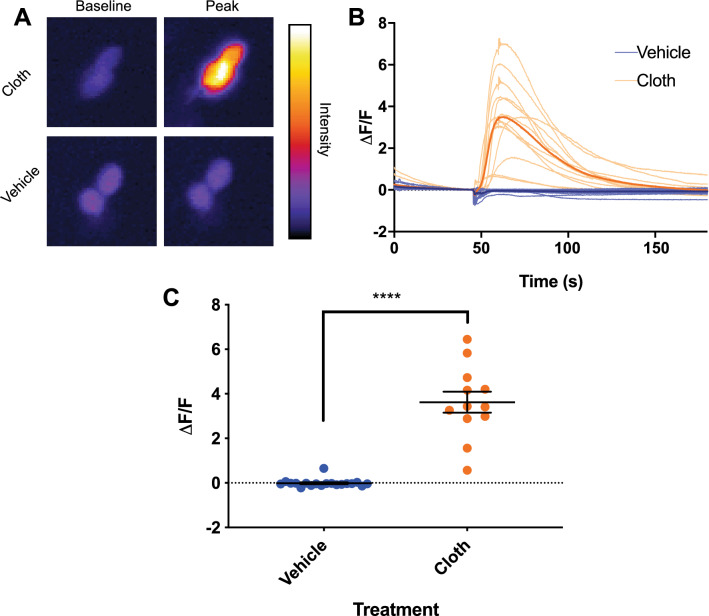
To examine whether neonicotinoids affect PDF-expressing circadian clock neurons, we next measured the Ca2+ responses of Drosophila l-LNv neurons. Intact whole-brain explants expressing the GCaMP5G Ca2+ indicator in the PDF+ clock neurons were imaged for Ca2+-dependent changes in fluorescence intensity in the l-LNv wake-promoting neurons upon exposure to either clothianidin or vehicle. We applied clothianidin because mass spectroscopy indicated an accumulation of clothianidin, as a metabolite of thiamethoxam, in bee brains when bees consumed thiamethoxam-dosed food (Supplemental Fig. 1). Exposure of isolated brains to clothianidin resulted in a ca. 350% increase in Ca2+ fluorescence in the l-LNv PDF+ clock neurons (ΔF/F, vehicle, − 0.01 ± 0.04, Clothianidin, 3.62 ± 0.47, p < 0.0001, Fig. 4A–C). These results are consistent with findings that Drosophila l-LNv neurons are excited directly by nicotinic cholinergic stimulation32,33, and likely express nicotinic acetylcholine receptors that are targeted by neonicotinoids. Recent results from Tasman et al. (2020, PREPRINT) demonstrate effects of chronic ingestion of neonicotinoids on Drosophila clock neurons as well as circadian rhythms and sleep34.
Figure 4.
Clothianidin induces excitatory increases in [Ca2+]i in Drosophila l-LNv clock neurons. (A) Representative images showing the baseline and peak fluorescence in Drosophila PDF+ l-LNv neurons expressing GCaMP5G fluorescent calcium indicator, in response to 4.1 ppb clothianidin (cloth, top) or vehicle (bottom). (B) Fluorescence intensity traces showing individual responses of Drosophila PDF+ l-LNv neurons to clothianidin (pale orange) or vehicle (pale blue). Averaged traces are shown for explants exposed to clothianidin (orange) and vehicle (blue). (C) Average peak fluorescence responses of neurons in explants exposed to vehicle (blue) or clothianidin (orange). Error bars represent SEM. ****Indicates p < 0.0001.
Discussion
This study demonstrates that neonicotinoid ingestion disrupts circadian rhythms in exposed honey bees. In addition, neonicotinoids alter the rhythmic behavior of bees that remain rhythmic following ingestion, delaying activity offset and extending activity into the night. Neonicotinoids also impair honey bee sleep, reducing total sleep and the number of sleep bouts.
The effects of neonicotinoids on honey bee circadian rhythms are enhanced by light input. In the presence of light cycles (LD) or constant light (LL), neonicotinoids induce loss of circadian behavioral rhythms in a dose-dependent manner. However, in the absence of light cycles (DD), neonicotinoids did not significantly disrupt honey bee circadian rhythms at similar exposure levels. In the field, forager honey bees experience the daily light cycle, and thus the effects of neonicotinoids measured in LD cycles are most relevant outside the laboratory.
In Drosophila, the PDF + l-LNv wake-promoting arousal neurons receive nicotinic cholinergic input from the compound eyes28,35,36. Neonicotinoid ingestion in bees mimics the effects of experimental overstimulation of Drosophila l-LNvs, extending activity into the night, increasing night-time activity, and dramatically reducing sleep28. Our results suggest that neonicotinoids aberrantly excite nicotinic cholinergic light input pathways to overstimulate homologous wake-promoting clock neurons in bees, leading to disruption and alteration of circadian rhythms and sleep. The neonicotinoid stimulation of Drosophila l-LNv neurons we observed (Fig. 4), and the previously observed delay of honey bee circadian rhythms in response to PDF injection into the putative bee clock nuclei10, support this proposed mechanism. It is also notable that chronic neonicotinoid ingestion also affects honey bee locomotor responses to light (phototaxis), and acute exposure can induce increased locomotor activity37.
Chronic ingestion of neonicotinoids resulted in three principal effects in our studies—disruption of behavioral circadian rhythms, shifts in the alignment of circadian activity with the light cycle, and impairment of sleep. Each of these effects on the honey bee circadian system have the potential for deleterious impacts on critical honey bee foraging behaviors which could ultimately impact hive health and survival. The abrogation of circadian rhythms that we found in significant proportions of bees that ingested neonicotinoids, could be expected to disrupt forager time-compensated sun-compass navigation, as it does in butterflies where the critical clocks are in the antennae38. Compromised circadian rhythms would also be expected to disrupt circadian clock-dependent time-memory for food source availability (Zeitgedächtnis). Each of these effects would significantly decrease the efficiency of honey bee foraging.
Ingestion of neonicotinoids delayed activity offset in LD, and this delay persisted when bees were then transitioned into DD, suggesting that neonicotinoids alter the timing of the honey circadian clock. The timing of activity offset relative to lights off has been shown to be a key marker of the entrainment, of honey bee circadian rhythms with the daily light cycle39, which is a dominant cue defining local environmental time. Thus, shifts in the alignment of circadian locomotor rhythm relative to the daily light cycle, as we observed in honey bees that remained rhythmic following neonicotinoid ingestion, would be expected to induce navigational errors due to changes in alignment of clock-based time-compensation for sun-compass orientation16,40. Neonicotinoid consumption induced ca. 3-h delays in locomotor activity alignment of dosed bees compared with the light cycle. Shifts of this magnitude are predicted to engender large navigational errors of ca. 45°, based on the known sun-compass time compensation of 15°/h41. Indeed, acute exposure to neonicotinoids impairs navigation and homing in honey bees12, 13, as do chronic exposures42, and quantitative models of hive viability predict that the observed rates of forager loss due to the neonicotinoid exposure can negatively impact hive survival12,43.
Interestingly, catch-move-release experiments following chronic neonicotinoid exposures showed that many of the neonicotinoid-exposed bees became lost and did not return to their hive due to mis-orientation of initial vector return flight paths42. This deficiency is consistent with dysregulation of circadian clock dependent sun-compass orientation predicted by the neonicotinoid effects reported here. Moreover, the disruption and modification of forager honey bee circadian rhythms may affect the overall bee colony temporal structure through disruption of social entrainment. Previous studies have reported that the rhythms of honey bee foragers serve to synchronize the rhythms of the hive-bound nurse bees44,45. In addition to possible indirect effects on nurse bee circadian rhythms by neonicotinoid disruption of forager rhythms, it would also be of interest to determine if neonicotinoid ingestion directly disrupts the circadian rhythms of nurse bees.
We found that chronic ingestion of neonicotinoids impaired honey bee sleep as well as circadian locomotor rhythms. Sleep deprivation has been widely demonstrated to disrupt neuro-cognitive functions such as learning, memory, and social communication in a wide variety of species. In honey bees, sleep deprivation specifically impairs critical navigational memory consolidation, also reducing the probability of forager bees returning to the hive17. In addition, sleep deprivation reduces the precision of the waggle dance by which foragers communicate the location of food sources18, degrading the social communication critical to foraging. Taken together, the sleep-disrupting effects of neonicotinoid ingestion may also compromise key neuro-behavioral mechanisms supporting honey bee foraging and hive survival.
Our results show dramatic impacts of neonicotinoids on circadian rhythms and sleep in honey bees. However, two limitations of our studies stem from the laboratory setting in which they were performed—(1) experiments were conducted over a relatively short study period (up to about 1 week), and (2) we measured the circadian rhythms of individuals, not entire hives. The relatively short duration of these experiments may actually underestimate the impact of long-term environmental neonicotinoids on the honey bee circadian system, as in the field exposures may be more prolonged and may include developmental exposures as well. It would be of interest in the future to measure circadian aspects of whole honey bee colony behavior in the field experiments such as studies that have demonstrated reduced hive productivity and whole hive survival due to exposure to neonicotinoids3,4.
Overall, we find that chronic ingestion of neonicotinoids disrupts circadian behavioral rhythms and sleep in forager honey bees, and that the predicted downstream neurobehavioral impacts of disrupted rhythms and sleep are potentially deleterious to hive health. Future studies are needed to identify and characterize the fundamental neural substrates that generate circadian and sleep behaviors in honey bees, and that synchronize bee circadian rhythms to local time through the daily light cycle. The cholinergic signaling neural circuits by which light and neonicotinoids impact honey bee locomotor rhythms also need to be identified. With the future development of additional experimental tools for honey bee neuroscience, new lines of research may elucidate the mechanism behind the interaction between pesticides and light signaling. The understanding of these pathways would open the way to elucidating the mechanisms by which neonicotinoids impact the circadian rhythms of honey bees, and suggest possible means to limit their disruptive impact.
Materials and methods
Honey bees
Honey bees (Apis mellifera ligustica) were maintained in standard Langstroth 10-frame boxes (Kelly Beekeeping, Clarkson, KY) on the Vanderbilt campus. All bees used for experiments came from healthy colonies with a mated queen. Vanderbilt does not use neonicotinoid pesticides on its property, and forager honey bees from our hives did not show detectable baseline levels of thiamethoxam or clothianidin, the two neonicotinoids that we used in our study (Supplemental Fig. 1).
Locomotor activity monitoring experiments
Foragers were caught outside the hive entrance upon their return from foraging trips, confirmed by the presence of pollen on their hind legs. Foragers were individually housed in plastic tubes containing either dosed bee candy (ground white cane sugar and honey) containing a specified amount of thiamethoxam, clothianidin, or control bee candy with no pesticide. Tubes were loaded into Locomotor Activity Monitors (LAMs, Trikinetics) and housed in an environmental chamber with a controlled light:dark cycle, constant temperature of 35 °C and humidity of 60%. A filter paper wick supplied water from a reservoir to each tube19. Data recording (infrared beam interruptions) began immediately after loading the monitors, and data beginning with the first full day of activity was used for analysis. Experiments of 12:12 light:dark cycle (LD) for 4 days, followed by 4 days of constant darkness (DD), took place in the spring/summer of 2017. Experiments of 250 lx constant light (LL) and constant darkness (DD) were performed during the spring/summer of 2019. In the case of the LL experiments, bees were placed into the incubator during the daytime and lights were not turned off for the duration of the experiment. For DD, foragers were placed in the incubator in the daytime and lights were turned off at 19:00 CDT that day and not turned back on for the duration of the experiment. For the LD experiments, locomotor activity rhythms and sleep data were analyzed from bees that survived in the activity monitors a minimum of 4 days, and up to 8 days of data were used. For the DD and LL experiments only bees that survived 5 full days of the experiment were used for data analysis.
Behavioral metrics (data analysis)
Locomotor activity rhythms are displayed as double plotted actograms (Fig. 1A–D). Free-running period calculations were calculated using autocorrelation analysis (Fig. 1L, Supplementary Fig. 5) generated using the previously published MATLAB toolboxes46. Individuals were scored as rhythmic or arrhythmic by a combination of eye inspection of each activity record, the absence of a significant peak in chi-square periodogram, and evaluation of the waveform (showing a significant and cyclical pattern) of the correlogram produced by the autocorrelation analysis (Fig. 1E,F, Fig. 2). The individual activity onset and offsets for each day were obtained using ClockLab (Actimetrics) with manual corrections as needed (Fig. 1I–L). Average activity duration was calculated by subtracting the onsets from the offsets of each day (offset time–onset time) and then averaging (Supplemental Fig. 4A,B). The proportion of activity occurring in the dark was calculated by dividing the number of infrared beam breaks recorded throughout the dark phase of a particular day by the number of beam breaks recorded throughout both the light and dark phases of that day. For each bee, all days were then averaged and these values grouped by treatment (Supplemental Fig. 4C,D). Total activity counts for LD portion of the experiment shown as total activity counts over 4 days of light: dark cycle (Supplemental Fig. 4E,F).
Sleep was analyzed from the activity data collected from the LAMs. Several studies have shown that lack of movement (beam interruptions) for five consecutive minutes serves as a reliable measure for sleep in honey bees29,45,46. Consistent with these studies we defined sleep as 5 min or longer bouts of inactivity. Data was analyzed using a custom toolbox for MATLAB that was originally developed to measure sleep in flies31. For each individual, we calculated sleep in 30 min bins, total sleep duration throughout the day, and the number of average sleep episodes in 24 h. (Fig. 3, Supplemental Fig. 6).
LC–MS/MS analysis
Sample analyses were carried out by the Vanderbilt Mass Spectrometry Core Lab using a Vanquish ultrahigh performance liquid chromatography (UHPLC) system interfaced to a Q Exactive HF quadrupole/orbitrap mass spectrometer (Thermo Fisher Scientific) equipped with an Ion Max API source, a HESI-II electrospray probe, and a 50 um ID stainless steel capillary. Chromatographic separation was performed with a reverse-phase Acquity HSS C18 column (1.7 µm, 2.1 × 50 mm, Waters, Milford, MA) at a flow rate of 300 µL/min. Mobile phases were made up of (A) 0.2% HCOOH in H2O and (B) 0.2% HCOOH in CH3CN. Gradient conditions were as follows: 0–1 min, B = 0%; 1–6 min, B = 0–100%; 6–6.5 min, B = 100%; 6.5–7.0 min, B = 100–0%; 7.0–10 min, B = 0%. The total chromatographic run time was 10 min; the sample injection volume was 10 µL. A software-controlled divert valve was used to transfer eluent from 0 to 1.5 min and from 5 to 10 min of each chromatographic run to waste. The instrument was calibrated weekly over a mass range of m/z 195 to m/z 1822 with a mixture of caffeine, MRFA, and Ultramark-1621 using the manufacturer’s HF-X tuning software. The mass spectrometer was operated in positive ion mode. The following optimized source parameters were used for the detection of analyte and internal standard: N2 sheath gas 40 psi; N2 auxiliary gas 10 psi; spray voltage 5.0 kV; capillary temperature 320 °C; vaporizer temperature 100 °C. Quantitation was based on parallel reaction monitoring (PRM) using a precursor isolation width of 2.0 m/z and the following MS/MS transitions.
| Analyte | Precursor mass (m/z) | Product mass (m/z) | HCD collision energy | S-lens RF level |
|---|---|---|---|---|
| Clothianidin | 250.0160 | 169.05450 | 30 | 60 |
| Clothianidin-d3 | 253.0348 | 172.0733 | 30 | 60 |
| Thiamethoxam | 292.0266 | 211.06510 | 30 | 60 |
| Thiamethoxam-d3 | 295.0454 | 214.0839 | 30 | 60 |
HCD product ion spectra were acquired in the Orbitrap at a resolving power of 30,000, an AGC target of 2e5, and a maximum injection time of 100 ms. Data acquisition and quantitative spectral analysis were done using Xcalibur version 2.2 sp1 and LCQuan version 2.7.0, respectively. Calibration curves were constructed for clothianidin and thiamethoxam by plotting peak area ratios (analyte / internal standard) against analyte concentrations for a series of seven calibrants, ranging in concentration from 2 pg to 1 ng. A weighting factor of 1/Ct2 was applied in the linear least-squares regression analysis to maintain homogeneity of variance across the concentration range (% error ≤ 20% for at least four out of every five standards). Foragers were exposed to either 0 ng/g, 25 ng/g or 70 ng/g of thiamethoxam and kept in individual chambers for five days. Individuals from each group were taken at 2 h, 26h, 50 h, 74 h, and 98 h after exposure to the pesticide food and flash-frozen in liquid nitrogen and kept at – 80 °C until processed. Brains were dissected in 100% ETOH chilled with dry ice. Each brain was homogenized in 5% 1:1 MeOH:H2O solution, acetonitrile. Internal standards for d3-thiamethoxam and d3-clothianidin were added at a concentration of 250 nM in solution. Homogenate was spun at 3000 rpm for 3 min after which supernatant was decanted and dried. Dried samples were stored at − 20 °C until processed for LC–MS. The dried residue was reconstituted in 100 µL of H2O/CH3OH (3:1), vigorously vortexed, and transferred to 200-µL silanized autosampler vials equipped with Teflon-lined bonded rubber septa.
Calcium imaging
Calcium imaging was performed as previously described47. Adult male flies (4–7 days post-eclosion) from the cross PDF-Gal4 > 20XUAS-IVS-GCamp5G 41 were immobilized on 300 mm petri dishes in 3 mL physiological saline containing 128 mM NaCl, 2 mM KCl, 4 mM MgCl2, 35.5 mM sucrose, 5 mM HEPES, and 1.8 mM Ca2+, pH 7.2. Labeled PDF soma were identified under halogen lamp illumination and allowed to rest for 2–3 min to photo bleach soma prior to baseline recording. l-LNv neurons we distinguished by soma size and position. Soma were imaged with a 40 × water-immersion objective with maximal pinhole aperture on a Zeiss LSM510Meta laser-scanning confocal microscope, using 256 × 256 resolution and a region of interest (ROI) box to achieve ~ 186 ms scans. Baseline fluorescence was recorded for 250 frames, followed by direct application of given drug, with a main recording for 1,000 total frames. To confirm that explants were viable even when no response to treatment/vehicle was observed, 50 μL of 2.5 M KCl was applied after 1,000 frames to confirm a normal calcium response to high potassium.
Supplementary information
Acknowledgements
This study was supported by an NSF Graduate Research Fellowship 0909667 and NIH F31 NS096813-02 to M.C.T., NIH R01 GM117650, a Vanderbilt Discovery grant, and Velux Stiftung Project Number 1389 to D.G.M. The authors would like to thank Kai-Wen Chiang, Emma Rushton, Chad R. Jackson, Tugrul Giray, Terry Jo Bichell, David Bichell and M. Wade Calcutt for discussions.
Author contributions
M.C.T. and D.G.M. conceived of the project. M.A.G.G. and M.C.T. performed pesticide/honey bee behavioral experiments. M.A.G.G., E.S.P. and M.C.T. performed honey bee LL experiments, DD experiments. M.A.G.G., M.C.T., E.S.P. analyzed the data. C.A.D, M.C.T., and M.A.G.G., performed Drosophila Ca2+ imaging experiments. M.C.T, M.A.G.G., C.A.D., D.M., and D.G.M. designed experiments. D.G.M., M.C.T. and M.A.G.G. wrote the paper. J.A.R., K.B., and D.M. provided editing, comments, and experimental guidance.
Competing interests
The authors declare no competing interests.
Footnotes
Publisher's note
Springer Nature remains neutral with regard to jurisdictional claims in published maps and institutional affiliations.
These authors contributed equally: Michael C. Tackenberg and Manuel A. Giannoni-Guzmán.
Supplementary information
is available for this paper at 10.1038/s41598-020-72041-3.
References
- 1.Ferrier, P. M., Rucker, R. R., Thurman, W. N. & Burgett, M. Economic Effects and Responses to Changes in Honey Bee Health. www.ers.usda.gov (2018).
- 2.Goulson D, Nicholls E, Botías C, Rotheray EL. Bee declines driven by combined Stress from parasites, pesticides, and lack of flowers. Science. 2015;347:1255957–1255957. doi: 10.1126/science.1255957. [DOI] [PubMed] [Google Scholar]
- 3.Tsvetkov N, et al. Chronic exposure to neonicotinoids reduces honey bee health near corn crops. Science. 2017;356:1395–1397. doi: 10.1126/science.aam7470. [DOI] [PubMed] [Google Scholar]
- 4.Woodcock BA, et al. Country-specific effects of neonicotinoid pesticides on honey bees and wild bees. Science. 2017;356:1393–1395. doi: 10.1126/science.aaa1190. [DOI] [PubMed] [Google Scholar]
- 5.Moore D. Honey bee circadian clocks: behavioral control from individual workers to whole-colony rhythms. J. Insect Physiol. 2001;47:843–857. [Google Scholar]
- 6.Bloch G, Bar-Shai N, Cytter Y, Green R. Time is honey: circadian clocks of bees and flowers and how their interactions may influence ecological communities. Philos. Trans. R. Soc. B. 2017;372:20160256. doi: 10.1098/rstb.2016.0256. [DOI] [PMC free article] [PubMed] [Google Scholar]
- 7.Eban-Rothschild A, Bloch G. Honeybee Neurobiology and Behavior. Dordrecht: Springer; 2012. Circadian rhythms and sleep in honey bees; pp. 31–45. [Google Scholar]
- 8.Lehmann M, Gustav D, Galizia CG. The early bee catches the flower - circadian rhythmicity influences learning performance in honey bees, Apis mellifera. Behav. Ecol. Sociobiol. 2011;65:205–215. doi: 10.1007/s00265-010-1026-9. [DOI] [PMC free article] [PubMed] [Google Scholar]
- 9.Helfrich-Förster C. Light input pathways to the circadian clock of insects with an emphasis on the fruit fly Drosophila melanogaster. J. Comp. Physiol. A. 2020;206:259–272. doi: 10.1007/s00359-019-01379-5. [DOI] [PMC free article] [PubMed] [Google Scholar]
- 10.Beer K, et al. Pigment-dispersing factor-expressing neurons convey circadian information in the honey bee brain. Open Biol. 2018;8:170224. doi: 10.1098/rsob.170224. [DOI] [PMC free article] [PubMed] [Google Scholar]
- 11.Fuchikawa T, et al. Neuronal circadian clock protein oscillations are similar in behaviourally rhythmic forager honeybees and in arrhythmic nurses. Open Biol. 2017;7:170047. doi: 10.1098/rsob.170047. [DOI] [PMC free article] [PubMed] [Google Scholar]
- 12.Henry M, et al. a common pesticide decreases foraging success and survival in honey bees. Science. 2012;336:348–350. doi: 10.1126/science.1215039. [DOI] [PubMed] [Google Scholar]
- 13.Fischer J, et al. Neonicotinoids interfere with specific components of navigation in honeybees. PLoS ONE. 2014;9:e91364. doi: 10.1371/journal.pone.0091364. [DOI] [PMC free article] [PubMed] [Google Scholar]
- 14.Zhang E, Nieh JC. The neonicotinoid imidacloprid impairs honey bee aversive learning of simulated predation. J. Exp. Biol. 2015;218:3199–3205. doi: 10.1242/jeb.127472. [DOI] [PubMed] [Google Scholar]
- 15.Siviter H, Koricheva J, Brown MJF, Leadbeater E. Quantifying the impact of pesticides on learning and memory in bees. J. Appl. Ecol. 2018;55:2812–2821. doi: 10.1111/1365-2664.13193. [DOI] [PMC free article] [PubMed] [Google Scholar]
- 16.Cheeseman JF, et al. General anesthesia alters time perception by phase shifting the circadian clock. Proc. Natl. Acad. Sci. 2012;109:7061–7066. doi: 10.1073/pnas.1201734109. [DOI] [PMC free article] [PubMed] [Google Scholar]
- 17.Beyaert L, Greggers U, Menzel R. Honeybees consolidate navigation memory during sleep. J. Exp. Biol. 2012;215:3981–3988. doi: 10.1242/jeb.075499. [DOI] [PubMed] [Google Scholar]
- 18.Klein BA, Klein A, Wray MK, Mueller UG, Seeley TD. Sleep deprivation impairs precision of waggle dance signaling in honey bees. Proc. Natl. Acad. Sci. 2010;107:22705–22709. doi: 10.1073/pnas.1009439108. [DOI] [PMC free article] [PubMed] [Google Scholar]
- 19.Giannoni-Guzmán MA, et al. Measuring individual locomotor rhythms in honey bees, paper wasps and other similar-sized insects. J. Exp. Biol. 2014;217:1307–1315. doi: 10.1242/jeb.096180. [DOI] [PMC free article] [PubMed] [Google Scholar]
- 20.Stewart SD, et al. Potential exposure of pollinators to neonicotinoid insecticides from the use of insecticide seed treatments in the mid-southern United States. Environ. Sci. Technol. 2014;48:9762–9769. doi: 10.1021/es501657w. [DOI] [PubMed] [Google Scholar]
- 21.Codling G, Al Naggar Y, Giesy JP, Robertson AJ. Concentrations of neonicotinoid insecticides in honey, pollen and honey bees (Apis mellifera L.) in central Saskatchewan, Canada. Chemosphere. 2016;144:2321–2328. doi: 10.1016/j.chemosphere.2015.10.135. [DOI] [PubMed] [Google Scholar]
- 22.Krupke CH, Hunt GJ, Eitzer BD, Andino G, Given K. Multiple routes of pesticide exposure for honey bees living near agricultural fields. PLoS ONE. 2012;7:e29268. doi: 10.1371/journal.pone.0029268. [DOI] [PMC free article] [PubMed] [Google Scholar]
- 23.Botías C, et al. Neonicotinoid residues in wildflowers, a potential route of chronic exposure for bees. Environ. Sci. Technol. 2015;49:12731–12740. doi: 10.1021/acs.est.5b03459. [DOI] [PubMed] [Google Scholar]
- 24.Nauen R, Ebbinghaus-Kintscher U, Salgado VL, Kaussmann M. Thiamethoxam is a neonicotinoid precursor converted to clothianidin in insects and plants. Pest. Biochem. Physiol. 2003;76:55–69. [Google Scholar]
- 25.Benzidane Y, et al. Effect of thiamethoxam on cockroach locomotor activity is associated with its metabolite clothianidin. Pest. Manag. Sci. 2010;66:1351–1359. doi: 10.1002/ps.2022. [DOI] [PubMed] [Google Scholar]
- 26.Moore D, Rankin MA. Circadian locomotor rhythms in individual honeybees. Physiol. Entomol. 1985;10:191–197. [Google Scholar]
- 27.Fernandez MDLP, et al. Impaired clock output by altered connectivity in the circadian network. Proc. Natl. Acad. Sci. 2007;104:5650–5655. doi: 10.1073/pnas.0608260104. [DOI] [PMC free article] [PubMed] [Google Scholar]
- 28.Sheeba V, et al. Large ventral lateral neurons modulate arousal and sleep in drosophila. Curr. Biol. 2008;18:1537–1545. doi: 10.1016/j.cub.2008.08.033. [DOI] [PMC free article] [PubMed] [Google Scholar]
- 29.Eban-Rothschild A, Bloch G. The colony environment modulates sleep in honey bee workers. J. Exp. Biol. 2015;218:404–411. doi: 10.1242/jeb.110619. [DOI] [PubMed] [Google Scholar]
- 30.Huber R, et al. Sleep homeostasis in drosophila melanogaster. Sleep. 2004;27:628–639. doi: 10.1093/sleep/27.4.628. [DOI] [PubMed] [Google Scholar]
- 31.Donelson N, et al. High-resolution positional tracking for long-term analysis of drosophila sleep and locomotion using the “tracker” program. PLoS ONE. 2012;7:e37250. doi: 10.1371/journal.pone.0037250. [DOI] [PMC free article] [PubMed] [Google Scholar]
- 32.Lelito KR, Shafer OT. Reciprocal cholinergic and GABAergic modulation of the small ventrolateral pacemaker neurons of Drosophila ’s circadian clock neuron network. J. Neurophysiol. 2012;107:2096–2108. doi: 10.1152/jn.00931.2011. [DOI] [PMC free article] [PubMed] [Google Scholar]
- 33.McCarthy EV, et al. Synchronized bilateral synaptic inputs to drosophila melanogaster neuropeptidergic rest/arousal neurons. J. Neurosci. 2011;31:8181–8193. doi: 10.1523/JNEUROSCI.2017-10.2011. [DOI] [PMC free article] [PubMed] [Google Scholar]
- 34.Tasman K, Hidalgo S, Buhl E, Rands SA, Hodge JJL. Neonicotinoids disrupt memory, circadian behaviour and sleep. bioRxiv Pharmacol. Toxicol. 2020 doi: 10.1101/2020.04.07.030171. [DOI] [PMC free article] [PubMed] [Google Scholar]
- 35.Schlichting M, Menegazzi P, Rosbash M, Helfrich-Förster C. A distinct visual pathway mediates high light intensity adaptation of the circadian clock in Drosophila. J. Neurosci. 2019;39:1497–1518. doi: 10.1523/JNEUROSCI.1497-18.2018. [DOI] [PMC free article] [PubMed] [Google Scholar]
- 36.Depetris-Chauvin A, et al. Adult-specific electrical silencing of pacemaker neurons uncouples molecular clock from circadian outputs. Curr. Biol. 2011;21:1783–1793. doi: 10.1016/j.cub.2011.09.027. [DOI] [PMC free article] [PubMed] [Google Scholar]
- 37.Tosi S, Nieh JC. A common neonicotinoid pesticide, thiamethoxam, alters honey bee activity, motor functions, and movement to light. Sci. Rep. 2017;7:15132. doi: 10.1038/s41598-017-15308-6. [DOI] [PMC free article] [PubMed] [Google Scholar]
- 38.Guerra PA, Merlin C, Gegear RJ, Reppert SM. Discordant timing between antennae disrupts sun compass orientation in migratory monarch butterflies. Nat. Commun. 2012;3:958. doi: 10.1038/ncomms1965. [DOI] [PMC free article] [PubMed] [Google Scholar]
- 39.Moore D, Rankin MA. Light and temperature entrainment of a locomotor rhythm in honeybees. Physiol. Entomol. 1993;18:271–278. [Google Scholar]
- 40.Cheeseman JF, et al. Way-finding in displaced clock-shifted bees proves bees use a cognitive map. Proc. Natl. Acad. Sci. 2014;111:8949–8954. doi: 10.1073/pnas.1408039111. [DOI] [PMC free article] [PubMed] [Google Scholar]
- 41.Lindauer M. Time-compensated sun orientation in bees. Cold Spring Harb. Symp. Quant. Biol. 1960;25:371–377. doi: 10.1101/sqb.1960.025.01.039. [DOI] [PubMed] [Google Scholar]
- 42.Tison L, et al. Honey bees’ behavior is impaired by chronic exposure to the neonicotinoid thiacloprid in the field. Environ. Sci. Technol. 2016;50:7218–7227. doi: 10.1021/acs.est.6b02658. [DOI] [PubMed] [Google Scholar]
- 43.Magal P, Webb GF, Wu Y. An environmental model of honey bee colony collapse due to pesticide contamination. Bull. Math. Biol. 2019;81:4908–4931. doi: 10.1007/s11538-019-00662-5. [DOI] [PubMed] [Google Scholar]
- 44.Bloch G. The social clock of the honeybee. J. Biol. Rhythms. 2010;25:307–317. doi: 10.1177/0748730410380149. [DOI] [PubMed] [Google Scholar]
- 45.Fuchikawa T, Eban-Rothschild A, Nagari M, Shemesh Y, Bloch G. Potent social synchronization can override photic entrainment of circadian rhythms. Nat. Commun. 2016;7:11662. doi: 10.1038/ncomms11662. [DOI] [PMC free article] [PubMed] [Google Scholar]
- 46.Levine JD, Funes P, Dowse HB, Hall JC. Signal analysis of behavioral and molecular cycles. BMC Neurosci. 2002;3:1. doi: 10.1186/1471-2202-3-1. [DOI] [PMC free article] [PubMed] [Google Scholar]
- 47.Doll CA, Broadie K. Activity-dependent FMRP requirements in development of the neural circuitry of learning and memory. Development. 2015;142:1346–1356. doi: 10.1242/dev.117127. [DOI] [PMC free article] [PubMed] [Google Scholar]
Associated Data
This section collects any data citations, data availability statements, or supplementary materials included in this article.


