Abstract
Background: Nephroptosis is a clinical condition characterized by symptoms related to an abnormal caudal movement of the kidney. During the past decade, the availability of laparoscopic surgery has led to a revival of interest in nephroptosis. Most of the traditional surgical techniques aim to achieve kidney fixation by placing triangulation sutures between the abdominal wall and the renal capsule. These sutures are often difficult to tie because of the confined working space.
Case Presentation: We herein present a case of a 31-year-old female patient who presented with symptomatic right-sided nephroptosis and was managed effectively by laparoscopic nephropexy. We have applied a technical modification to facilitate laparoscopic fixation by utilizing suture and nonabsorbable polymer clips (“sliding clip” technique).
Conclusion: Laparoscopic nephropexy is a safe and effective procedure for the management of symptomatic nephroptosis. The “sliding clip” technique is a modification familiar to most urologists that facilitates intracorporeal suturing and adequate renal fixation.
Keywords: laparoscopy, mobile kidney, nephropexy, polymer clips
Introduction and Background
Nephroptosis (“Ren Mobilis”) is defined as renal descent of more than two vertebral bodies (>5 cm) when the patient moves from supine to erect.1 The management of symptomatic nephroptosis aims to achieve fixation of the kidney (nephropexy). Open nephropexy has been largely discredited because of low success rates and significant morbidity. With the introduction of more accurate diagnostic criteria and the armamentarium of minimally invasive urology, surgical treatment of nephroptosis has received renewed interest.2 We herein describe a technical modification for laparoscopic nephropexy using the “sliding clip” principle.
Presentation of Case
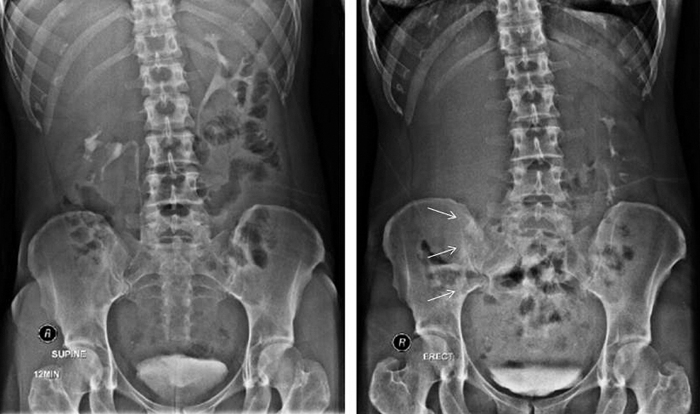
A 31-year-old healthy female patient presented with right-sided chronic pain refractory to analgesics. She denied fever, lower urinary tract symptoms, or hematuria. The patient's past history was unremarkable for urinary tract issues. Intravenous urogram confirmed the presence of right-sided nephroptosis (Fig. 1) and the patient elected to undergo laparoscopic transperitoneal nephropexy.
FIG. 1.
Preoperative intravenous urogram. Intravenous urography in supine (left) and standing (right) position, confirming right-sided nephroptosis (arrows).
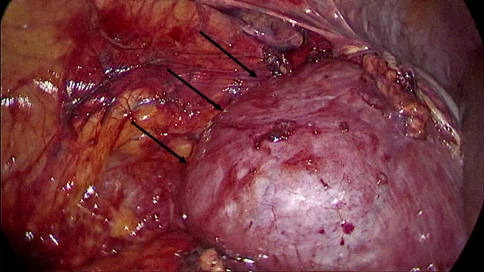
The patient was positioned in the lateral position and access was achieved through three 5-mm ports. The kidney appeared freely movable in a medial and caudal direction. The right colon and mesentery were deflected medially, allowing identification of the right ureter near the lower pole of the right kidney. The posterior peritoneum was incised and dissected from Gerota's fascia, the perirenal fat was dissected and the kidney was mobilized completely (Fig. 2).
FIG. 2.
Intraoperative image. The existing adhesions between the kidney, the peritoneum, and the colon were released to completely mobilize the kidney (nephrolysis).
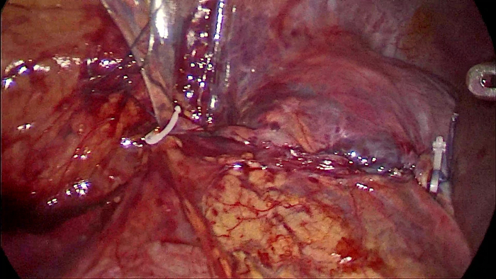
The ptotic kidney was fixed in the correct position using three separate stitches (Fig. 3). The sutures were passed through the superior and lateral surface of the kidney and the psoas and quadratus lumborum muscles and were fixed using Hem-o-Lok clips (Pilling Weck; Teleflex Medical, Markham, ON, Canada), according to the “sliding clip” technique. In particular, double knots were made 2 cm from the short tail of the suture (length 15 cm; 2-0 Prolene) and a Hem-o-Lok clip was applied proximally to the knots. Once the suture was passed through the kidney surface and the muscle, another Hem-o-Lok clip was applied and slided using the needle holder. Finally, an additional Hem-o-Lok clip secured the tension of the suture. After completion of kidney fixation, the right colon flexure was attached to the lateral abdominal wall and the kidney was secured under the right liver lobe (Fig. 4).
FIG. 3.
Renal fixation using the “sliding clip” technique. The sutures were passed through the superior and lateral surface of the kidney and the psoas and quadratus lumborum muscles (left), and were fixed using Hem-o-Lok clips, according to the “sliding clip” technique (right).
FIG. 4.
Restoration of peritoneal support. Attaching the right colon to the lateral abdominal wall, covering the kidney with the right colonic flexure and securing the kidney under the right liver lobe represent essential parts of an effective nephropexy.
The total operative time was 72 minutes. The early postoperative course was uneventful. Postoperatively the patient had bed rest for 24 hours to allow initial scarring and fixation by avoiding gravitational forces. The urethral catheter was removed on the second postoperative day. The patient was discharged on postoperative day 3. Intravenous urogram performed 3 months later confirmed the absence of right nephroptosis, whereas the patient remained asymptomatic.
Discussion and Literature Review
Nephroptosis is often present in young and slim women (male-to-female ratio ∼3:100) and usually involves the right kidney.1 In the vast majority of patients, the condition is asymptomatic and nephroptosis is a relatively common finding on routine Intravenous urography (incidence of 20% in healthy women).2 The pathophysiology of nephroptosis is not completely understood, but deficient support from perinephric structures is the key mechanism, whereas some degree of insufficient rotation of the colon might contribute as well.1 Symptoms secondary to nephroptosis (such as pain and hematuria) are attributed to various underlying mechanisms but acute ureteral obstruction is probably the most significant factor and is a consistent finding on imaging studies.1,2
The diagnosis of nephroptosis requires a high index of suspicion and imaging confirmation. Intravenous urography in supine and standing positions represents the historical mainstay for diagnosis. Ultrasonography with color Doppler imaging allows noninvasive determination of reduced renal perfusion.1 Mercaptoacetyltriglycine-3 scintigraphy in supine and erect position demonstrates typical findings such as decreased renal perfusion and obstructed excretion curve.2
The mainstay of treatment for symptomatic nephroptosis is surgical renal fixation. Conservative measures (weight gain, abdominal wall exercises, frequent supine rests, and abdominal corset) are neither practical nor efficient.1 For patients with nephroptosis, decision making for intervention involves two questions: whether surgery should be performed, and, if so, by what method? Rassweiler et al.2 proposed surgical intervention in cases with confirmed renal ptosis combined with either two objective symptoms or one objective and one subjective symptom.
Surgical methods aim to achieve fixation of the kidney in a cephalad retroperitoneal position, ensure that the lower pole remains lateral and prevent tension on the vascular pedicle or the ureter. Surgical approaches have ranged from open surgery to laparoscopy to percutaneous treatment.1
Various approaches for open nephropexy have been described, but its limitations, including high morbidity and poor outcomes, have led to its demise. Percutaneous endoscopic renal fixation offers some advantages, such as short procedure time and technical simplicity.1 Fixation is achieved by scar formation around the kidney after percutaneous access and insertion of a drain, balloon catheter, circle (U) nephrostomy tube or polyglactin suture to anchor the kidney.1,2 There are some technical and clinical limitations, however, such as inadequate visualization of nearby structures and risk of damage during the procedure, as well as the inability to address colonic malrotation, which appears to be of importance in terms of pathogenesis.1 Compared with laparoscopy, percutaneous nephropexy appears to necessitate longer hospital stay and postoperative bed rest, whereas long-term efficacy data are lacking.1,2
Because of its high efficacy and low morbidity, laparoscopic nephropexy is the most widely used approach.1–4 Laparoscopic renal fixation can be performed with either a transperitoneal or a retroperitoneal approach. Most surgeons prefer the transperitoneal approach because it provides a larger working space for suturing.3,4 The retroperitoneal technique allows quicker access to the posterior renal surface for suture anchoring, as well as decreased risk of postoperative adhesions between the kidney, peritoneum, and colon.2
Three main techniques for laparoscopic fixation have been used, including suture anchoring of the capsule to the posterior abdominal wall muscles, fixation with fascial flaps or muscle bands, and fixation with foreign material, such as mesh or tissue adhesive.1–4 Anchoring the lateral concavity (transperitoneal access) or the posterior surface (retroperitoneal access) of the kidney to the psoas and/or quadratus lumborum muscle, using one to three nonabsorbable sutures represents a relatively straightforward approach.2–4 Additional measures to achieve anchoring include the use of a polyglactin or polypropylene mesh, reinforcement with tissue adhesive to promote adhesions or the application of a tension-free vaginal tape to attach the kidney to the dorsal abdominal wall.1
Apart from securing the kidney in a cephalad position, some additional steps can be applied. According to Rassweiler et al.,2 complete mobilization of the kidney (nephrolysis) is an important step, aiming to release existing adhesions to the peritoneum and especially the colon, which may cause insufficient renal support. According to other authors, an incomplete rotation of the colon could represent a contributing pathogenic mechanism. Based on this assumption, attaching the right colon to the lateral abdominal wall and covering the kidney with the right colonic flexure is an essential part of an effective nephropexy.1,2
There is a relative paucity of scientific evidence in regard to the ideal approach for nephropexy. Only a few prospective studies have been performed, comparative data are lacking, and long-term outcomes have yet to be published. According to the only retrospective comparison of laparoscopic (23 patients) vs open nephropexy (12 patients),3 efficacy was comparable. Laparoscopy resulted in faster recovery, less postoperative pain, and shorter hospital stay, but was more time-consuming and more expensive. According to available data, success rates for laparoscopic nephropexy are in the 80% to 100% range.1 Transperitoneal laparoscopic nephropexy resulted in resolution of symptoms in 80% of patients at 3-year follow-up, according to a large series,4 whereas similar outcomes have been reported from centers applying a retroperitoneal approach (resolution of pain in 83%–84%).2
From a technical standpoint, the principles of laparoscopic nephropexy were applied in this case in a relatively straightforward manner, including complete nephrolysis, 3-point anchoring of the lateral concavity of the kidney to the posterior abdominal wall, and repositioning of a malrotated colonic flexure. We believe that one of the key issues in achieving effective nephropexy is to prevent renal pedicle kinking, which results in renal impairment. One must also take extra care to avoid injury to the kidney or the ilio-hypogastric or genitofemoral nerves during suturing.
Nephropexy is considered a technically demanding procedure. One major difficulty is the need to place tight triangulation sutures between the abdominal wall and the renal capsule in a confined working space. We applied a simple point of technique to facilitate suturing and avoid excessive tension on renal parenchyma. The “sliding clip” technique is widely used for renorrhaphy during partial nephrectomy and most urologists are familiar with its principle.
Conclusions
Symptomatic nephroptosis constitutes a rare but distinct clinical entity. Careful patient assessment and a more stringent approach to imaging will help to identify candidates for surgical intervention. Laparoscopic nephropexy is a safe and effective procedure, whereas the sliding clip method is a technical modification familiar to most urologists that facilitates intracorporeal suturing and adequate renal fixation.
Disclosure Statement
No competing financial interests exist.
Funding Information
No funding was received for this article.
Cite this article as: Philippou P, Michalakis A, Ioannou K, Miliatou M (2020) Laparoscopic nephropexy: the sliding clip technique, Journal of Endourology Case Reports 6:3, 224–227, DOI: 10.1089/cren.2020.0057.
References
- 1. Srirangam SJ, Pollard AJ, Adeyoju AA, O'Reilly PH. Nephroptosis: Seriously misunderstood? BJU Int 2009;103:296–300 [DOI] [PubMed] [Google Scholar]
- 2. Rassweiler JJ, Frede T, Recker F, Stock C, Seemann O, Alken P. Retroperitoneal laparoscopic nephropexy. Urol Clin North Am 2001;28:137–144 [DOI] [PubMed] [Google Scholar]
- 3. Fornara P, Doehn C, Jocham D. Laparoscopic nephropexy: 3-year experience. J Urol 1997;158:1679–1683 [DOI] [PubMed] [Google Scholar]
- 4. McDougall EM, Afane JS, Dunn MD, Collyer WC, Clayman RV. Laparoscopic nephropexy: Long-term follow-up—Washington University experience. J Endourol 2000;14:247–250 [DOI] [PubMed] [Google Scholar]


