

Graphical abstract

Keywords: Myxoma, Carney complex, Tuberculosis, Pyrexia
Highlights
-
•
Cardiac myxoma can present with prolonged fever and pleural effusion.
-
•
Multichamber myxoma should raise the suspicion of Carney complex.
-
•
Echocardiography plays a pivotal role for diagnosis, treatment planning, and follow-up.
-
•
Compatible with almost normal life expectancy if diagnosed, treated, and monitored.
-
•
Postsurgery recurrence of cardiac myxoma is common, demanding lifelong follow-up.
Introduction
Tuberculosis (TB) is a common diagnosis in developing countries and is a common cause of prolonged pyrexia and pleural effusion.1,2 Sometimes treatment for TB is offered rather empirically in a resource-constraint environment, and other times, cardiac myxoma is a well-documented cause of prolonged pyrexia and pyrexia of unknown origin.3 We describe a case of a patient with prolonged fever and pleural effusion who was on antitubercular therapy. Echocardiography refuted the working diagnosis and revealed multichamber myxoma; finally, a diagnosis of Carney complex was made.
Case Presentation
A 35-year-old mother of one child presented with high-grade fever, cough, New York Heart Association class III dyspnea, and cachexia for 3 months. Physical examination revealed multiple facial lentigines (Figure 1), leukonychia, dependent edema, and raised jugular venous pressure. Her pulse was 104/minute, her blood pressure was 90/60 mm Hg, and she had a grade 3/4 diastolic murmur in the mitral area. Diagnostic work-up at the local facility included a chest x-ray noting bilateral pleural effusions, more on the right side, and electrocardiography displaying sinus tachycardia (Figure 2). She had total leukocyte count 10,000/ mm3, platelet count 180,000/mm3, and hemoglobin (Hb) 11.0 g/dL. Renal and hepatic functions, serum electrolytes, thyroid stimulating hormone, and serum albumin were normal, and blood culture was negative. Her erythrocyte sedimentation rate (ESR) was raised to 90 mm in the first hour, and C-reactive protein to 24 mg/L. Analysis of pleural fluid revealed protein 3.8 g/dL, lymphocytes 90%, and negative GeneXpert test for Mycobacterium tuberculosis. Sputum for GeneXpert test for Mycobacterium tuberculosis was also negative. However, considering the exudative nature of the pleural fluid, raised ESR, and C-reactive protein in a patient with prolonged pyrexia in an endemic zone, she was administered anti-TB therapy for 1 month.
Figure 1.
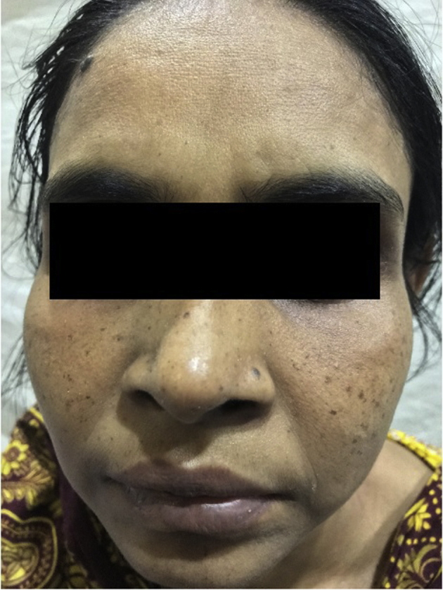
Multiple pigmented lentigines distributed symmetrically in the face of the patient.
Figure 2.
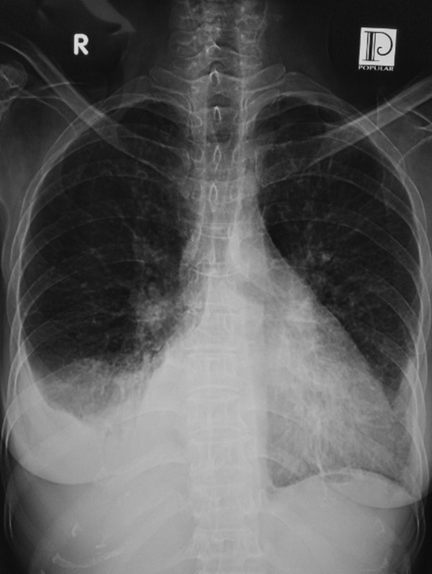
Chest x-ray posteroanterior view showing bilateral pleural effusion, with more on the right side.
As her condition did not improve, she underwent transthoracic echocardiography, which revealed multiple lobulated masses of mixed echo-density, within the left atrium, left ventricle (LV), and right ventricle (RV). The largest mass was occupying the left atrial cavity and was attached to the basal interatrial septum with a stalk. The LV mass was related to the posterior mitral leaflet, while the RV mass was attached to the anterior tricuspid leaflet. The LV and RV systolic function was good, and there were mild mitral regurgitation and mild tricuspid regurgitation, with the pulmonary artery systolic pressure of 52 mm Hg (Figure 3, Videos 1 and 2). A diagnosis of multichamber myxoma was made. Due to the probability of Carney complex, a careful search for other components connected the multiple pigmented lesions in the face to a new diagnosis. However, her thyroid and pituitary function tests and adrenal imaging were normal. She was referred for further evaluation and management. She underwent emergent excision of the cardiac masses. Histopathologic examination of all three masses showed features compatible with myxoma with hemorrhage and necrosis (Figure 4). Postoperative recovery was slow and uneventful. There was moderate mitral regurgitation, but no residual tumor in follow-up echocardiography (Video 3). The patient's first-degree relatives were screened by transthoracic echocardiography. Her 6-year-old son was found to have single myxoma in the right atrium (Figure 5, Video 4, Video 5, Video 6), which was later confirmed histopathologically after surgical excision. Accordingly, with the diagnosis of Carney complex, both the mother and the son were kept on annual follow-up with skin evaluation; echocardiography; serum growth hormone, prolactin, urinary free cortisol, and thyroid stimulating hormone assays; ultrasonography of thyroid gland, ovaries (mother), and testes (son); and growth monitoring and pubertal staging of the son.
Figure 3.

Preoperative echocardiography. (A) Parasternal long-axis view showing a large left atrial myxoma. (B) Apical 4-chamber view showing multicentric myxoma in the LA, LV, and RV.
Figure 4.

Histopathological examination. (A) (lower) and (B) (higher) magnification images showing abundant loose myxoid stroma with scattered round, polygonal, or stellate cells.
Figure 5.

Echocardiography of the patient's son. (A) Apical four-chamber view and (B) right ventricular inflow view showing myxoma in the right atrium.
Discussion
Carney complex is a rare multiple neoplasia syndrome, characterized by pigmented lesions of the skin and mucosa, cardiac and extracardiac myxomatous tumors, and multiple endocrine and nonendocrine neoplasms.4 Previously described as LAMB (lentigines, atrial myxomas, myxoid neurofibromas, and ephelide) syndrome and NAME (nevi, atrial myxoma, blue nevi) syndrome, these are now reclassified as Carney complex.5 Carney complex was first described in 1985 in a series of 40 patients by J. Aidan Carney, a pathologist at the Mayo Clinic, as the complex of myxomas, spotting pigmentation, and endocrine overreactivity.6 Its exact prevalence is unknown; around 750 cases have been reported worldwide so far.7 It is inherited as an autosomal-dominant disorder in three-fourths of cases, while in the remaining one-fourth, it occurs sporadically as a result of a de novo genetic mutation.8 The disease is caused by inactivating mutations or large deletions of the PRKAR1A gene located at 17q22-24 coding for the regulatory subunit type I alpha of protein kinase A gene.5
Cardiac myxoma is an important manifestation of Carney complex and may present with a triad of obstructive, embolic, and constitutional manifestations. The most common symptoms are associated with obstruction due to the size and location of the tumor. The next most common symptoms are associated with pulmonary and systemic embolization. Constitutional symptoms include malaise, anorexia, fever, arthralgia, and weight loss, and it often mimics connective tissue disorders. Our patient mainly presented with constitutional symptoms including pyrexia, the differential diagnoses of which include disseminated TB in developing countries. Investigation findings, especially high ESR, anemia, leukocytosis, and exudative pleural effusion, led to the empirical diagnosis of TB. Establishing the tubercular etiology in pleural effusion is not straightforward. Despite high specificity (e.g., 98.6%), the GeneXpert test for Mycobacterium tuberculosis in case of pleural effusion suffers from low sensitivity (e.g., 51.4%).9 On the other hand, sputum may be bacteriologically smear and/or culture positive in less than 1/3 of cases with tubercular pleural effusion.10
Cardiac myxoma associated with Carney complex is often multicentric, occurs at a younger age and at unusual sites, and shows a higher tendency of recurrence after resection in comparison to solitary ones. Our patient had three myxomas in three different chambers of the heart, while her 6-year-old son had one solitary myxoma in the right atrium. Myxomatous tumor also occurs in skin and breast. Lentigines, blue nevus, and cutaneous myxoma are the common skin manifestations of Carney complex. Lentigines are flat small brown to black macules preferentially distributed over the face, lips, genital area, and mucosa. Our patient had lentigines over the face (Figure 1). Endocrinopathies due to the adrenal, pituitary, or testicular tumors are the most frequent systemic manifestations of Carney complex. Primary pigmented nodular adrenocortical disease is the most frequently observed endocrine tumor, occurring in an estimated 25% of affected individuals.11 Thyroid nodules and adenomas are common, but most patients are euthyroid. Our patient had no symptoms suggestive of endocrinopathies, and the endocrine laboratory workup was within normal limits. For the child of our patient, the diagnosis of myxoma was only made by family screening. Cardiac myxoma may rarely present with pleural effusion12; however, pleural effusion associated with Carney complex has not been reported previously. As in our case, presence of constitutional symptoms and pleural effusion may lead to the erroneous diagnosis of TB.
The Carney complex is diagnosed by the criteria defined by Stratakis et al.4 (Table 1). Diagnosis is made by the presence of two or more major criteria (total 12) or, alternatively, one major criterion and one supplemental criterion (total 2). Our patient had two major criteria, that is, spotty skin pigmentation and histopathologically proven cardiac myxoma, and one supplemental criterion, that is, affected first-degree relative. A search for an inactivating mutation of the PRKAR1A gene could not be done due to lack of a facility.
Table 1.
Diagnostic criteria for Carney complex
|
For a diagnosis of Carney complex, either (1) two of the 12 manifestations of the disease listed or (2) one of these 12 manifestations and one of the supplemental criteria is required.
Histologic confirmation needed.
Today, genetic testing for mutations in the PRKAR1A gene is increasingly used for diagnostic certainty. In addition, genetic screening of potentially affected family members is recommended.
Once Carney complex is diagnosed, the patient should be managed accordingly. Each specific manifestation including neoplasms should be addressed separately. Cardiac myxomas need surgical excision often on an emergency basis to avoid grave complications. Primary pigmented nodular adrenocortical disease is best managed surgically, by bilateral adrenalectomy.7 Pituitary adenomas may be treated with somastotatin analogs or removed surgically.13
Cardiac myxoma in the setting of Carney complex has a high recurrence rate after surgery. The reported recurrence rates were 10%, 21%, and 33% in familial variety, Carney complex, and multiple myxomas, respectively. The usual life expectancy of Carney complex is 50-55 years, but with careful surveillance, it may be almost normal. The most common causes of death are related to complications of cardiac myxoma, such as embolic strokes, postoperative cardiomyopathy and cardiac arrhythmias, and metastatic psammomatous melanotic schwannomas, pancreatic, and other cancers.5
Patients with Carney complex need lifelong yearly follow-up consisting of (1) echocardiogram, (2) skin evaluation, (3) blood tests for endocrine surveillance, for example, serum growth hormone, prolactin, and insulin-like growth factor 1, urinary free cortisol, thyroid function tests, (4) ultrasound of thyroid gland, (5) computed tomographic scanning of adrenal glands and magnetic resonance imaging of pituitary, (6) clinical and ultrasonographic examination of testes and ovaries, and (7) growth monitoring and pubertal staging of children.8
Conclusion
Carney complex is a rare genetic disorder of multiple neoplasia syndrome. Sometimes, it may be associated with atypical presentation. Over the past decades, there has been significant advancement in different aspects of the disease, including genetics. Physicians should have appropriate preparedness to diagnose and manage this condition.
Clinical Pearls
-
•
Cardiac myxoma can rarely present with prolonged fever and pleural effusion leading to chances of misdiagnosis.
-
•
Multichamber myxoma should raise the suspicion of Carney complex, other components of which should be looked for.
-
•
Echocardiography plays a pivotal role for diagnosis, treatment planning, and follow-up.
-
•
First-degree relatives should be screened for.
-
•
Compatible with almost normal life expectancy if diagnosed, treated, and monitored properly.
-
•
Postsurgery recurrence of cardiac myxoma is common, demanding lifelong follow-up.
Footnotes
Conflicts of interest: The authors reported no actual or potential conflicts of interest relative to this document.
Supplementary data related to this article can be found at https://doi.org/10.1016/j.case.2020.05.010.
Supplementary Data
Preoperative two-dimensional echocardiography, parasternal long-axis view showing echogenic mass in the left atrium.
Preoperative two-dimensional echocardiography, apical four-chamber view showing echogenic mass in the left atrium, LV, and RV.
Postoperative echocardiography two-dimensional guided color Doppler imaging, apical four-chamber view showing moderate mitral regurgitation.
Two-dimensional echocardiography of the patient's son, apical four-chamber view showing large polypoid mass in the right atrium.
Two-dimensional echocardiography of the patient's son, subcostal view showing no relationship of the right atrial mass in relation with the inferior vena cava.
Two-dimensional echocardiography of the patient's son, right ventricular inflow view showing the large myxoma in the right atrium.
References
- 1.Tan X.Y., He Q.Y. [Chinese literature review of etiology distribution of adult patients with fever of unknown origin from 1979 to 2012][Article in Chinese] Zhonghua Nei Ke Za Zhi. 2013 Dec;52(12):1013–1017. [PubMed] [Google Scholar]
- 2.Bofinger J.J., Schlossberg D. Fever of unknown origin caused by tuberculosis. Infect Dis Clin North Am. 2007;21:947–962, viii. doi: 10.1016/j.idc.2007.08.001. [DOI] [PubMed] [Google Scholar]
- 3.Gavrielatos G., Letsas K.P., Pappas L.K., Dedeilias P., Sioras E., Kardaras F. Large left atrial myxoma presented as fever of unknown origin: a challenging diagnosis and a review of the literature. Cardiovasc Pathol. 2007;16:365–367. doi: 10.1016/j.carpath.2007.01.001. [DOI] [PubMed] [Google Scholar]
- 4.Stratakis C.A., Kirschner L.S., Carney J.A. Clinical and molecular features of the Carney complex: diagnostic criteria and recommendations for patient evaluation. J Clin Endocrinol Metab. 2001;86:4041–4046. doi: 10.1210/jcem.86.9.7903. [DOI] [PubMed] [Google Scholar]
- 5.Correa R., Salpea P., Stratakis C.A. Carney complex: an update. Eur J Endocrinol. 2015;173:M85–M97. doi: 10.1530/EJE-15-0209. [DOI] [PMC free article] [PubMed] [Google Scholar]
- 6.Carney J.A., Gordon H., Carpenter P.C., Shenoy B.V., Go V.L. The complex of myxomas, spotty pigmentation, and endocrine overactivity. Medicine (Baltimore) 1985;64:270–283. doi: 10.1097/00005792-198507000-00007. [DOI] [PubMed] [Google Scholar]
- 7.Vindhyal M.R., Elhomsy G. StatPearls [Internet] StatPearls Publishing; Treasure Island, FL: 2020. Carney complex.https://www.ncbi.nlm.nih.gov/books/NBK507877/ Available at: [Google Scholar]
- 8.Kirschner L.S., Carney J.A., Pack S.D., Taymans S.E., Giatzakis C., Cho Y.S. Mutations of the gene encoding the protein kinase A type I-alpha regulatory subunit in patients with the Carney complex. Nat Genet. 2000;26:89–92. doi: 10.1038/79238. [DOI] [PubMed] [Google Scholar]
- 9.Sehgal I.S., Dhooria S., Aggarwal A.N., Behera D., Agarwal R. Diagnostic performance of Xpert MTB/RIF in tuberculous pleural effusion: systematic review and meta-analysis. J Clin Microbiol. 2016;54:1133–1136. doi: 10.1128/JCM.03205-15. [DOI] [PMC free article] [PubMed] [Google Scholar]
- 10.Chaudhuri A.D., Bhuniya S., Pandit S., Dey A., Mukherjee S., Bhanja P. Role of sputum examination for acid fast bacilli in tuberculous pleural effusion. Lung India. 2011;28:21–24. doi: 10.4103/0970-2113.76296. [DOI] [PMC free article] [PubMed] [Google Scholar]
- 11.Stratakis C.A., Raygada M. Carney Complex. In: Adam M.P., Ardinger H.H., Pagon R.A., Wallace S.E., Bean L.J.H., Stephens K., editors. GeneReviews® [Internet] University of Washington; Seattle: 1993-2020. https://www.ncbi.nlm.nih.gov/books/NBK1286/ Available at: [Google Scholar]
- 12.Cakar M.A., Arslan C., Yildiz A., Vatan M.B., Gunduz H. Left atrial myxoma with pleural effusion. J Clin Med Res. 2009;1:297–299. doi: 10.4021/jocmr2009.11.1269. [DOI] [PMC free article] [PubMed] [Google Scholar]
- 13.Watson J.C., Stratakis C.A., Bryant-Greenwood P.K., Koch C.A., Kirschner L.S., Nguyen T. Neurosurgical implications of Carney complex. J Neurosurg. 2000;92:413–418. doi: 10.3171/jns.2000.92.3.0413. [DOI] [PubMed] [Google Scholar]
Associated Data
This section collects any data citations, data availability statements, or supplementary materials included in this article.
Supplementary Materials
Preoperative two-dimensional echocardiography, parasternal long-axis view showing echogenic mass in the left atrium.
Preoperative two-dimensional echocardiography, apical four-chamber view showing echogenic mass in the left atrium, LV, and RV.
Postoperative echocardiography two-dimensional guided color Doppler imaging, apical four-chamber view showing moderate mitral regurgitation.
Two-dimensional echocardiography of the patient's son, apical four-chamber view showing large polypoid mass in the right atrium.
Two-dimensional echocardiography of the patient's son, subcostal view showing no relationship of the right atrial mass in relation with the inferior vena cava.
Two-dimensional echocardiography of the patient's son, right ventricular inflow view showing the large myxoma in the right atrium.