Abstract
Wolbachia is an intracellular bacterial symbiont of arthropods notorious for inducing many reproductive manipulations that foster its dissemination. Wolbachia affects many aspects of host biology, including metabolism, longevity and physiology, being described as a nutrient provisioning or metabolic parasite, depending on the host-microbe association. Sirtuins (SIRTs) are a family of NAD+-dependent post-translational regulatory enzymes known to affect many of the same processes altered by Wolbachia, including aging and metabolism, among others. Despite a clear overlap in control of host-derived pathways and physiology, no work has demonstrated a link between these two regulators. We used genetically tractable Drosophila melanogaster to explore the role of sirtuins in shaping signaling pathways in the context of a host-symbiont model. By using transcriptional profiling and metabolic assays in the context of genetic knockouts/over-expressions, we examined the effect of several Wolbachia strains on host sirtuin expression across distinct tissues and timepoints. We also quantified the downstream effects of the sirtuin x Wolbachia interaction on host glucose metabolism, and in turn, how it impacted Wolbachia titer. Our results indicate that the presence of Wolbachia is associated with (1) reduced sirt-4 expression in a strain-specific manner, and (2) alterations in host glutamate dehydrogenase expression and ATP levels, key components of glucose metabolism. We detected high glucose levels in Wolbachia-infected flies, which further increased when sirt-4 was over-expressed. However, under sirt-4 knockout, flies displayed a hypoglycemic state not rescued to normal levels in the presence of Wolbachia. Finally, whole body sirt-4 over-expression resulted in reduced Wolbachia ovarian titer. Our results expand knowledge of Wolbachia-host associations in the context of a yet unexplored class of host post-translational regulatory enzymes with implications for conserved host signaling pathways and bacterial titer, factors known to impact host biology and the symbiont’s ability to spread through populations.
Author summary
Here we utilize Drosophila genetic tools to dissect how Wolbachia interacts with a class of host enzymes known as sirtuins, widely recognized as key regulators of host biological processes including metabolism, longevity, stress response, and others. By focusing on the sirt-4 gene, we demonstrate that the presence of Wolbachia is associated with a consistent reduction in sirt-4 transcriptional levels across multiple tissues and timepoints. The bacterium is also associated with alterations in the expression of glutamate dehydrogenase and total ATP levels, key components of the glucose metabolism pathway. Finally, we show that the Wolbachia-sirt-4 interaction is associated with the modulation of host glucose homeostasis and, that sirt-4 negatively affects the growth of the bacteria in the host reproductive tissues. Our results further expand knowledge of host-microbe interactions in the context of host physiological manipulation by intracellular bacteria, offering new ways to interpret Wolbachia’s successful dissemination in insect populations in nature.
Introduction
Wolbachia is a genus of gram-negative maternally inherited obligate bacterial endosymbiont of nematodes and arthropods. These bacteria are present in at least 40% of all known insect species [1]. Wolbachia can induce a range of reproductive manipulations including male killing, genetic feminization, parthenogenesis and cytoplasmic incompatibility (CI), to facilitate its spread [2,3]. Despite evidence pointing towards horizontal transfer of Wolbachia among species [4], this bacterium, and most heritable bacterial symbionts of arthropods, are primarily transmitted through the female germline [5]. This poses selection pressure to increase the proportion of females that are infected. However, the existence of reproductive manipulations are not sufficient to explain Wolbachia’s increase in infection prevalence and efficiency of spread through insect populations. For example, both wAu in D. simulans and wSuz in Drosophila suzukii spread despite its imperfect maternal transmission rate and no induction of CI [6,7]. Considering that Wolbachia has been shown to induce fitness costs in Aedes aegypti mosquitoes [8,9], it has been proposed that for this host that the infection frequency must be above an unstable equilibrium threshold in order for the bacterial infection to be sustained [10]. Under this scenario, a bacterial variant that increases host fitness will likely have an advantage over existing strains [11], or a benefit over uninfected hosts [12], (see [13] for an example of such benefit documented in another maternally-inherited organism).
Wolbachia can increase its likelihood of spread by evolving mutualistic relationships with its host, hence benefiting both itself, and the host. This is often documented in the form of nutrient provisioning from the bacteria to its host, as exemplified in the bedbug Cimex lectularius and vitamin B provisioning by Wolbachia [14], and also present in many insect:symbiont systems [15,16]. The conflation of both reproductive parasitism and mutualism, derived from a “host context-specific” scenario, can lead to the emergence of a symbiotic relationship termed Jekyll and Hyde, as to represent the “many faces” of Wolbachia’s impact on host biology [17]. In this context, host reproduction is manipulated while also providing fitness benefits to its host. For instance, in the planthopper Laodelphax striatellus, Wolbachia induces strong CI [18], while provisioning the host with the vitamin B members biotin [19] and riboflavin. The enzymes able to synthesize the latter are also shared amongst the genome of distinct Wolbachia strains[20]. In this agricultural pest, Wolbachia has also been associated with an increase in fecundity [21], an effect observed in field-collected Drosophila simulans as well [22] (see [15,16] for an extensive review on the topic).
In keeping with the Jekyll side of Wolbachia:host interactions, there is evidence to suggest that the symbiont can also be a drain on host resources. Genomic studies focused on wMel [23,24], and nutrient competition assays in mosquitoes infected with the wMelPop [25], indicate that Wolbachia relies on host amino acids to support its energetic requirements. It has also been noted that the wMel Wolbachia strain has limited carbohydrate metabolism capabilities [23,24]. Apart from interfering with reproduction and metabolite availability, Wolbachia is also known to impact other aspects of host physiology [16,26] by still emerging mechanisms. In the mutually exclusive association between Wolbachia and filarial nematodes, evidence indicates that Wolbachia plays a role in heme provisioning [27,28], while directly relying on host pyruvate production, through glycolysis, for its own survival. Removal of Wolbachia via antibiotic treatment led to increased host levels of glucose and glycogen [29,30].
In D. melanogaster, glucose metabolism is managed by a series of genetic networks and signaling pathways. These can act both locally in metabolically active tissues, as well as via hormonal signals, thus maintaining homeostasis through interorgan communication [31]. In mammals, the pancreatic islands play a key role in the regulation of glucose metabolism, where glucose acts to depolarize β-cells membrane potential, stimulating mitochondrial ATP production, which in turn shuts down the ATP-sensitive potassium channels, opening the voltage-dependent Ca2+ channels to finally release insulin (see [32] for an in-depth review). In flies, this process is mainly dictated by the insulin-producing cells (IPCs), located in the fly brain, responsible for secreting Insulin-like peptides (ILPs) (see [33] for an in-depth review). The Drosophila genome encodes for eight known ILPs (dILPs 1–8) and one known insulin receptor (dInR) [34–36]. Distinct dILPs are produced and secreted by multiple tissues in a spatiotemporal manner during larval growth and in the adult fly. For instance, dILP6 is known to be secreted by the fat body (see [31,34–36] for more details on dILPs and the many layers by which secretion is regulated). In mammals, the adipose tissue not only has a role in energy storage, but it also acts as an endocrine organ. Interesting enough, the same holds true for D. melanogaster, in which the fat body, a tissue infected by Wolbachia [37], acts as a key regulatory organ of glucose metabolism, coupling sensing of nutrients such as amino acids, fats and sugars to IPC signaling and systemic hormone activity [31,33]. Previous work in larvae demonstrated that the fat body-localized amino acid transporter Slimfast (Slif) activates the Target of Rapamycin kinase complex 1 (TORC1), leading to fat body signaling to brain IPCs and dILPs release into circulation, promoting fly metabolic activity and growth [38]. A similar mechanism highlighting the importance of the fat body for insulin signaling has been proposed in adult flies, in which a ligand for the JAK/STAT pathway called Unpaired 2 (Upd2) is produced by this tissue in response to diets high in fat and sugar, indirectly activating IPCs and dILPs release via interaction with GABAergic neurons [39].
Wolbachia and sirtuins have overlapping effects on host processes, that range from impacts on host glucose and amino acid metabolism, to broader traits such as host longevity and regulation of immune responses. However, no work to date has demonstrated a link between these two master regulators of host biology. For instance, in the context of glucose metabolism, Wolbachia infection has been associated with the upregulation of the insulin/IGF-like signaling pathway [40] in D. melanogaster. Mutants for this pathway display a plethora of extreme detrimental traits in flies, ranging from significant reduction in body size, full sterility and dramatic reduction in lifespan. However, in the presence of Wolbachia, all of these effects become mild, limiting any reductions in body size, fecundity, and extension in lifespan [40]. Also in flies, exciting work has shown that a yeast-enriched diet suppresses ovarian Wolbachia titer, while a sucrose-based diet (latter also expanded to galactose, lactose, maltose and trehalose [41]) increased bacterial load, a dietary effect mediated by both the somatic TORC1 and insulin signaling pathways [42], but for which the precise mechanistic factors up and downstream of such modulation of Wolbachia density remains elusive.
Silent information regulators, commonly known as sirtuins, compose a family of highly conserved host post-translational deacetylase and ADP-ribosyltransferase regulatory enzymes that use nicotinamide adenine dinucleotide (NAD+) as a co-substrate [43]. The genome of Drosophila melanogaster encodes for five sirtuins: SIRT-1, SIRT-2, SIRT-4, SIRT-6, and SIRT-7, named after their mammalian orthologs. SIRT-1 is both nuclear and cytoplasmic, while SIRT-2 is mainly cytoplasmic but can translocate to the nucleus upon external triggers such as ionizing radiation [44], and SIRT-6 and SIRT-7 are primarily found in the nucleus [45,46]. In contrast, Drosophila spp. SIRT-4 is the only sirtuin imported to the mitochondria. The segregation of sirtuins into various cell compartments is associated with the specific regulation of many biological processes in the host that often overlap, including, but not limited to, immunity, lifespan, metabolism, epigenetics, and stress responses; see [46,47] for a review on the many processes regulated by each of these enzymes.
In both mammalian cells and Drosophila [48,49], mitochondria-translocated sirtuins have been implicated in regulation of insulin signaling, fatty acid oxidation, amino acid catabolism and ATP/ADP ratio [50,51], among other functions (S1 Fig). Upregulation of sirt-4 decreases oxidative processes in the mitochondria that serve as initiators of the tricarboxylic acid (TCA) cycle, resulting in inhibition of insulin secretion. One of the components involved in oxidation is glutamate dehydrogenase (gdh), an enzyme encoded in the nucleus and translocated to the mitochondria that catalyzes the conversion of the glutamine-derived molecule glutamate to α-ketoglutarate under the negative regulation of ATP (an organic compound also known to directly impact the mTOR pathway [52]) and positive regulation of ADP/leucine [53]. Previous work found that SIRT-4 directly binds GDH and inhibits its activity [54]. This causes inhibition of glutamine metabolism and a decline in ATP/ADP ratio. Reduced ATP production is associated with a decrease in insulin secretion and increase in host glycemia due to accumulation of free circulating glucose [54,55]. In parallel to GDH-dependent ATP production, SIRT-4 also acts through the inner mitochondrial transmembrane protein adenine nucleotide translocator 2 (ANT2), an ATP/ADP translocator [56], and methylcrotonyl-CoA carboxylase enzyme (MCCC), involved in leucine catabolism [57] in order to mediate cellular ATP homeostasis.
Here, to gain insight into the nature of interactions between Wolbachia and sirtuins, we took advantage of the genetically tractable system of D. melanogaster, which allows for the systematic and unbiased study of host pathways. In particular, we focused on understanding Wolbachia’s impact on host glucose metabolism in light of sirtuins and how host sirtuins, in turn, affect Wolbachia. By performing transcriptional profiling analyses coupled with metabolic assays on whole body, fat body and ovaries of distinct wild type and mutant lines of D. melanogaster, we demonstrate for the first time, that Wolbachia presence is associated with alterations in sirtuin transcript levels, and that this has downstream consequences on host glucose metabolism and its associated effectors. Finally, we show that alterations in sirtuin expression is associated with changes in bacterial density. Our findings greatly contribute towards understanding the manipulation of key host physiological processes, with implications for alterations in bacterial titer, factors known to impact overall host biology as well as the symbiont’s ability to spread through insect populations.
Results
Wolbachia infection is associated with decreased sirt-4 transcript levels in distinct timepoints and host tissues
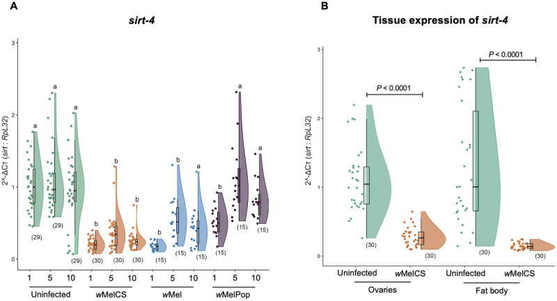
We quantified the transcriptional levels of all five Drosophila sirtuin genes, namely sirt-1, sirt-2, sirt-4, sirt-6, and sirt-7, in virgin female flies infected with either wMel, wMelCS or wMelPop (Fig 1A and S2 Fig). In 5-day-old flies, none of the Wolbachia strains tested were associated with alterations in mRNA levels in four out of five sirtuin genes—sirt-1, sirt-2, sirt-6 and sirt-7- relative to expression in the Wolbachia-free group (S2 Fig and S3 Table). In contrast, expression of sirt-4 varied significantly across tested groups at day 1 (Kruskal-Wallis H = 43.9, P<0.0001), 5 (Kruskal-Wallis H = 30.1, P<0.0001) and 10 (Kruskal-Wallis H = 24.6, P<0.0001) (Fig 1A). The wMel strain was associated with a significant reduction in the expression of sirt-4 at 1 (Mann-Whitney Dunn’s corrected test, df = 1, P<0.0001), 5 (Mann-Whitney Dunn’s corrected test, df = 1, P = 0.0019) but not 10 (Mann-Whitney Dunn’s corrected test, df = 1, P = 0.0605) day-old flies, relative to the uninfected counterpart. For this strain, the greatest reduction in sirt-4 expression was seen in 1-day-old flies (82.5% median reduction). In contrast, wMelPop had no significant effect on sirt-4 levels at either day 1 (Mann-Whitney Dunn’s corrected test, df = 1, P = 0.2684), day 5 (Mann-Whitney Dunn’s corrected test, df = 1, P>0.9999) or day 10 (Mann-Whitney Dunn’s corrected test, df = 1, P>0.9999). wMelCS presence was associated with a significant reduction in sirt-4 expression in 1-day old flies relative to its uninfected counterpart (Mann-Whitney Dunn’s corrected test, df = 1, P<0.0001; 81% median reduction). Additionally, sirt-4 expression was still significantly lower in both 5 (Mann-Whitney Dunn’s corrected test, df = 1, P<0.0001; 65% median reduction) and 10 days-old flies (Mann-Whitney Dunn’s corrected test, df = 1, P = 0.0005; 77% median reduction) relative to Wolbachia-free females (Fig 1A)—although this effect was still not as strong as the 81% median reduction in sirt-4 levels observed for wMelCS-infected vs. uninfected flies. Interestingly, day 1 excluded, wMelCS infection was associated with the most consistent reduction in sirt-4 transcriptional levels (relative to uninfected group) across the timepoints tested, when compared to other strains (S4 Table, Fig 1A). To check if variations in sirt-4 transcript levels were associated with the distinct Wolbachia strains tested, as well as the fly age, and the interaction between Wolbachia strain x fly age, we performed a generalized linear model of regression (GLM). Our GLM approach indicated that except for the interaction between Wolbachia strain x fly age (GLM, df = 4, P = 0.1753); both fly age (GLM, df = 2, P<0.0001) and Wolbachia strain (GLM, df = 2, P<0.0001) were significant factors associated with differences in sirt-4 expression.
Fig 1. Wolbachia presence is associated with reduced sirt-4 transcript levels.
(A) whole wildtype Wolbachia-free (uninfected—green) and wildtype infected (wMel—orange, wMelCS—blue and wMelPop—purple) virgin female flies were collected at 1, 5 and 10 days of adulthood, had their RNA extracted and levels of sirt-4 quantified relative to host RpL32 using SYBR Green. wMel and wMelCS-infected flies displayed significantly lower relative sirt-4 levels than uninfected flies at all three time points. wMelPop-infected flies had reduced relative sirt-4 levels only at 1 day of adulthood. Data represent a maximum of two biological replicate experiments of randomly sampled flies. Raincloud plots depict median relative sirt-4 levels with P-values determined via Kruskal-Wallis on entire dataset followed by Mann-Whitney Dunn’s-corrected test for pairwise comparisons. Each dot represents a single whole fly. Sample size is depicted in parenthesis for each group. (B) One-day old uninfected (green) and wMelCS-infected (orange) virgin female flies had their ovaries and fat body dissected, RNA extracted and levels of sirt-4 quantified relative to host RpL32 using SYBR Green. Tissue-specific relative levels of sirt-4 were significantly lower in both tissues of wMelCS-infected flies. Data represent two biological replicate experiments. Raincloud plots depict median relative sirt-4 levels with P-values determined for all pairwise comparisons via Unpaired T-test with Brown-Forsythe or Welch's correction for the ovaries and Mann-Whitney U test for fat body. Each dot represents a pool of 5 pairs of ovaries or 5 carcasses without gut and Malpighian tubules. Sample size is depicted in parenthesis for each group.
Next, we selected the wMelCS strain to further determine if the pattern of reduced expression in sirt-4 was consistent across distinct tissues of 1-day old flies. Both the ovaries (Unpaired T-test with Welch's correction, W = 9.06, df = 33.99, F = 11.54, P<0.0001) and fat body (Mann-Whitney U test, U = 17, df = 1, P<0.0001) had significantly lower sirt-4 levels in wMelCS-infected flies compared to uninfected controls (Fig 1B), demonstrating that the association between wMelCS presence and reduction in sirt-4 transcript levels is conserved across the tissues examined here.
Wolbachia infection is associated with increased transcript levels of gdh, a target of sirt-4 involved in glucose homeostasis
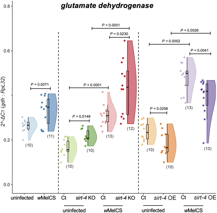
We focused on host glucose homeostasis to investigate any potential sirt-4-related effect of Wolbachia on host metabolism. To this end, we began by quantifying the expression of gdh (Fig 2), a direct target of SIRT-4 involved in glutamine metabolism and ATP homeostasis [54]. There was a 26% mean increase in gdh expression in wildtype wMelCS-infected flies compared to wildtype uninfected controls (Unpaired T-test with Welch's correction, W = 3.14, df = 14, F = 5.17, P = 0.0071). We next utilized a sirt-4 knockout line of flies and found that genetic manipulation of sirt-4 in the context of Wolbachia infection significantly altered gdh expression (Brown-Forsythe ANOVA, F = 34.26, df = 3, P<0.0001). In the absence of Wolbachia, there was a significant 31.7% mean increase in gdh expression in sirt-4 KO flies compared to controls (Unpaired T test Dunnett’s corrected, P = 0.0148). As for sirt-4 KO wMelCS-infected flies, we detected a significant 40.6% mean increase in gdh expression relative to control (Unpaired T test Dunnett’s corrected, P = 0.0233) (Fig 2). Comparisons of both control and sirt-4 KO groups also revealed that Wolbachia’s presence caused substantial alterations in gdh expression (One-way ANOVA, F = 29.74, P<0.0001). For instance, we observed an 88% mean increase between control groups (Unpaired T test Welch’s corrected, P<0.0001), and a 100% mean increase for the sirt-4 KO group (Unpaired T test Dunnett’s corrected, P<0.0001).
Fig 2. Wolbachia presence is associated with increased gdh expression in a sirt-4-dependent manner.
Whole 1-day old virgin female flies from wildtype Wolbachia-free (uninfected–light blue), wildtype infected (wMelCS–dark blue), sirt-4 knockout—KO uninfected (“ct”—control–light green: FM6/ sirt-4 KO vs. sirt-4 KO–dark green: sirt-4 KO/sirt-4 KO), wMelCS-infected (“ct”—control–light red vs. sirt-4 KO–dark red), sirt-4 overexpression—OE uninfected (“ct”—control–light orange: “Act5cGAL4 >“ vs. sirt-4 OE–dark orange: “Act5cGAL4 > UAS sirt-4 OE”) and wMelCS-infected (“ct”—control–light purple vs. sirt-4 OE–dark purple) scenarios were collected. Female flies had their RNA extracted and levels of gdh quantified relative to host RpL32 using SYBR Green. Wolbachia alone increased gdh expression. A condition that peaked when sirt-4 was knocked-out. Meanwhile, sirt-4 OE decreased gdh expression, with a less pronounced effect under Wolbachia presence. Data represent a maximum of two biological replicate experiments of randomly sampled flies. Raincloud plots depict median relative gdh levels with P-values determined for all comparisons via One-way ANOVA followed by unpaired T-test with Brown-Forsythe or Welch's correction for wildtype flies, One-way ANOVA followed by Dunnett’s T3 multiple pairwise comparison correction for sirt-4 KO experiment and Kruskal-Wallis on entire dataset followed by Mann-Whitney Dunn’s-corrected test for pairwise comparisons in the sirt-4 OE experiment. Each dot represents a single whole fly. Sample size is depicted in parenthesis for each group.
In flies in which sirt-4 was overexpressed (12-fold mean increase in sirt-4 expression), we also found a significant alteration in gdh expression (Kruskal-Wallis, H = 33.72, df = 4, P<0.0001). In Wolbachia-free flies, we observed a significant 29% median reduction in gdh expression (Mann-Whitney Dunn’s corrected test, df = 1, P = 0.0256). The same effect of sirt-4 OE was also observed in the presence wMelCS, however to a reduced extent when compared to the reduction observed in uninfected flies, with a significant 16.1% median reduction in gdh expression (Mann-Whitney Dunn’s corrected test, df = 1, P = 0.0041). Nonetheless, as observed in the sirt-4 KO scenario, when sirt-4 expression was not manipulated, Wolbachia presence was associated with a significant increase in gdh transcriptional levels by a median of 113.4% (Mann-Whitney Dunn’s corrected test, df = 1, P = 0.0002; control groups). Remarkably, this substantial increase in gdh expression associated with the presence of Wolbachia was also observed between groups where sirt-4 was overexpressed (Mann-Whitney Dunn’s corrected test, df = 1, P = 0.0026; experimental sirt-4 OE groups; 151.8% median increase), demonstrating that in scenarios where Wolbachia is present, overall gdh expression is elevated, regardless of sirt-4 genetic manipulation.
Altered host ATP levels in the presence of Wolbachia and genetic modulation of sirt-4
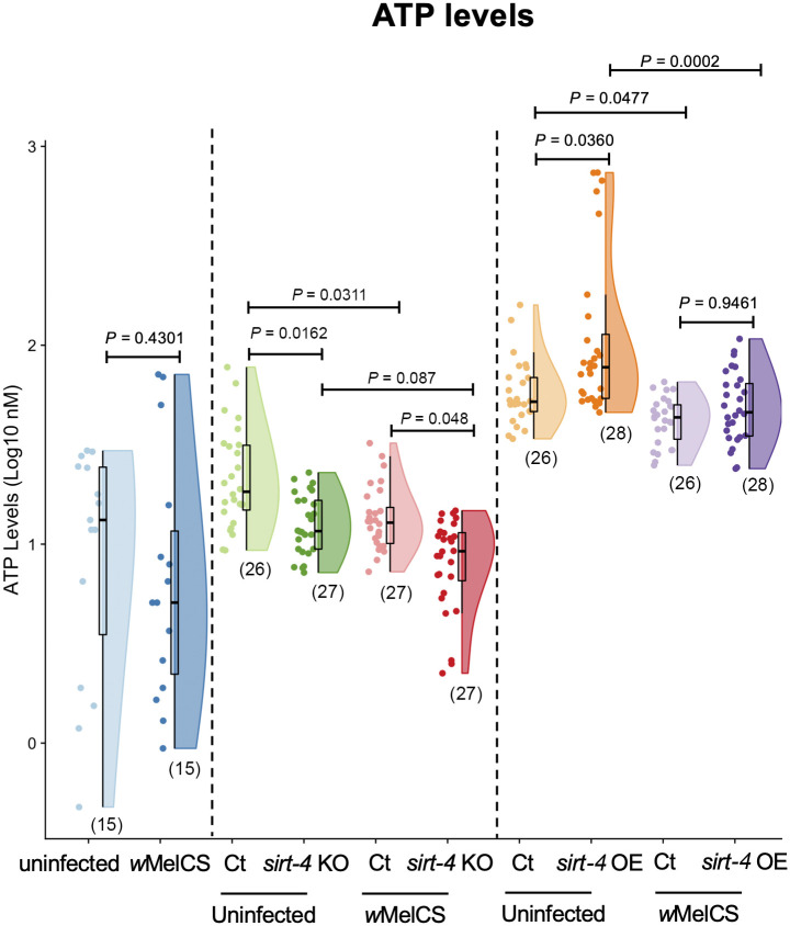
After exploring the interactions between Wolbachia, sirt-4 and gdh expression, and given the interplay between glycolysis and ATP production in the cell, we sought to check the ATP levels of the flies in scenarios of both sirt-4 KO and OE in the context of Wolbachia infection (Fig 3). In wildtype flies, there was no difference in ATP levels due to the presence of the bacterium (Mann-Whitney U test, df = 1, P = 0.4301). In sirt-4 KO flies, alterations in ATP levels were associated with the presence of Wolbachia (Kruskal-Wallis, H = 29.47, P<0.0001). Here, we detected significantly lower ATP levels in both Wolbachia-free (Mann-Whitney Dunn’s corrected test, df = 1, P = 0.0162) and infected scenarios (Mann-Whitney Dunn’s corrected test, df = 1, P = 0.0482), with a greater reduction (control vs. sirt-4 KO) observed in uninfected flies (median decrease of 34.3% vs. 26.8%). Interestingly, in contrast to the comparison between Wolbachia-infected and uninfected wildtype flies, when comparing the controls for sirt-4 KO lines (having wildtype sirt-4 expression), the presence of Wolbachia was associated with a significant 29% median reduction in ATP levels (Mann-Whitney Dunn’s corrected test, df = 1, P = 0.0311; control groups). However, we detected no significant difference in sirt-4 KO flies (Mann-Whitney Dunn’s corrected test, df = 1, P = 0.087; sirt-4 KO groups).
Fig 3. Wolbachia effects on host total ATP levels are sirt-4-independent.
Whole 1-day old virgin female flies from wildtype Wolbachia-free (uninfected–light blue), wildtype infected (wMelCS–dark blue), sirt-4 knockout—KO uninfected (“ct”—control–light green: FM6/ sirt-4 KO vs. sirt-4 KO–dark green: sirt-4 KO/sirt-4 KO), wMelCS-infected (control–light red vs. sirt-4 KO–dark red), sirt-4 overexpression—OE uninfected (“ct”—control–light orange: “Act5cGAL4 >“ vs. sirt-4 OE–dark orange: “Act5cGAL4 > UAS sirt-4 OE”) and wMelCS-infected (“ct”—control–light purple vs. sirt-4 OE–dark purple) scenarios were collected. Female flies were pooled and total ATP levels enzymatically quantified. Wolbachia alone led to a statistically significant decrease in ATP levels only when comparing both control and experimental groups in the sirt-4 KO and OE scenarios. A decrease in ATP levels due to sirt-4 KO was observed in both Wolbachia-infected and uninfected groups, while sirt-4 OE induced the opposite effect. Data represent a maximum of two biological replicate experiments of randomly sampled flies. Raincloud plots depict median total ATP levels with P-values determined via Kruskal-Wallis on entire dataset followed by Mann-Whitney Dunn’s-corrected test for pairwise comparisons for wildtype flies and both sirt-4 KO and OE experiments. Each dot represents a pool of 5 whole flies. Sample size is depicted in parenthesis for each group.
We observed that the presence of Wolbachia was also associated with significant alterations in host ATP levels when sirt-4 was overexpressed (Kruskal-Wallis, H = 32.64, P<0.0001). Similar to our observations for gdh expression, sirt-4 OE flies displayed the opposite effect of sirt-4 KOs, with higher levels of total ATP in Wolbachia-free flies (Mann-Whitney Dunn’s corrected test, df = 1, P = 0.036; 49% median increase). However, when the bacterium was present and sirt-4 expression was genetically elevated, we observed no difference in total ATP (Mann-Whitney Dunn’s corrected test, df = 1, P = 0.9461). Consistent with our observations in sirt-4 KO experiments, Wolbachia presence was associated with a significant reduction in ATP levels for both controls (Mann-Whitney Dunn’s corrected test, df = 1, P = 0.0477) and sirt-4 OE (Mann-Whitney Dunn’s corrected test, df = 1, P = 0.0002) groups, with a stronger reduction observed when sirt-4 was overexpressed (16.6% for controls vs. 36.5% for sirt-4 OE). These results indicate that sirt-4 plays a role in ATP production. However, Wolbachia’s potential interaction with sirt-4 seems unlikely to be the only factor contributing to the observed reductions in total availability of this molecule.
Reduced transcript levels of sirt-4 in Wolbachia-infected flies is associated with alterations in host glucose levels
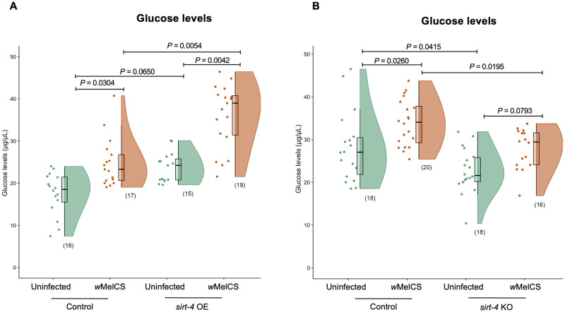
We measured free glucose levels in sirt-4 OE and sirt-4 KO flies both uninfected and wMelCS-infected (Fig 4). We observed a significant alteration in median free glucose levels associated with the presence of Wolbachia (Kruskal-Wallis, H = 38.37, P< 0.0001). In this scenario, we detected an increase in median free glucose levels by 38.7% in the control group (Mann-Whitney Dunn’s corrected test, df = 1, P = 0.0304) and 49.9% in sirt-4 OE flies (Mann-Whitney Dunn’s corrected test, df = 1, P = 0.0042) when wMelCS was present. sirt-4 OE was associated with a significant increase in median free glucose levels by 67.4% in wMelCS-infected flies (Mann-Whitney Dunn’s corrected test, df = 1, P = 0.0054), promoting a hyperglycemic state. sirt-4 OE uninfected flies displayed a 30.7% median increase in free glucose levels (Mann-Whitney Dunn’s corrected test, df = 1, P = 0.065). As for sirt-4 KO, the presence of Wolbachia was also associated with a significant alteration in host glucose levels (One-way ANOVA, F = 10.8, P<0.0001). Here, we observed a hypoglycemic state in both uninfected (Unpaired T test Tukey’s corrected, P = 0.0415; 19.4% reduction) and wMelCS-infected flies (Unpaired T test Tukey’s corrected, P = 0.0195; 18% reduction). In control flies, where sirt-4 expression was kept intact, wMelCS presence was associated with a significant increase in mean free glucose levels by 20.2% compared to uninfected controls (Unpaired T test Tukey’s corrected, P = 0.0256). Finally, in sirt-4 KO flies where wMelCS was present, mean glucose levels were not significantly different from uninfected flies (Unpaired T test Tukey’s corrected, P = 0.0793), suggesting that the hyperglycemic state detected before in the presence of the bacteria was lost.
Fig 4. Wolbachia x sirt-4 interaction is associated with altered host glycemic levels.
Whole 1-day old virgin uninfected and wMelCS-infected female flies in both (A) sirt-4 overexpression—OE (control–green: “Act5cGAL4 >“ vs. sirt-4 OE–orange: “Act5cGAL4 > UAS sirt-4 OE”) and (B) sirt-4 knockout—KO (control–green: FM6/ sirt-4 KO vs. sirt-4 KO–orange: sirt-4 KO/sirt-4 KO) scenarios were collected and the total glucose levels measured. Wolbachia presence alone induced a significant increase in median glucose levels in both control and sirt-4 OE groups. Overexpressing sirt-4 in uninfected flies caused no statistically significant increase in median glucose levels, despite a 30.7% increase, while the same overexpression construct in the presence of Wolbachia induced a hyperglycemic stage. sirt-4 KO induced the opposite effect, with flies becoming hypoglycemic in both Wolbachia-infected and uninfected groups. Wolbachia alone induced an increase in mean glucose levels between control groups, however, the presence of the bacterium did not cause the same increase when sirt-4 was knocked out, with the bacterium being unable to induce a shift in host glycemic levels. Data represent a maximum of two biological replicate experiments of randomly sampled flies. Raincloud plots depict median glucose levels with P-values determined for all pairwise comparisons via Kruskal-Wallis on entire dataset followed by Mann-Whitney Dunn’s-corrected test for pairwise comparisons in the sirt-4 OE experiment. For the sirt-4 KO experiment, P-values were determined by One-Way ANOVA followed by Unpaired T test Tukey’s-corrected pairwise comparison test. Each dot represents a pool of 5 whole flies. Sample size is depicted in parenthesis for each group.
Sirt-4 overexpression is associated with reduced Wolbachia density in the ovaries
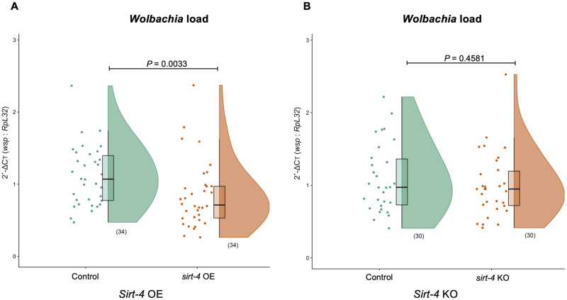
We tested if sirt-4 KO was associated with changes on Wolbachia density. We examined the ovaries of 1-day old virgin female flies with wMelCS infection. Overall, sirt-4 KO did not alter the relative median bacterial density (Mann-Whitney U test, U = 399, P<0.4581. Strikingly though, sirt-4 OE was associated with a significant decrease on relative median levels of Wolbachia in the ovaries by 33.6% (Mann-Whitney U test, U = 341, P<0.0033) (Fig 5). Furthermore, there were no significant differences in host RpL32 ovarian DNA abundance between control and sirt-4 mutants for both sirt-4 OE (Mann-Whitney U test, df = 1, P = 0.1777) and sirt-4 KO scenarios (Mann-Whitney U test, df = 1, P = 0.3538), confirming the effects we observed on Wolbachia density in sirt-4 mutants (S3 Fig).
Fig 5. sirt-4 overexpression is associated with reduced Wolbachia density in the ovaries.
One-day old virgin wMelCS-infected female flies had their ovaries dissected, DNA extracted and levels of Wolbachia quantified for the wsp gene relative to host RpL32 using SYBR Green in both (A) sirt-4 overexpression—OE (control–green: “Act5cGAL4 >“ vs. sirt-4 OE–orange: “Act5cGAL4 > UAS sirt-4 OE”) and (B) sirt-4 knockout—KO (control–green: FM6/ sirt-4 KO vs. sirt-4 KO–orange: sirt-4 KO/sirt-4 KO) scenarios. sirt-4 overexpression, but not the knockout, significantly reduced Wolbachia levels. Data represent two biological replicate experiments of randomly sampled flies. Raincloud plots depict median relative wsp levels with P-values determined for all pairwise comparisons via Kruskal-Wallis on entire dataset followed by Mann-Whitney Dunn’s-corrected test for pairwise comparisons. Each dot represents a pool of 5 pairs of ovaries. Sample size is depicted in parenthesis for each group.
Discussion
Wolbachia’s wide distribution across distinct arthropod hosts and the ramifications associated with its presence [3] make it an interesting model of host-microbe interactions. More specifically, the Jekyll and Hyde “host context-dependent” association, involving nutrient provisioning or scavenging [16], provides an unique opportunity to study the mechanisms at the intersection between host and endosymbiont metabolic processes. Here, aided by the power of fly genetics, we combined transcriptional profiling and metabolic assays to explore the interaction between two regulators of host biology–Wolbachia and sirtuins. Our data provide the first evidence that the presence of distinct Wolbachia strains is associated with a decrease in the transcriptional levels of D. melanogaster sirt-4. In subsequent experiments, focused on the wMelCS strain, we observed that this effect was consistent across distinct host tissues, namely the ovaries and fat body, and timepoints (1, 5 and 10 days of age).
The consistent reduction in sirt-4 associated with wMelCS-infected flies but not flies infected with the over-replicating wMelPop or wMel strains (for which we detected a reduction in sirt-4 transcript levels in both 1 and 5 but not 10-day old flies for the latter) was interesting, as it suggests that bacterial density is likely not a driving factor for our observations since wMelCS is known to achieve densities double that observed for wMel but twenty times lower than wMelPop [58]. Nonetheless, this hypothesis should be further validated, given that we did not explicitly measure Wolbachia titers in the same flies that were used for our experiments. We highlight the importance of future experiments directly exploring the association between distinct bacterial strains, their intrinsic replicative ability, and the transcriptional level of sirt-4, focusing on individuals of the same age, sex and host tissue, as Wolbachia density has been shown to affected by these factors [59,60].
Although evidence indicates that the non-repeat regions of the wMelCS and wMelPop genomes are identical, there is a triplication of a ~19-kb region composed by eight genes spanning from WD_RS02245 to WD_RS06080 (old locus tags: WD0507 to WD0514) in wMelPop, not present in wMelCS [61]. The region, known as Octomom, contain genes coding for distinct ankyrin-proteins, as well as reverse transcriptases [61,62]. It has been shown that higher Octomom copy number results in increased bacterial density [62]. Additionally, the region also encompasses the putative transcriptional regulator WD_RS02250 and the gene WD_RS02810 associated with DNA repair (old locus tags: WD0508 and WD0625, respectively), both part of the Eukaryotic association module of prophage WO, in which expression of WD_RS02250 has been associated with increased bacterial titer [63]. The differences in sirt-4 expression observed between these two strains could be related to this genomic region. Another potential explanation is that the observed differences are due to epigenetic changes. Recent work in parasitoid wasps observed a series of host genes that were differently methylated in the presence of Wolbachia [64], similarly to what has been reported in Aedes aegypti mosquitoes infected with the wMelPop strain [65] and the reproductive tissues of male Drosophila infected with wMel [66]. Both represent intriguing hypotheses to be tested in future studies.
Next, we sought to explore factors involved in regulating host carbohydrate metabolism, particularly those known to impact the monosaccharide glucose, highlighted in S1 Fig (figure is not intended to cover all factors involved in the insulin signaling pathway, only those for which sirt-4 plays a role). Sirt-4 has an important role in the mitochondria regulating energy homeostasis through changes in the ATP/ADP ratio. This process is in part modulated by GDH, which facilitates glutamine metabolism and ATP production, hence influencing insulin secretion and glucose homeostasis [50].
In fruit flies, both RNA-Seq and proteomic data indicate a high expression and production of gdh in all host tissues, with a peak in expression in 1 day-old females (FlyBase ID: FBgn0001098) [67]. The Wolbachia genome also codes for a gdh gene [68], however, despite its presence in both organisms, GDH function is remarkably different. In prokaryotes, GDH activity is anabolic, synthetizing amino acids from basic precursors, but since eukaryotes depend on exogenous sources of amino acids, GDH activity is catabolic, oxidizing amino acids for protein synthesis [53]. Given the disparity in function, we specifically designed a primer set for the Drosophila gdh, as to avoid unspecific amplification of Wolbachia’s gdh.
It has been shown that Wolbachia acts as a parasite when it comes to reliance on certain host amino acids for energy production, as evidenced by studies involving multiple Wolbachia strains [23–25]. In calorie-sufficient scenarios, SIRT-4 has been shown to inhibit GDH activity, limiting insulin secretion induced by glutamine. However, during calorie restriction, there is an increase in GDH activity, leading to an increase in insulin secretion in response to glutamine and leucine [54]. Our work is the first to observe increased gdh transcript levels associated with the presence of Wolbachia infection. This Such correlation coupled with (1) the predicted presence of amino acid uptake transporters and their associated metabolic pathways (glutamine included) in the genome of distinct Wolbachia strains [23,24], and (2) the observation that SIRT-4 (known to directly inhibit GDH activity) protein levels peak in a nutrient rich environment [69], together suggests that Wolbachia might be acting as a nutrient scavenger, depleting the host of key nutrients. This depletion might then mimic a calorie restriction scenario driving the downregulation of host sirt-4, and leading to upregulation of its direct target gdh in order to compensate for such energetic loss.
Given our current data, we cannot identify the exact mechanism behind our aforementioned hypothesis, and whether our gdh results are a direct or indirect effect of an interaction with Wolbachia. Our hypothesis linked to Wolbachia opens the door for future research into this particularly interesting topic given how little we know about the nuances of distinct Wolbachia strains and its host association in the context of nutrient parasitism. For instance, the genome of Wolbachia is populated by an unusual high number of genes encoding ankyrin domain (ANK) repeats, with counts varying in a strain- and supergroup-specific manner [70,71]. Microbial-derived ANKS have been associated with the modulation of host gene transcription via chromatin interaction (see [72] for more details on ANKs). Additionally, the genome of symbiotic and pathogenic bacteria are known to encode a series of machineries that allow for the translocation of DNA or molecules mediating host-microbe interactions, such as the Type 4 secretion system (T4SS) [73]. The genome of many Wolbachia strains is known to encode a T4SS [74,75], and furthermore, ANK-containing effectors of endosymbiotic bacteria related to Wolbachia were found to be translocated by T4SS [72].
The core balance for energy production resides within the mitochondria and its machinery for ATP production, a process directly regulated by sirtuin [76]. Our data indicate that although sirt-4 plays a role in ATP production, as evidenced by other authors [77], Wolbachia’s potential interaction with sirt-4 seems unlikely to be the only factor contributing to the observed reduction in ATP, highlighting how this molecule can be generated by multiple pathways in the cell [78]. These results match the overall sirt-4-dependent changes in total ATP levels observed in mice, in which SIRT-4 is proposed to act by controlling the efficiency by which ATP is produced [77]. In that study, the authors saw a decrease in ATP production both in vitro and in vivo as a result of sirt-4 KO, with opposing effects on ATP levels when sirt-4 was overexpressed. According to the authors, removal of SIRT-4 mimics a starvation condition that initiates a homeostatic response involving the enzyme AMP-dependent kinase (AMPK) and the peroxisome proliferator-activated receptor gamma coactivator 1-alpha (PGC1α). AMPK works as a sensor of cellular energy status by tracking changes in the AMP/ATP ratio [79]. In extreme conditions such as nutrient deprivation, AMPK blocks malonyl-coA production by the enzyme acetyl-coA-carboxylase, and phosphorylates PGC1α, leading to an increase in mitochondrial processes such as fatty acid oxidation [80], ultimately decreasing ATP production.
The data here, although in support of the findings discussed from mice, present a conflict with a previous study done in D. melanogaster where the authors saw no difference in total ATP levels in either whole individuals or eviscerated abdomens of SIRT-4 KO flies [49]. One potential explanation for such discrepancy may relate to an issue commonly overlooked by the Drosophila community: the presence of Wolbachia. The authors did not explicitly account for the presence of the bacterium in the Drosophila stocks they used. Data indicate that at least 30% of all Bloomington Drosophila stocks are infected with Wolbachia [81].
A recent study demonstrated a positive correlation between Wolbachia and mitochondrial titers in the ovarian tissue of distinct Drosophila and Wolbachia genotypes, with uninfected individuals displaying similar mitochondrial titers as infected flies [82]. Additionally, Wolbachia titers were unaffected by a decrease in mitochondrial titer, as evidenced by knockdown of mitochondrial genes. However, these experiments were measured in the context of the low replicative strain wMel. In fact, the positive correlation between both mitochondria and Wolbachia titers were disrupted in the presence of a high replicating strain of the bacteria, namely wMelCS. This indicates, as mentioned by the authors of this study, that distinct strains of Wolbachia differ in their ability to modify the environment in which they are inserted, with wMelCS likely creating a more competitive environment and thus impacting mitochondria. In our work, we detected a trend towards decreased ATP levels in wildtype wMelCS-infected flies, in comparison to its uninfected counterpart. Additionally, comparisons between both control groups in the sirt-4 KO and OE experiments indicated a significantly lower level of total ATP in wMelCS-infected flies. Further work exploring the impact of manipulating distinct mitochondrial genes on Wolbachia density, in the context of sirt-4 would be of great importance, given the existence of multiple factors affecting mitochondrial energetics in the host [83]. For instance, the work of Henry [82] differs from a previous study in which knockdown of the mitochondrial gene NADH dehydrogenase, an enzyme responsible for the conversion of NADH to its oxidized form NAD+ (a key substrate in which sirtuin activity relies on), led to a significant reduction in Wolbachia load [84].
The insulin/IGF signaling pathway is conserved across all multi-cellular organisms, responding to external changes in the environment by modulating organism growth, metabolic homeostasis, lifespan and reproduction. In flies, dILPs are produced and released by the IPCs in the brain, in response to signals originating from endocrine organs such as the fat body, in an intricate interorgan communication process, affecting, among other processes, host glucose metabolism [31,33,85].
Overall, our results of host glycemia under sirt-4 OE and KO are in agreement with observations in other organisms, in which sirt-4 upregulation has a direct negative impact on insulin secretion and therefore glucose homeostasis [50,77,86,87]. Previous work in D. melanogaster has shown that Wolbachia presence leads to increased insulin/IGF-like signaling [40]. By demonstrating that wMelCS flies displayed reduced mRNA levels of sirt-4, including in the fat body, and that the genetic manipulation of this gene was associated with modulation of fly glycemic levels, our work expands on the current knowledge of Wolbachia’s manipulation of host metabolism. More specifically, through the data here shown, we propose that the previous reported upregulation of insulin secretion in Wolbachia-infected flies is potentially mediated by sirt-4.
Finally, by studying host metabolism in the context of sirt-4, and coupling our results on glucose metabolism to the observed reduction in Wolbachia density under a sirt-4 OE scenario, our work is able to point out another potential piece in the intriguing puzzle that is the process of regulating Wolbachia titers in the host. Previous work has shown that yeast-enriched diet resulted in reduced ovarian Wolbachia titer via TORC1, in which the upstream effectors remain to be discovered [42]. Additionally, exciting recent work has shown that GDH inhibition via SIRT-4 leads to mTORC1 activation [69]. As such, by linking previous work to our data presented here, we propose that the observed reduction in Wolbachia density detected in flies reared on a yeast-enriched diet is potentially the result of the upstream effector SIRT-4. We must point out that both notions are based on our observations of alterations in host metabolism and gene expression associated with scenarios in which Wolbachia was present. As we cannot yet demonstrate the mechanism (direct or indirect) by which such regulation would occur, we cannot make an explicit causal link.
We must stress that the regulation of glucose metabolism does not rely solely on SIRT-4 and therefore could explain why our results on glucose-related processes, such as gdh expression and ATP levels, indicate a potential additive effect of Wolbachia on the former and a partial association on the latter. For instance, SIRT-1 (homologous to sirt-2 in Drosophila) is also known to regulate gluconeogenesis [88], glycolysis [89] and insulin secretion [90], working as a SIRT-4 antagonist. In addition to its role in fatty acid production, PGC1α is also known to be directly deacetylated by SIRT-1 under calorie restriction, leading to decreased expression of genes involved in glycolysis while also causing an increase in glucose production [89].
Critical to our scientific questions related to modulation of the host glycemic state, Wolbachia has also been shown to have a strong impact on the composition of the host microbiota [91,92]. Microbiota composition has been directly linked to host metabolic homeostasis, affecting processes such as insulin signaling, glucose balance, and triglyceride levels [93–95]. It has been shown that Wolbachia affects the abundance of Acetobacter, a genus commonly present in Drosophila fly stocks [96] that can modulate host glycemia [94,95]. In subsequent studies, it would be interesting to screen the microbiota diversity of Wolbachia-infected flies in the context of sirtuin expression, an unexplored venue that might explain some of the results observed here.
Considering the current impact of Wolbachia on viral load within the host (please refer to [97] for a detailed review on Wolbachia in the context of viral infection), and the worldwide deployment of Wolbachia as a tool against arboviral transmission [98], it would also be relevant to test if cells infected with Wolbachia display similar alterations in glycemia and how this modulation might by affected by the presence of a viral agent, since an increase in glucose uptake and glycolytic flux is one of the metabolic signatures of viral infection [99], dengue included [100].
Sirtuins have been implicated in defense against human viral pathogens with sirtuin inhibition shown to be beneficial for the replication of influenza A virus (RNA virus), herpes simplex virus 1, adenovirus type 5, and human cytomegalovirus (all DNA viruses). Sirtuin activation however, led to reduced viral titers of both influenza A and human cytomegalovirus. Resveratrol is a well-known powerful activator of sirtuins [101]. Recent work demonstrated that addition of this compound to cell culture pre- and post- exposure to ZIKV reduced the viral load from 30% to 90%, respectively, showing another promising venue of sirtuins as viral inhibiting agents [102]. The bacterial sirtuin CobB, a homolog of Escherichia coli sirtuin negatively impacted the growth of both bacteriophages T4 and λ [103,104]. This all points to sirtuins as another area of study yet to be explored in the context of Wolbachia-mediated pathogen blocking.
In accordance with empirical models of reduced genome size in symbiont bacteria [105], most Wolbachia strains (wFol excluded [106]) underwent significant gene loss, displaying variable genome sizes in distinct strain/host interactions [107]. These losses often occur in metabolic pathways, with the bacterium relying on their hosts to acquire key metabolic components such as amino acids and lipids [25,108]. This reliance on host processes has led to the link of many metabolic pathways as essential in regulating Wolbachia density within the host. Despite the initial belief that consumption of host amino acids by Wolbachia was via ERAD-driven proteolysis [84], recent work suggests that amino acids are obtained from the core proteasome by bacteria strategically positioned between the ER and the Golgi [109,110]. This is only one of many ways by which Wolbachia seems to be interacting with host metabolic processes. For instance, a whole genome screening in D. melanogaster identified 8% of genes from a total of 14,024 (covering 80% of D. melanogaster Release 6 genome) to effectively impact Wolbachia density, including the identification of sirt-2 whose knockdown reduced bacterial density, among many other genes with unknown function [110]. The identification of sirt-4 as a factor associated with alterations in Wolbachia density in our work expands the list of potential candidates capable of modulating the bacterium population within the host.
In summary, here we used a transcriptional approach coupled with metabolic assays to characterize for the first time, the interaction between Wolbachia and host sirtuins. Our initial focus was on the impact of the bacterium on expression of all known Drosophila sirtuins. This characterization led us to identify a novel significant association between Wolbachia and sirt-4, a gene that when upregulated, was associated with reduced levels of Wolbachia in the ovaries. By investigating the sirt-4-dependent mitochondrial pathway that modulates glucose metabolism in the host, we characterized the expression profile of glutamate dehydrogenase, a key enzyme in the TCA cycle, which we found to be upregulated in scenarios where Wolbachia was present. Finally, we found that the presence of Wolbachia was associated with alterations in both total ATP levels as well as the glycemic state of the fly in a sirt-4-related manner. To conclude, we postulate that through yet elusive mechanisms, Wolbachia presence is associated with altered sirt-4 expression, which is, in turn, associated with alterations in the glycemic state of its host. Future work aiming at understanding how this metabolic interaction affects viral infection would be important not only for future studies to inform the use of Wolbachia as a viral control agent, but also basic biological questions such as cell colonization [111,112], and how the modulation of host physiology impacts the symbiont’s ability to spread through insect populations [10,11,113].
Materials and methods
Fly stocks and husbandry
The D. melanogaster stocks utilized in this study are listed in S1 Table. Flies were maintained in an incubator at 25°C under a 12 h light:dark cycle regime with 60% relative humidity. Flies were reared on a cornmeal-yeast-molasses-agar diet supplemented with dry yeast pellets. More specifically, the food consisted of: 312g of active dry yeast, 756g of cornmeal, 112g of agar, 756mL of molasses, 80mL of propionic acid, 231mL of Tegosep (106.6g of methyl 4-hydroxybenzoate in 1L of ethyl alcohol), in 9.6L of water. The initial survey of Wolbachia affecting sirtuin expression levels (S1 Fig) were performed in Drosophila melanogaster with the yellow white background of either uninfected or infected with wMel, wMelCS, or wMelPop Wolbachia strains. These stocks were generated by 5 generations of backcrossing as to have uninfected and infected fly lines containing the same genetic background. The stocks used here included the Bloomington line 8840—sirt-4 KO (sirt-4white+1 homologous recombination deletion allele), line 22029—sirt-4 OE (Sirt-4 P[Mae-UAS.6.11] transposable element insertion) and line 3954 with the ubiquitously expressed GAL4 system (transposable element P[Act5C-GAL4] insertion) (S1 Table). Wolbachia infection status in stocks was confirmed prior to starting experiments via PCR for the detection of the Wolbachia surface protein (wsp) gene.
Nucleic acid extraction
Fly samples were stored at -80°C, and total DNA and RNA were extracted using the TRIzol reagent and phenol:chloroform:isoamyl alcohol (ThermoFisher Scientific) according to manufacturer’s instructions. Samples were homogenized in 200μL-1000μL (for whole flies or a pool of dissected tissues, respectively) of TRIzol using a motor-driven pellet pestle mixer (Sigma-Aldrich). Total DNA and RNA was quantified using the NanoDrop One spectrophotometer system (ThermoFisher Scientific). To each RNA sample, a mix of 1μL of DNase I recombinant enzyme and 5μL of buffer (ROCHE) were added and incubated at 37°C for 50 min. Prior to initiating experiments, a subset of samples were tested via qPCR, in a reaction without the reverse transcriptase enzyme, to ensure no genomic DNA contamination. DNA and RNA samples were then diluted to 50ng/μL in nuclease-free water and stored at -80°C (RNA) and -20°C (DNA) until tested.
Gene expression analysis
Wolbachia, sirtuin and gdh target genes were quantified in technical duplicates for each sample collected. Target gene expression levels were quantified relative to the Drosophila ribosomal gene RpL32 (protein S32), which served as endogenous control. Total volume was 10μL per reaction, each containing: 5μL of PowerUp SYBR Green Master Mix (ThermoFisher Scientific), 0.2μL of each forward and reverse primers (10μM), 0.25μL of SuperScript III Reverse Transcriptase (except in reactions involving DNA), 0.35μL of nuclease-free water and 200ng of template RNA (Wolbachia quantification used the same amount of DNA). Thermocycling conditions were as follows: an initial reverse transcription step at 50°C for 5 min; RT inactivation/initial denaturation at 95°C for 2 min, and 40 cycles of 95°C for 15 sec and 60°C for 1 min using an ABI 7900ht Real-Time PCR system (ThermoFisher Scientific). Cycling conditions were similar for Wolbachia quantification, without the addition of the initial reverse transcription step.
Wolbachia expression levels were quantified using wolbachia surface protein (wsp) gene, while primer sequences for host sirtuins 1–7 used in the assays were designed using NCBI’s Primer-BLAST (http://www.ncbi.nlm.nih.gov/tools/primer-blast/) and both MFEPrimer 3.0 (http://mfeprimer.igenetech.com/) and IDT’s OligoAnalyzer tool (http://www.idtdna.com) for quality control. Expression levels of ghd were measured using primers available at the Harvard Medical School DRSC Functional Genomics Resources website (http://www.flyrnai.org/flyprimerbank) (S2 Table).
Prior to use in experiments, each primer pair for a specific target gene designed in this study was examined for both specificity and amplification efficiency as recommended [114]. Specificity analysis was performed by melt curve analysis, with all pairs displaying a single peak, while efficiency analysis was achieved by examining the amplification performance under a series of sample template dilutions. All primer pairs displayed an efficiency of between 90–110% at the dilution used in the experiments described below.
Sample collection for Wolbachia, sirtuins and glutamate dehydrogenase quantification
Wolbachia
We looked at the effects of sirt-4 KO and OE on bacterial density in the ovaries of 1-day old virgin females. Collections consisted of a set of 15–25 samples per replicate, each sample consisting of a pool of 5 pairs of ovaries. Flies were anesthetized on a CO2 pad and dissections carried out on a glass plate with fresh sterile 1X phosphate buffered saline solution, replaced between each individual dissection. Tubes were kept in dry ice during dissections and immediately transferred to-80°C once collection was completed. This was used as a standard fly-handling procedure for all experiments described below.
Sirtuins
Sample collection for sirtuin expression levels also comprised two parts. For the first part, up to 15 whole individual 5-days old virgin wildtype female flies per Wolbachia infection status group (uninfected, wMel, wMelCS, wMelPop) per replicate were collected, had their RNA extracted and analyzed for differences in expression of sirt-1-7. Collections for only sirt-4-related experiments also included adult female flies spanning from 1 to 10 days of adulthood prior to total RNA extraction and analysis, chosen based on the lifetime transcriptional profile for this gene observed in the FlyAtlas RNA-Seq dataset (FlyBase ID: FBgn0029783). Part two focused on the wMelCS strain, where sirt-4 expression was analyzed in the ovaries and fat body of 1-day old virgin wildtype female flies compared to Wolbachia-free individuals. This timepoint and bacterial strain were chosen given that 1) RNA-Seq data shows that the absolute expression of sirt-4 peaks at day 1 in adult females (FlyBase ID: FBgn0029783), therefore maximizing confidence in the impact of sirt-4 in all conditions tested, and 2) of all bacterial strains tested, wMelCS caused the most consistent reduction in sirt-4 expression in whole body and individual tissues in 1-day old females. Collection consisted of a set of 15 samples per replicate with each sample consisting of a pool of 5 pairs of ovaries or 5 fly carcasses minus the gut and Malpighian tubules (as a proxy for fat body). The ovary tissue was selected for study because it represents a key organ for Wolbachia tropism in the host, as it is essential for high frequencies of maternal transmission of Wolbachia [112,115]. In addition to its immune function [116], the fat body is critical for regulating host physiology. It not only stores and generates energy, but also actively synthesizes most of the metabolites and proteins present in the hemolymph [117], acting as an coordinator of nutrient sensing (particularly amino acids [38]) and activator (endocrine activity) of many local and systemic responses, including insulin signaling [85]. Previous work has shown that Wolbachia actively infects the fat body of distinct insect species, including mosquitoes and fruit flies, where it plays a role in host immunity and bioenergetics [37]. Last, it has been shown that overexpression of sirt-4 in the fat body increases D. melanogaster lifespan [118], a trait also affected by Wolbachia.
Glutamate dehydrogenase (gdh)
Collection consisted of 6–7 whole individual 1-day old virgin wildtype uninfected, wildtype wMelCS-infected, and sirt-4 KO and sirt-4 OE (uninfected and wMelCS-infected) female flies per replicate, per group. RNA extraction and gene expression analyses for all experiments were conducted as described in the “gene expression analysis section”.
Metabolite quantification assays
Plate assays for total glucose and ATP levels were performed as reported elsewhere [49,119] using the Glucose Hexokinase Reagent kit (Sigma-Aldrich) and the ATP Determination Kit (ThermoFisher Scientific). A total of 10–15 samples per replicate, per group (wildtype uninfected, wildtype wMelCS-infected, and sirt-4 KO and sirt-4 OE uninfected and wMelCS-infected) were collected. Each sample consisted of a pool of 5 whole 1-day old virgin female flies.
Statistical analysis
Datasets were first assayed for normality using the D’Agostino & Pearson omnibus test. Non-normally distributed data of more than 2 groups were analyzed using a Kruskal–Wallis followed by individual Mann-Whitney Dunn’s-corrected multiple comparisons. Both analyses used a level of significance set at P<0.05. Normally distributed datasets were compared using a standard One-way ANOVA, followed by Tukey’s multiple comparison test, or a pairwise comparison using Unpaired T-test with Brown-Forsythe and Welch's correction in order to correct for groups with significantly unequal variances or sample sizes. Comparisons between multiple non-parametric distributed groups were performed using the One-way ANOVA followed by Dunnett’s multiple comparisons analysis. All analyses used a level of significance set at P<0.05. To test for the impact of experimental factors on sirt-4 transcript levels, we used JMP Pro 14 (SAS) to perform a generalized linear regression model (GLM) under a Poisson distribution assumption, with sirt-4 transcript levels set as test variable and days of adulthood, bacterial strain and the interaction between days of adulthood and bacterial strains as the explanatory variables. Details on specific statistical tests performed in each dataset are present within the legend of each experimental figure. All statistical analyses (GLM model excluded) were performed using Prism 8.1.1 (Graphpad) and the graphs made using Rstudio 1.1.463 (Rstudio) with the raincloud plot visualization package [120].
Supporting information
Scheme representing the main SIRT-4-dependent factors regulating insulin secretion, based on the literature. Our work shows that Wolbachia downregulates the expression of sirt-4. The expression levels of MCCC, IDE and ANT2, were not taken into account in this work.
(TIF)
Whole wildtype Wolbachia-free (uninfected—red) and wildtype infected (wMelCS—yellow, wMel—green and wMelPop—blue) virgin female flies were collected at 5 days of adulthood, had their RNA extracted and levels of sirt-1, sirt-2, sirt-6 and sirt-7 quantified relative to host RpL32 using SYBR Green. None of the strain tested significantly affected the relative expression of the sirtuin genes tested when compared to the uninfected group. Data represent one biological replicate experiment of randomly sampled flies. Raincloud plots depict median relative sirtuin levels with P-values determined via Kruskal-Wallis on entire dataset followed by Mann-Whitney Dunn’s-corrected test for pairwise comparisons. Each dot represents a single whole fly. Sample size is depicted in parenthesis for each group.
(TIF)
One-day old virgin wMelCS-infected female flies had their ovaries dissected, DNA extracted and CT values for Drosophila melanogaster ovarian RpL32 endogenous control gene quantified using SYBR Green in both (A) sirt-4 overexpression—OE (control–green: “Act5cGAL4 >“ vs. sirt-4 OE–orange: “Act5cGAL4 > UAS sirt-4 OE”) and (B) sirt-4 knockout—KO (control–green: FM6/ sirt-4 KO vs. sirt-4 KO–orange: sirt-4 KO/sirt-4 KO) scenarios. There was no statistically significant difference in CT values for host endogenous control gene between controls and sirt-4 OE and KO mutants. Data represent two biological replicate experiments of randomly sampled flies. Scatter plots depict median Ct values with P-values determined for all pairwise comparisons via Mann-Whitney U test on a non-parametric dataset. Each dot represents a pool of 5 pairs of ovaries. Sample size is depicted in parenthesis for each group.
(TIFF)
Drosophila species and their corresponding Wolbachia strains, source, Bloomington stock center reference number (when applicable) and target construct used in experimental procedures are listed.
(DOCX)
(DOCX)
(DOCX)
(DOCX)
Acknowledgments
The authors are grateful for all the assistance provided by undergraduate students involved in the weekly preparation of fly food and vials for work. We are also grateful for the efforts of Dr. Kim McCall and Dr. Elizabeth McGraw towards the publication of the manuscript. Finally, we would like to thank the editor and anonymous reviewers for providing extremely valuable feedback towards improving the manuscript.
Data Availability
All relevant data are within the manuscript and its Supporting Information files.
Funding Statement
HLCD was a Latin American postdoctoral fellow in the Biomedical Sciences, supported by the Pew Charitable Trusts - www.pewtrusts.org MAD and HF were supported by internal funds from Boston University. The funders had no role in study design, data collection and analysis, decision to publish, or preparation of the manuscript.
References
- 1.Zug R, Hammerstein P. Still a host of hosts for Wolbachia: analysis of recent data suggests that 40% of terrestrial arthropod species are infected. PLOS One. 2012;7: e38544 10.1371/journal.pone.0038544 [DOI] [PMC free article] [PubMed] [Google Scholar]
- 2.McGraw EA, O’Neill SL. Beyond insecticides: new thinking on an ancient problem. Nat Rev Microbiol. 2013;11: 181–93. 10.1038/nrmicro2968 [DOI] [PubMed] [Google Scholar]
- 3.Werren JH, Baldo L, Clark ME. Wolbachia: master manipulators of invertebrate biology. Nat Rev Microbiol. 2008;6: 741–51. 10.1038/nrmicro1969 [DOI] [PubMed] [Google Scholar]
- 4.Vavre F, Fleury F, Lepetit D, Fouillet P, Boulétreau M. Phylogenetic evidence for horizontal transmission of Wolbachia in host-parasitoid associations. Mol Biol Evol. 1999;16: 1711–1723. 10.1093/oxfordjournals.molbev.a026084 [DOI] [PubMed] [Google Scholar]
- 5.Raychoudhury R, Baldo L, Oliveira DCSG, Werren JH. Modes of acquisition of Wolbachia: horizontal transfer hybrid introgression, and codivergence in the Nasonia species complex. Evolution (NY). 2009;63: 165–183. [DOI] [PubMed] [Google Scholar]
- 6.Hamm CA, Begun DJ, Vo A, Smith CCR, Saelao P, Shaver AO, et al. Wolbachia do not live by reproductive manipulation alone: infection polymorphism in Drosophila suzukii and D. subpulchrella. Mol Ecol. 2014;23: 4871–4885. 10.1111/mec.12901 [DOI] [PMC free article] [PubMed] [Google Scholar]
- 7.Kriesner P, Hoffmann AA. Rapid spread of a Wolbachia infection that does not affect host reproduction in Drosophila simulans cage populations. Evolution (NY). 2018;72: 1475–1487. [DOI] [PubMed] [Google Scholar]
- 8.Yeap HL, Mee P, Walker T, Weeks AR, O’Neill SL, Johnson P, et al. Dynamics of the “popcorn” Wolbachia infection in outbred Aedes aegypti informs prospects for mosquito vector control. Genetics. 2011;187: 583–95. 10.1534/genetics.110.122390 [DOI] [PMC free article] [PubMed] [Google Scholar]
- 9.Walker T, Johnson PH, Moreira LA, Iturbe-Ormaetxe I, Frentiu FD, McMeniman CJ, et al. The wMel Wolbachia strain blocks dengue and invades caged Aedes aegypti populations. Nature. 2011;476: 450–453. 10.1038/nature10355 [DOI] [PubMed] [Google Scholar]
- 10.Turelli M. Cytoplasmic incompatibility in populations with overlapping generations. Evolution. 2010;64: 232–41. 10.1111/j.1558-5646.2009.00822.x [DOI] [PubMed] [Google Scholar]
- 11.Turelli M. Evolution of incompatibility-inducing microbes and their hosts. Evolution (NY). 1994;48: 1500–1513. [DOI] [PubMed] [Google Scholar]
- 12.Turelli M, Cooper BS, Richardson KM, Ginsberg PS, Peckenpaugh B, Antelope CX, et al. Rapid Global Spread of wRi-like Wolbachia across Multiple Drosophila. Curr Biol. 2018;28: 963–971.e8. 10.1016/j.cub.2018.02.015 [DOI] [PMC free article] [PubMed] [Google Scholar]
- 13.Himler AG, Adachi-Hagimori T, Bergen JE, Kozuch A, Kelly SE, Tabashnik BE, et al. Rapid spread of a bacterial symbiont in an invasive whitefly is driven by fitness benefits and female bias. Science. 2011;332: 254–6. 10.1126/science.1199410 [DOI] [PubMed] [Google Scholar]
- 14.Hosokawa T, Koga R, Kikuchi Y, Meng X-YX-Y, Fukatsu T. Wolbachia as a bacteriocyte-associated nutritional mutualist. Proc Natl Acad Sci. 2010;107: 769–774. 10.1073/pnas.0911476107 [DOI] [PMC free article] [PubMed] [Google Scholar]
- 15.Zug R, Hammerstein P. Bad guys turned nice? A critical assessment of Wolbachia mutualisms in arthropod hosts. Biol Rev Camb Philos Soc. 2014;49. [DOI] [PubMed] [Google Scholar]
- 16.Newton ILG, Rice DW. The Jekyll and Hyde Symbiont: Could Wolbachia Be a Nutritional Mutualist? J Bacteriol. 2019;202: e00589–19. [DOI] [PMC free article] [PubMed] [Google Scholar]
- 17.Jiggins FM, Hurst GDD. Rapid Insect Evolution by Symbiont Transfer. Science (80-). 2011;332. [DOI] [PubMed] [Google Scholar]
- 18.Noda H. Cytoplasmic incompatibility in a rice planthopper. J Hered. 1984;75: 345–348. [Google Scholar]
- 19.Ju J-F, Bing X-L, Zhao D-S, Guo Y, Xi Z, Hoffmann AA, et al. Wolbachia supplement biotin and riboflavin to enhance reproduction in planthoppers. ISME J. 2020;14: 676–687. 10.1038/s41396-019-0559-9 [DOI] [PMC free article] [PubMed] [Google Scholar]
- 20.Moriyama M, Nikoh N, Hosokawa T, Fukatsu T. Riboflavin Provisioning Underlies Wolbachia’s Fitness Contribution to Its Insect Host. MBio. 2015;6: 1–8. [DOI] [PMC free article] [PubMed] [Google Scholar]
- 21.Guo Y, Hoffmann AA, Xu X-Q, Zhang X, Huang H-J, Ju J-F, et al. Wolbachia -induced apoptosis associated with increased fecundity in Laodelphax striatellus (Hemiptera: Delphacidae). Insect Mol Biol. 2018;27: 796–807. 10.1111/imb.12518 [DOI] [PubMed] [Google Scholar]
- 22.Weeks AR, Turelli M, Harcombe WR, Reynolds KT, Hoffmann AA. From Parasite to Mutualist: Rapid Evolution of Wolbachia in Natural Populations of Drosophila. PLOS Biol. 2007;5: e114 10.1371/journal.pbio.0050114 [DOI] [PMC free article] [PubMed] [Google Scholar]
- 23.Jiménez NE, Gerdtzen ZP, Olivera-Nappa Á, Salgado JC, Conca C. A systems biology approach for studying Wolbachia metabolism reveals points of interaction with its host in the context of arboviral infection. PLOS Negl Trop Dis. 2019;13: e0007678 10.1371/journal.pntd.0007678 [DOI] [PMC free article] [PubMed] [Google Scholar]
- 24.Wu M, Sun LV, Vamathevan J, Riegler M, Deboy R, Brownlie JC, et al. Phylogenomics of the reproductive parasite Wolbachia pipientis wMel: a streamlined genome overrun by mobile genetic elements. PLOS Biol. 2004;2: E69 10.1371/journal.pbio.0020069 [DOI] [PMC free article] [PubMed] [Google Scholar]
- 25.Caragata EP, Rancès E, O’Neill SL, McGraw EA. Competition for Amino Acids Between Wolbachia and the Mosquito Host, Aedes aegypti. Microb Ecol. 2014;67: 205–218. 10.1007/s00248-013-0339-4 [DOI] [PubMed] [Google Scholar]
- 26.Negri I. Wolbachia as an “infectious” extrinsic factor manipulating host signaling pathways. Front Endocrinol. 2011;2: 115. [DOI] [PMC free article] [PubMed] [Google Scholar]
- 27.Lentz CS, Halls V, Hannam JS, Niebel B, Strübing U, Mayer G, et al. A Selective Inhibitor of Heme Biosynthesis in Endosymbiotic Bacteria Elicits Antifilarial Activity In Vitro. Chem Biol. 2013;20: 177–187. 10.1016/j.chembiol.2012.11.009 [DOI] [PubMed] [Google Scholar]
- 28.Wu B, Novelli J, Foster J, Vaisvila R, Conway L, Ingram J, et al. The heme biosynthetic pathway of the obligate Wolbachia endosymbiont of Brugia malayias a potential anti-filarial drug target. PLOS Negl Trop Dis. 2009;3: 1–10. [DOI] [PMC free article] [PubMed] [Google Scholar]
- 29.Voronin D, Schnall E, Grote A, Jawahar S, Ali W, Unnasch TR, et al. Pyruvate produced by Brugia spp. via glycolysis is essential for maintaining the mutualistic association between the parasite and its endosymbiont, Wolbachia. PLOS Pathog. 2019;15: e1008085 10.1371/journal.ppat.1008085 [DOI] [PMC free article] [PubMed] [Google Scholar]
- 30.Voronin D, Bachu S, Shlossman M, Unnasch TR, Ghedin E, Lustigman S. Glucose and Glycogen Metabolism in Brugia malayi Is Associated with Wolbachia Symbiont Fitness. PLOS One. 2016;11: e0153812 10.1371/journal.pone.0153812 [DOI] [PMC free article] [PubMed] [Google Scholar]
- 31.Droujinine IA, Perrimon N. Interorgan Communication Pathways in Physiology: Focus on Drosophila. Annu Rev Genet. 2016;50: 539–570. 10.1146/annurev-genet-121415-122024 [DOI] [PMC free article] [PubMed] [Google Scholar]
- 32.MacDonald PE, Joseph JW, Rorsman P. Glucose-sensing mechanisms in pancreatic beta-cells. Philos Trans R Soc Lond B Biol Sci. 2005;360: 2211–25. 10.1098/rstb.2005.1762 [DOI] [PMC free article] [PubMed] [Google Scholar]
- 33.Nässel DR, Kubrak OI, Liu Y, Luo J, Lushchak OV. Factors that regulate insulin producing cells and their output in Drosophila. Front Physiol. 2013;4: 1–12. [DOI] [PMC free article] [PubMed] [Google Scholar]
- 34.Garrelli A, Andersen DS, Léopold P. Imaginal Discs Secrete Insulin-Like Peptide 8 to Mediate Plasticity of Growth and Maturation. Science (80-). 2012;336: 579–582. [DOI] [PubMed] [Google Scholar]
- 35.Grönke S, Clarke D-F, Broughton S, Andrews TD, Partridge L. Molecular Evolution and Functional Characterization of Drosophila Insulin-Like Peptides. PLOS Genet. 2010;6: e1000857 10.1371/journal.pgen.1000857 [DOI] [PMC free article] [PubMed] [Google Scholar]
- 36.Colombani J, Andersen DS, Léopold P. Secreted Peptide Dilp8 Coordinates Drosophila Tissue Growth with Developmental Timing. Science (80-). 2012;336: 582–585. [DOI] [PubMed] [Google Scholar]
- 37.Pietri JE, DeBruhl H, Sullivan W. The rich somatic life of Wolbachia. Microbiologyopen. 2016; 1–14. [DOI] [PMC free article] [PubMed] [Google Scholar]
- 38.Colombani J, Raisin S, Pantalacci S, Radimerski T, Montagne J, Léopold P. A nutrient sensor mechanism controls Drosophila growth. Cell. 2003;114: 739–749. 10.1016/s0092-8674(03)00713-x [DOI] [PubMed] [Google Scholar]
- 39.Rajan A, Perrimon N. Drosophila Cytokine Unpaired 2 Regulates Physiological Homeostasis by Remotely Controlling Insulin Secretion. Cell. 2012;151: 123–137. 10.1016/j.cell.2012.08.019 [DOI] [PMC free article] [PubMed] [Google Scholar]
- 40.Ikeya T, Broughton S, Alic N, Grandison R, Partridge L. The endosymbiont Wolbachia increases insulin/IGF-like signalling in Drosophila. Proc R Soc B Biol Sci. 2009;276: 3799–3807. [DOI] [PMC free article] [PubMed] [Google Scholar]
- 41.Camacho M, Oliva M, Serbus LR. Dietary saccharides and sweet tastants have differential effects on colonization of Drosophila oocytes by Wolbachia endosymbionts. Biol Open. 2017;6: 1074–1083. 10.1242/bio.023895 [DOI] [PMC free article] [PubMed] [Google Scholar]
- 42.Serbus LR, White PM, Silva JP, Rabe A, Teixeira L, Albertson R, et al. The Impact of Host Diet on Wolbachia Titer in Drosophila. PLOS Pathog. 2015;11: e1004777 10.1371/journal.ppat.1004777 [DOI] [PMC free article] [PubMed] [Google Scholar]
- 43.Michan S, Sinclair D. Sirtuins in mammals: insights into their biological function. Biochem J. 2007;404: 1–13. 10.1042/BJ20070140 [DOI] [PMC free article] [PubMed] [Google Scholar]
- 44.Inoue T, Hiratsuka M, Osaki M, Yamada H, Kishimoto I, Yamaguchi S, et al. SIRT2, a tubulin deacetylase, acts to block the entry to chromosome condensation in response to mitotic stress. Oncogene. 2007;26: 945–957. 10.1038/sj.onc.1209857 [DOI] [PubMed] [Google Scholar]
- 45.Kaczyński A. Sirtuins in Biology and Disease Acta Mechanica et Automatica. Frontiers Media SA; 2013. [Google Scholar]
- 46.Nakagawa T, Guarente L. Sirtuins at a glance. J Cell Sci. 2011;124: 833–838. 10.1242/jcs.081067 [DOI] [PMC free article] [PubMed] [Google Scholar]
- 47.Giblin W, Skinner ME, Lombard DB. Sirtuins: Guardians of mammalian healthspan. Trends Genet. 2014;30: 271–286. 10.1016/j.tig.2014.04.007 [DOI] [PMC free article] [PubMed] [Google Scholar]
- 48.Mathias RA, Greco TM, Oberstein A, Budayeva HG, Chakrabarti R, Rowland EA, et al. Sirtuin 4 Is a Lipoamidase Regulating Pyruvate Dehydrogenase Complex Activity. Cell. 2014;159: 1615–1625. 10.1016/j.cell.2014.11.046 [DOI] [PMC free article] [PubMed] [Google Scholar]
- 49.Wood JG, Schwer B, Wickremesinghe PC, Hartnett DA, Burhenn L, Garcia M, et al. Sirt4 is a mitochondrial regulator of metabolism and lifespan in Drosophila melanogaster. Proc Natl Acad Sci. 2018;115: 201720673. [DOI] [PMC free article] [PubMed] [Google Scholar]
- 50.Min Z, Gao J, Yu Y. The Roles of Mitochondrial SIRT4 in Cellular Metabolism. Front Endocrinol. 2019;9: 1–8. [DOI] [PMC free article] [PubMed] [Google Scholar]
- 51.van de Ven RAH, Santos D, Haigis MC. Mitochondrial Sirtuins and Molecular Mechanisms of Aging. Trends Mol Med. 2017;23: 320–331. 10.1016/j.molmed.2017.02.005 [DOI] [PMC free article] [PubMed] [Google Scholar]
- 52.Dennis PB, Jaeschke A, Saitoh M, Fowler B, Kozma SC, Thomas G. Mammalian TOR: a homeostatic ATP sensor. Science. 2001;294: 1102–5. 10.1126/science.1063518 [DOI] [PubMed] [Google Scholar]
- 53.Stanley CA. Regulation of glutamate metabolism and insulin secretion by glutamate dehydrogenase in hypoglycemic children. Am J Clin Nutr. 2009;90: 862–866. [DOI] [PMC free article] [PubMed] [Google Scholar]
- 54.Haigis MC, Mostoslavsky R, Haigis KM, Fahie K, Christodoulou DC, Murphy AJJ, et al. SIRT4 Inhibits Glutamate Dehydrogenase and Opposes the Effects of Calorie Restriction in Pancreatic β Cells. Cell. 2006;126: 941–954. 10.1016/j.cell.2006.06.057 [DOI] [PubMed] [Google Scholar]
- 55.Csibi A, Fendt SM, Li C, Poulogiannis G, Choo AY, Chapski DJ, et al. The mTORC1 pathway stimulates glutamine metabolism and cell proliferation by repressing SIRT4. Cell. 2013;153: 840–854. 10.1016/j.cell.2013.04.023 [DOI] [PMC free article] [PubMed] [Google Scholar]
- 56.Klingenberg M. The ADP and ATP transport in mitochondria and its carrier. Biochim Biophys Acta—Biomembr. 2008;1778: 1978–2021. [DOI] [PubMed] [Google Scholar]
- 57.Anderson KA, Huynh FK, Fisher-Wellman K, Stuart JD, Peterson BS, Douros JD, et al. SIRT4 Is a Lysine Deacylase that Controls Leucine Metabolism and Insulin Secretion. Cell Metab. 2017;25: 838–855.e15. 10.1016/j.cmet.2017.03.003 [DOI] [PMC free article] [PubMed] [Google Scholar]
- 58.Chrostek E, Marialva MSP, Esteves SS, Weinert L a, Martinez J, Jiggins FM, et al. Wolbachia Variants Induce Differential Protection to Viruses in Drosophila melanogaster: A Phenotypic and Phylogenomic Analysis. PLOS Genet. 2013;9: e1003896 10.1371/journal.pgen.1003896 [DOI] [PMC free article] [PubMed] [Google Scholar]
- 59.Amuzu HE, McGraw EA. Wolbachia-Based Dengue Virus Inhibition Is Not Tissue-Specific in Aedes aegypti. PLOS Negl Trop Dis. 2016;10: e0005145 10.1371/journal.pntd.0005145 [DOI] [PMC free article] [PubMed] [Google Scholar]
- 60.Kaur R, Martinez J, Rota-Stabelli O, Jiggins FM, Miller WJ. Age, tissue, genotype and virus infection regulate Wolbachia levels in Drosophila. Mol Ecol. 2020; mec.15462. [DOI] [PubMed] [Google Scholar]
- 61.Woolfit M, Iturbe-Ormaetxe I, Brownlie JC, Walker T, Riegler M, Seleznev A, et al. Genomic Evolution of the Pathogenic Wolbachia Strain, wMelPop. Genome Biol Evol. 2013;5: 2189–2204. 10.1093/gbe/evt169 [DOI] [PMC free article] [PubMed] [Google Scholar]
- 62.Chrostek E, Teixeira L. Mutualism Breakdown by Amplification of Wolbachia Genes. PLOS Biol. 2015;13: 1–22. [DOI] [PMC free article] [PubMed] [Google Scholar]
- 63.LePage DP, Metcalf JA, Bordenstein SR, On J, Perlmutter JI, Shropshire JD, et al. Prophage WO genes recapitulate and enhance Wolbachia-induced cytoplasmic incompatibility. Nature. 2017;543: 243–247. 10.1038/nature21391 [DOI] [PMC free article] [PubMed] [Google Scholar]
- 64.Wu X, Lindsey ARI, Chatterjee P, Werren JH, Stouthamer R, Yi SV. Distinct epigenomic and transcriptomic modifications associated with Wolbachia-mediated asexuality. PLOS Pathog. 2020;16: e1008397 10.1371/journal.ppat.1008397 [DOI] [PMC free article] [PubMed] [Google Scholar]
- 65.Ye YH, Woolfit M, Huttley GA., Rancès E, Caragata EP, Popovici J, et al. Infection with a Virulent Strain of Wolbachia Disrupts Genome Wide-Patterns of Cytosine Methylation in the Mosquito Aedes aegypti. PLOS One. 2013;8: e66482 10.1371/journal.pone.0066482 [DOI] [PMC free article] [PubMed] [Google Scholar]
- 66.LePage DP, Jernigan KK, Bordenstein SR. The relative importance of DNA methylation and Dnmt2-mediated epigenetic regulation on Wolbachia densities and cytoplasmic incompatibility. PeerJ. 2014;2:e678 10.7717/peerj.678 [DOI] [PMC free article] [PubMed] [Google Scholar]
- 67.Papadopoulou D, Louis C. The Glutamate Dehydrogenase Gene of Drosophila Melanogaster: Molecular Analysis and Expression. J Neurogenet. 2000;14: 125–143. 10.3109/01677060009083479 [DOI] [PubMed] [Google Scholar]
- 68.Baldridge GD, Baldridge AS, Witthuhn BA, Higgins L, Markowski TW, Fallon AM. Proteomic profiling of a robust Wolbachia infection in an Aedes albopictus mosquito cell line. Mol Microbiol. 2014;94: 537–556. 10.1111/mmi.12768 [DOI] [PMC free article] [PubMed] [Google Scholar]
- 69.Shaw E, Talwadekar M, Rashida Z, Mohan N, Acharya A, Khatri S, et al. Anabolic SIRT4 Exerts Retrograde Control over TORC1 Signaling by Glutamine Sparing in the Mitochondria. Mol Cell Biol. 2019;40. [DOI] [PMC free article] [PubMed] [Google Scholar]
- 70.Iturbe-Ormaetxe I, Burke GR, Riegler M, O’Neill SL. Distribution, Expression, and Motif Variability of Ankyrin Domain Genes in Wolbachia pipientis. J Bacteriol. 2005;187: 5136–5145. 10.1128/JB.187.15.5136-5145.2005 [DOI] [PMC free article] [PubMed] [Google Scholar]
- 71.Lindsey ARI, Werren JH, Richards S, Stouthamer R. Comparative genomics of a parthenogenesis-inducing Wolbachia symbiont. G3 Genes, Genomes, Genet. 2016;6: 2113–2123. [DOI] [PMC free article] [PubMed] [Google Scholar]
- 72.Al-Khodor S, Price CT, Kalia A, Abu Kwaik Y. Functional diversity of ankyrin repeats in microbial proteins. Trends Microbiol. 2010;18: 132–9. 10.1016/j.tim.2009.11.004 [DOI] [PMC free article] [PubMed] [Google Scholar]
- 73.Christie PJ, Atmakuri K, Krishnamoorthy V, Jakubowski S, Cascales E. Biogenesis, Architecture, and Function of Bacterial Type 4 secretion Systems. Annu Rev Microbiol. 2005;59: 451–485. 10.1146/annurev.micro.58.030603.123630 [DOI] [PMC free article] [PubMed] [Google Scholar]
- 74.Pichon S, Bouchon D, Cordaux R, Chen L, Garrett R a, Grève P. Conservation of the Type IV secretion system throughout Wolbachia evolution. Biochem Biophys Res Commun. 2009;385: 557–62. 10.1016/j.bbrc.2009.05.118 [DOI] [PubMed] [Google Scholar]
- 75.Rancès E, Voronin D, Tran-Van V, Mavingui P. Genetic and functional characterization of the type IV secretion system in Wolbachia. J Bacteriol. 2008;190: 5020–30. 10.1128/JB.00377-08 [DOI] [PMC free article] [PubMed] [Google Scholar]
- 76.Spinelli JB, Haigis MC. The multifaceted contributions of mitochondria to cellular metabolism. Nat Cell Biol. 2018;20: 745–754. 10.1038/s41556-018-0124-1 [DOI] [PMC free article] [PubMed] [Google Scholar]
- 77.Ho L, Titus AS, Banerjee KK, George S, Lin W, Deota S, et al. SIRT4 regulates ATP homeostasis and mediates a retrograde signaling via AMPK. Aging. 2013;5: 835–849. 10.18632/aging.100616 [DOI] [PMC free article] [PubMed] [Google Scholar]
- 78.Bonora M, Patergnani S, Rimessi A, De Marchi E, Suski JM, Bononi A, et al. ATP synthesis and storage. Purinergic Signal. 2012;8: 343–357. 10.1007/s11302-012-9305-8 [DOI] [PMC free article] [PubMed] [Google Scholar]
- 79.Hardie DG, Carling D. The AMP-activated protein kinase—fuel gauge of the mammalian cell? Eur J Biochem. 1997;246: 259–73. 10.1111/j.1432-1033.1997.00259.x [DOI] [PubMed] [Google Scholar]
- 80.Hardie DG. AMP-activated protein kinase: an energy sensor that regulates all aspects of cell function. Genes Dev. 2011;25: 1895–1908. 10.1101/gad.17420111 [DOI] [PMC free article] [PubMed] [Google Scholar]
- 81.Clark ME, Anderson CL, Cande J, Karr TL. Widespread Prevalence of Wolbachia in Laboratory Stocks and the Implications for Drosophila Research. Genetics. 2005;170: 1667–1675. 10.1534/genetics.104.038901 [DOI] [PMC free article] [PubMed] [Google Scholar]
- 82.Henry LP, Newton ILG. Mitochondria and Wolbachia titers are positively correlated during maternal transmission. Mol Ecol. 2018;27: 2634–2646. 10.1111/mec.14700 [DOI] [PubMed] [Google Scholar]
- 83.Wilson DF. Factors affecting the rate and energetics of mitochondrial oxidative phosphorylation. Med Sci Sports Exerc. 1994;26: 37–43. [PubMed] [Google Scholar]
- 84.White PM, Serbus LR, Debec A, Codina A, Bray W, Guichet A, et al. Reliance of Wolbachia on High Rates of Host Proteolysis Revealed by a Genome-Wide RNAi Screen of Drosophila Cells. Genetics. 2017;205: 1473–1488. 10.1534/genetics.116.198903 [DOI] [PMC free article] [PubMed] [Google Scholar]
- 85.Andersen DS, Colombani J, Léopold P. Coordination of organ growth: Principles and outstanding questions from the world of insects. Trends Cell Biol. 2013;23: 336–344. 10.1016/j.tcb.2013.03.005 [DOI] [PubMed] [Google Scholar]
- 86.Ahuja N, Schwer B, Carobbio S, Waltregny D, North BJ, Castronovo V, et al. Regulation of insulin secretion by SIRT4, a mitochondrial ADP-ribosyltransferase. J Biol Chem. 2007;282: 33583–33592. 10.1074/jbc.M705488200 [DOI] [PubMed] [Google Scholar]
- 87.Zaganjor E, Vyas S, Haigis MC. SIRT4 Is a Regulator of Insulin Secretion. Cell Chem Biol. 2017;24: 656–658. 10.1016/j.chembiol.2017.06.002 [DOI] [PubMed] [Google Scholar]
- 88.Wang F, Tong Q. SIRT2 Suppresses Adipocyte Differentiation by Deacetylating FOXO1 and Enhancing FOXO1’s Repressive Interaction with PPARγ. Mol Biol Cell. 2009;20: 801–808. 10.1091/mbc.e08-06-0647 [DOI] [PMC free article] [PubMed] [Google Scholar]
- 89.Rodgers JT, Puigserver P. Fasting-dependent glucose and lipid metabolic response through hepatic sirtuin 1. Proc Natl Acad Sci U S A. 2007;104: 12861–6. 10.1073/pnas.0702509104 [DOI] [PMC free article] [PubMed] [Google Scholar]
- 90.Liang F, Kume S, Koya D. SIRT1 and insulin resistance. Nat Rev Endocrinol. 2009;5: 367–373. 10.1038/nrendo.2009.101 [DOI] [PubMed] [Google Scholar]
- 91.Simhadri RK, Fast EM, Guo R, Schultz MJ, Vaisman N, Ortiz L, et al. The Gut Commensal Microbiome of Drosophila melanogaster Is Modified by the Endosymbiont Wolbachia. mSphere. 2017;2: e00287–17. 10.1128/mSphere.00287-17 [DOI] [PMC free article] [PubMed] [Google Scholar]
- 92.Dittmer J, Bouchon D. Feminizing Wolbachia influence microbiota composition in the terrestrial isopod Armadillidium vulgare. Sci Rep. 2018;8: 6998 10.1038/s41598-018-25450-4 [DOI] [PMC free article] [PubMed] [Google Scholar]
- 93.Shin SC, Kim S-H, You H, Kim B, Kim AC, Lee K-A, et al. Drosophila Microbiome Modulates Host Developmental and Metabolic Homeostasis via Insulin Signaling. Science (80-). 2011;334: 670–674. [DOI] [PubMed] [Google Scholar]
- 94.Newell PD, Douglas AE. Interspecies Interactions Determine the Impact of the Gut Microbiota on Nutrient Allocation in Drosophila melanogaster. Appl Environ Microbiol. 2014;80: 788–796. 10.1128/AEM.02742-13 [DOI] [PMC free article] [PubMed] [Google Scholar]
- 95.Chaston JM, Newell PD, Douglas AE. Metagenome-Wide Association of Microbial Determinants of Host Phenotype in Drosophila melanogaster. MBio. 2014;5. [DOI] [PMC free article] [PubMed] [Google Scholar]
- 96.Ye YH, Seleznev A, Flores HA, Woolfit M, McGraw EA. Gut microbiota in Drosophila melanogaster interacts with Wolbachia but does not contribute to Wolbachia-mediated antiviral protection. J Invertebr Pathol. 2017;143: 18–25. 10.1016/j.jip.2016.11.011 [DOI] [PubMed] [Google Scholar]
- 97.Lindsey A, Bhattacharya T, Newton I, Hardy R. Conflict in the Intracellular Lives of Endosymbionts and Viruses: A Mechanistic Look at Wolbachia-Mediated Pathogen-blocking. Viruses. 2018;10: 141. [DOI] [PMC free article] [PubMed] [Google Scholar]
- 98.Ryan PA, Turley AP, Wilson G, Hurst TP, Retzki K, Brown-Kenyon J, et al. Establishment of wMel Wolbachia in Aedes aegypti mosquitoes and reduction of local dengue transmission in Cairns and surrounding locations in northern Queensland, Australia. Gates Open Res. 2019;3: 1547 10.12688/gatesopenres.13061.2 [DOI] [PMC free article] [PubMed] [Google Scholar]
- 99.Ritter JB, Wahl AS, Freund S, Genzel Y, Reichl U. Metabolic effects of influenza virus infection in cultured animal cells: Intra- and extracellular metabolite profiling. BMC Syst Biol. 2010;4: 61 10.1186/1752-0509-4-61 [DOI] [PMC free article] [PubMed] [Google Scholar]
- 100.Fontaine KA, Sanchez EL, Camarda R, Lagunoff M. Dengue Virus Induces and Requires Glycolysis for Optimal Replication. Sandri-Goldin RM, editor. J Virol. 2015;89: 2358–2366. 10.1128/JVI.02309-14 [DOI] [PMC free article] [PubMed] [Google Scholar]
- 101.Villalba JM, Alcaín FJ. Sirtuin activators and inhibitors. BioFactors. 2012;38: 349–359. 10.1002/biof.1032 [DOI] [PMC free article] [PubMed] [Google Scholar]
- 102.Mohd A, Zainal N, Tan K, AbuBakar S. Resveratrol affects Zika virus replication in vitro. Sci Rep. 2019;9: 14336 10.1038/s41598-019-50674-3 [DOI] [PMC free article] [PubMed] [Google Scholar]
- 103.Koyuncu E, Budayeva HG, Miteva YV., Ricci DP, Silhavy TJ, Shenk T, et al. Sirtuins are evolutionarily conserved viral restriction factors. MBio. 2014;5: 1–10. [DOI] [PMC free article] [PubMed] [Google Scholar]
- 104.Budayeva HG, Rowland EA, Cristea IM. The intricate roles of mammalian sirtuins in defense against viral pathogens. J Virol. 2015;242: JVI.03220-14. [DOI] [PMC free article] [PubMed] [Google Scholar]
- 105.McCutcheon JP, Moran NA. Extreme genome reduction in symbiotic bacteria. Nature Reviews Microbiology. 2012. pp. 13–26. [DOI] [PubMed] [Google Scholar]
- 106.Kampfraath AA, Klasson L, Anvar SY, Vossen RHAM, Roelofs D, Kraaijeveld K, et al. Genome expansion of an obligate parthenogenesis-associated Wolbachia poses an exception to the symbiont reduction model. BMC Genomics. 2019;20: 106 10.1186/s12864-019-5492-9 [DOI] [PMC free article] [PubMed] [Google Scholar]
- 107.Lo WS, Huang YY, Kuo CH. Winding paths to simplicity: Genome evolution in facultative insect symbionts. FEMS Microbiology Reviews. 2016. pp. 855–874. 10.1093/femsre/fuw028 [DOI] [PMC free article] [PubMed] [Google Scholar]
- 108.Caragata EP, Rancès E, Hedges LM, Gofton AW, Johnson KN, O’Neill SL, et al. Dietary cholesterol modulates pathogen blocking by Wolbachia. PLOS Pathog. 2013;9: e1003459 10.1371/journal.ppat.1003459 [DOI] [PMC free article] [PubMed] [Google Scholar]
- 109.Fattouh N, Cazevieille C, Landmann F. Wolbachia endosymbionts subvert the endoplasmic reticulum to acquire host membranes without triggering ER stress. PLOS Negl Trop Dis. 2019;13: e0007218 10.1371/journal.pntd.0007218 [DOI] [PMC free article] [PubMed] [Google Scholar]
- 110.Grobler Y, Yun CY, Kahler DJ, Bergman CM, Lee H, Oliver B, et al. Whole genome screen reveals a novel relationship between Wolbachia levels and Drosophila host translation. PLOS Pathog. 2018;14: e1007445 10.1371/journal.ppat.1007445 [DOI] [PMC free article] [PubMed] [Google Scholar]
- 111.Toomey ME, Panaram K, Fast EM, Beatty C, Frydman HM. Evolutionarily conserved Wolbachia-encoded factors control pattern of stem-cell niche tropism in Drosophila ovaries and favor infection. Proc Natl Acad Sci U S A. 2013;110: 10788–93. 10.1073/pnas.1301524110 [DOI] [PMC free article] [PubMed] [Google Scholar]
- 112.Frydman HM, Li JM, Robson DN, Wieschaus E. Somatic stem cell niche tropism in Wolbachia. Nature. 2006;441: 509–512. 10.1038/nature04756 [DOI] [PubMed] [Google Scholar]
- 113.Zug R, Hammerstein P. Evolution of reproductive parasites with direct fitness benefits. Heredity. 2018;120: 266–281. 10.1038/s41437-017-0022-5 [DOI] [PMC free article] [PubMed] [Google Scholar]
- 114.Bustin S a, Benes V, Garson J a, Hellemans J, Huggett J, Kubista M, et al. The MIQE guidelines: minimum information for publication of quantitative real-time PCR experiments. Clin Chem. 2009;55: 611–22. 10.1373/clinchem.2008.112797 [DOI] [PubMed] [Google Scholar]
- 115.Ferree PM, Frydman HM, Li JM, Cao J, Wieschaus E, Sullivan W. Wolbachia utilizes host microtubules and Dynein for anterior localization in the Drosophila Oocyte. PLOS Pathog. 2005;1: 0111–0124. [DOI] [PMC free article] [PubMed] [Google Scholar]
- 116.Lemaitre B, Hoffmann J. The host defense of Drosophila melanogaster. Annu Rev Immunol. 2007;25: 697–743. 10.1146/annurev.immunol.25.022106.141615 [DOI] [PubMed] [Google Scholar]
- 117.Arrese EL, Soulages JL. Insect fat body: energy, metabolism, and regulation. Annu Rev Entomol. 2010;55: 207–225. 10.1146/annurev-ento-112408-085356 [DOI] [PMC free article] [PubMed] [Google Scholar]
- 118.Wood JG, Schwer B, Wickremesinghe PC, Hartnett DA, Burhenn L, Garcia M, et al. Sirt4 is a mitochondrial regulator of metabolism and lifespan in Drosophila melanogaster. Proc Natl Acad Sci U S A. 2018;115: 1564–1569. 10.1073/pnas.1720673115 [DOI] [PMC free article] [PubMed] [Google Scholar]
- 119.Tennessen JM, Barry WE, Cox J, Thummel CS. Methods for studying metabolism in Drosophila. Methods. 2014;68: 105–115. 10.1016/j.ymeth.2014.02.034 [DOI] [PMC free article] [PubMed] [Google Scholar]
- 120.Allen M, Poggiali D, Whitaker K, Marshall TR, Kievit RA. Raincloud plots: a multi-platform tool for robust data visualization. Wellcome Open Res. 2019;4: 63 10.12688/wellcomeopenres.15191.1 [DOI] [PMC free article] [PubMed] [Google Scholar]
Associated Data
This section collects any data citations, data availability statements, or supplementary materials included in this article.
Supplementary Materials
Scheme representing the main SIRT-4-dependent factors regulating insulin secretion, based on the literature. Our work shows that Wolbachia downregulates the expression of sirt-4. The expression levels of MCCC, IDE and ANT2, were not taken into account in this work.
(TIF)
Whole wildtype Wolbachia-free (uninfected—red) and wildtype infected (wMelCS—yellow, wMel—green and wMelPop—blue) virgin female flies were collected at 5 days of adulthood, had their RNA extracted and levels of sirt-1, sirt-2, sirt-6 and sirt-7 quantified relative to host RpL32 using SYBR Green. None of the strain tested significantly affected the relative expression of the sirtuin genes tested when compared to the uninfected group. Data represent one biological replicate experiment of randomly sampled flies. Raincloud plots depict median relative sirtuin levels with P-values determined via Kruskal-Wallis on entire dataset followed by Mann-Whitney Dunn’s-corrected test for pairwise comparisons. Each dot represents a single whole fly. Sample size is depicted in parenthesis for each group.
(TIF)
One-day old virgin wMelCS-infected female flies had their ovaries dissected, DNA extracted and CT values for Drosophila melanogaster ovarian RpL32 endogenous control gene quantified using SYBR Green in both (A) sirt-4 overexpression—OE (control–green: “Act5cGAL4 >“ vs. sirt-4 OE–orange: “Act5cGAL4 > UAS sirt-4 OE”) and (B) sirt-4 knockout—KO (control–green: FM6/ sirt-4 KO vs. sirt-4 KO–orange: sirt-4 KO/sirt-4 KO) scenarios. There was no statistically significant difference in CT values for host endogenous control gene between controls and sirt-4 OE and KO mutants. Data represent two biological replicate experiments of randomly sampled flies. Scatter plots depict median Ct values with P-values determined for all pairwise comparisons via Mann-Whitney U test on a non-parametric dataset. Each dot represents a pool of 5 pairs of ovaries. Sample size is depicted in parenthesis for each group.
(TIFF)
Drosophila species and their corresponding Wolbachia strains, source, Bloomington stock center reference number (when applicable) and target construct used in experimental procedures are listed.
(DOCX)
(DOCX)
(DOCX)
(DOCX)
Data Availability Statement
All relevant data are within the manuscript and its Supporting Information files.


