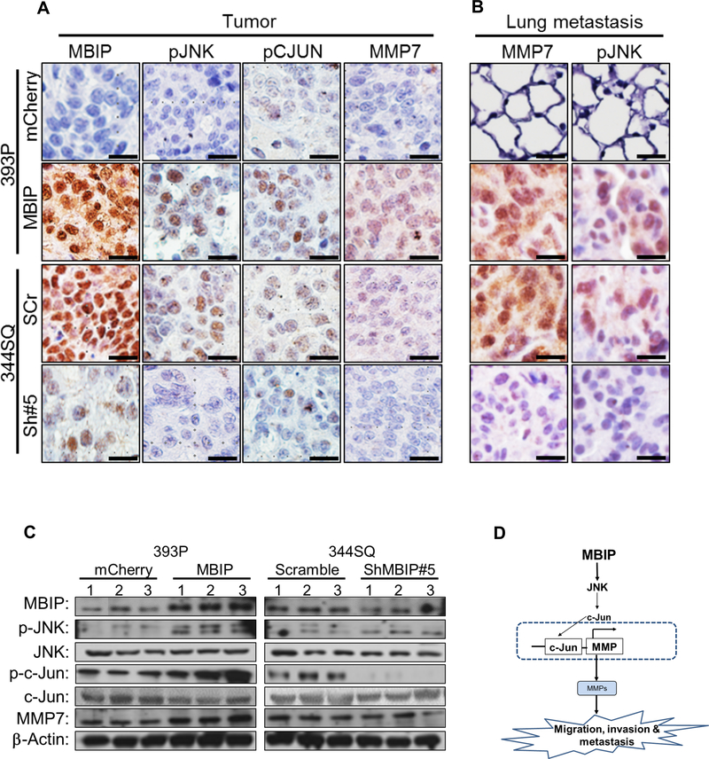
Figure 6. MBIP drives the JNK/c-JUN/MMP axis in vivo.
Representative IHC staining images from (A) Primary tumors or (B) metastatic lung lesions, from mice injected with MBIP-overexpressing (393-mCherry control and 393P-MBIP) and MBIP knockdown (344SQ-Scramble control and 344SQ-ShMBIP#5) cells, analyzed with antibodies against MBIP, p-JNK, p-c-Jun and MMP7. Scale bar = 50 μm. (C) Immunoblot analysis to determine expression levels of MBIP, p-JNK, total JNK, p-c-Jun, total c-Jun and MMP-7, in primary tumors formed by flank injection of MBIP-overexpressing (393-mCherry control and 393P-MBIP) and MBIP knockdown (344SQ-Scramble control and 344SQ-ShMBIP#5) cells as indicated. β-Actin was used as an internal loading control. (D) Schematic model: MBIP amplification leads to phosphorylation of JNK. Phosphorylated JNK actives c-JUN and AP-1 promoter which results into the transcription of MMPs, which is necessary for the increase in migration, invasion and metastasis.

