Abstract
Background
Adrenal vein sampling (AVS) is essential for diagnostics of primary aldosteronism, distinguishing unilateral from bilateral disease and determining treatment options. We reviewed the performance of AVS for primary aldosteronism at our center during first 15 years, comparing the initial period to the period after the introduction of a dedicated radiologist. Additionally, AVS outcomes were checked against CT findings and the proportion of operated patients with proven unilateral disease was estimated.
Patients and methods
A retrospective cross-sectional study conducted at the national endocrine referral center included all patients with primary aldosteronism who underwent AVS after its introduction in 2004 until the end of 2018. AVS was performed sequentially during Synacthen infusion. When the ratio of cortisol concentrations from adrenal vein and inferior vena cava was at least 5, AVS was considered successful.
Results
Data from 235 patients were examined (168 men; age 32–73, median 56 years; BMI 18–48, median 30.4 kg/ m2). Average number of annual AVS procedures increased from 7 in the 2004–2011 period to 29 in the 2012–2018 period (p < 0.001). AVS had to be repeated in 10% of procedures; it was successful in 77% of procedures and 86% of patients. The proportion of patients with successful AVS (92% in 2012–2018 vs. 66% in 2004–2011, p < 0.001) and of successful AVS procedures (82% vs. 61%, p < 0.001) was statistically significantly higher in the recent period.
Conclusions
Number of AVS procedures and success rate at our center increased over time. Introduction of a dedicated radiologist and technical advance expanded and improved the AVS practice.
Key words: angiography, adrenal gland, endocrine disorders, secondary hypertension
Introduction
Primary aldosteronism is the most common form of secondary hypertension, with a prevalence of 5.9% among hypertensive patients in primary care practice.1 Autonomous and excessive secretion of aldosterone from one or both adrenal glands in patients with primary aldosteronism causes significantly higher cardiovascular risk and more pronounced renal damage compared to equally severe essential hypertension.1, 2, 3 Amongst available targeted treatment, the preferred therapeutic option is unilateral laparoscopic adrenalectomy, which can normalize or decrease blood pressure in most patients with proven unilateral disease.4,5 Longterm medical treatment with mineralocorticoid receptor antagonists is not only more expensive and less convenient, but it might also have worse outcomes overall.4,6
Therefore, a crucial part of diagnostic workup in primary aldosteronism is to correctly determine which patients have unilateral disease and could pursue surgical cure. Adrenal computed tomography (CT) (or magnetic resonance imaging (MRI)) should be the first test in the subtype evaluation of primary aldosteronism and to exclude adrenocortical carcinoma.4 However, because of increasing prevalence of nonfunctioning adrenal incidentalomas, the reliability of CT in localizing unilateral disease (e.g. an aldosterone producing adenoma) declines with patient age.4,7 In most patients CT cannot accurately distinguish between unilateral and bilateral forms, and may even lead to inappropriate treatment of primary aldosteronism.8 The only exception are infrequent younger patients below 35 years of age with florid disease and a clear one-sided adrenal adenoma with normal contralateral gland.4,8,9
All other surgical candidates should proceed to adrenal vein sampling (AVS), which is regarded as the gold standard to demonstrate lateralization and to avoid unnecessary or inappropriate adrenalectomy. More than 50 years after its introduction, AVS remains controversial as an invasive, expensive and a technically challenging method with successful bilateral catheterization obtained in only about 75% of cases.4,10 Cannulation of the small and short right adrenal vein with direct drainage into the inferior vena cava (IVC) is often the main obstacle to a successful procedure, while the sampling from the left adrenal vein is relatively straightforward. There is substantial inconsistency in how AVS is performed and interpreted. When done by experienced radiologists, the complication rate is low at between 0.2 and 0.9%.11 Only a limited number of referral centers worldwide routinely carry out the procedure.10,12 Recently, the introduction of cone beam CT (CBCT) and other technical developments have further improved the AVS success rate and reduced the complications.13, 14, 15, 16
Primarily, we aimed to review the performance of AVS for primary aldosteronism at our center from its introduction in 2004 up to 2018. The initial period from 2004 to 2011 was compared to the period after the introduction of a dedicated radiologist in 2012. Our secondary objectives were to check the outcomes of AVS against CT findings and to estimate the proportion of patients with proven unilateral disease who ultimately had surgery.
Patients and methods
Study design
We conducted a retrospective cross-sectional study from AVS introduction in November 2004 to the end of 2018 at the Slovenian national tertiary endocrine referral center, which serves a country with a population of 2 million inhabitants. All the data originated from the Slovenian AVS database. The data collection and its analysis were approved by the National Medical Ethics Committee.
Patients
All patients with confirmed primary aldosteronism who underwent AVS at our center during the study period were suitable for enrollment. The diagnostic work-up for primary aldosteronism was done according to the established guidelines4,17, as previously detailed elsewhere.18
Radiological imaging
One to three months before the AVS, all patients but one had adrenal imaging with a dual-source computed tomography (CT) scanner (Somatom Dual Source, Siemens, Germany). Our pre-specified adrenal CT protocol included 1 mm axial slices through the abdomen before, and if necessary also after, the intravenous administration of 80–100 ml of iodinated contrast (370 mgJ/mL), injected at a rate of 3–4 ml/s via antecubital vein during breath-holding. Contrast-enhanced images were acquired after 60 seconds and 15 minutes. The standard scanning parameters included beam collimation of 64x0.6 mm, 16 slices and gantry rotation time of 0.5 s. Tube voltage was set at 120 kV, while the tube current was variable, optimized for body mass index and size, ranging between 160 and 210 mA. Source images of all phases were reconstructed on the axial plane at 5 mm, and on the coronal planes at 4 mm. Since 2017 a new-generation CT scanner (Somatom Force, Siemens, Germany) has been used, which allowed for more precise adaptation to the individual patient body characteristics. The scanning protocol remained essentially the same except for axial reconstructions at 2 mm. In 2014, interdisciplinary meetings dedicated to adrenal pathology were introduced where CT scans were meticulously reassessed with a radiologist, if both adrenals were described as normal. Finally, any thickening of at least 5 mm was deemed abnormal (Figure 1). The interventional radiologist also reviewed the images, in order to recognize the adrenal veins, especially on the right side.
Figure 1.
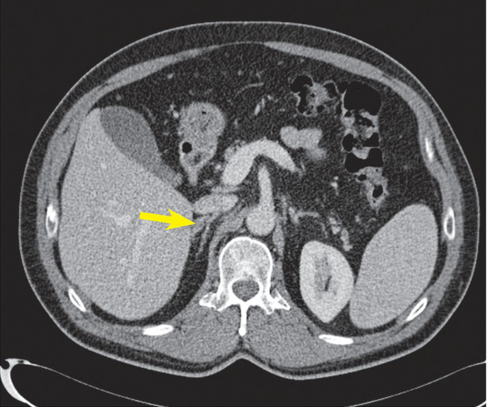
Tiny aldosterone-producing adenoma (8 mm) in lateral limb of the right adrenal gland (arrow) (CT scan).
Adrenal vein sampling
AVS was executed after an overnight fast between 8 and 9 AM. All patients were in the recumbent position for at least 1 hour before sampling. Infusion of synthetic adrenocorticotropic hormone (ACTH) Synacthen (50 μg/h) was started 30 min before AVS and continued throughout the procedure.
During local anesthesia, a 5 Fr sheath (Avanti+ Introducer, Cordis, USA in the first period; Radiofocus Introducer II, Terumo, Japan in the recent period) was introduced into the right femoral vein. AVS was performed sequentially with the right adrenal vein always being cannulated and sampled first, using a 5 Fr Mickelson catheter (Cook Medical Inc., USA) or a 5 Fr Cobra C2 catheter with open-ended tip and two side-holes (Cordis, USA) in the first period. In the recent period a 4 Fr Mickelson catheter (Cook Medical Inc., USA) was routinely used on the right side (Figure 2). Catheterization of the left renal vein then followed with the same catheter, which was used as a guide for a 2.7 Fr Progreat microcatheter (coaxial type with catheter and guidewire; Terumo Interventional Systems, USA) to cannulate the common trunk of the left inferior phrenic vein and the left adrenal vein. The corresponding blood sample was drawn either at the junction of these two veins or selectively from the left adrenal vein above the junction (Figure 3). Finally, a microcatheter was removed and the Mickelson catheter slightly pulled out to sample blood from the infra-renal IVC. On the other hand, a 4 Fr MPA 2 catheter with open-ended tip and two side-holes (Cordis, USA) was used on the left side in the first period. Standard 0.035-inch guidewire (J Tef Guidewire, Kimal, UK) was used in all cases. Additionally, 0.035-inch guidewire with J angled tip (Terumo, Japan) was used on the left side in the first period. Blood samples were drawn in 5 ml syringes and sent to laboratory for aldosterone and cortisol measurements. Hemostasis at the puncture site was ensured by manual compression.
Figure 2.
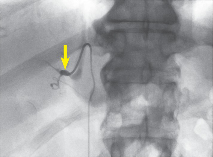
The right adrenal vein during sampling (arrow) (angiography).
Figure 3.
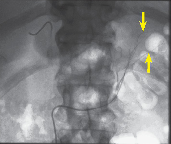
Branches of the left adrenal vein during sampling (arrows) (angiography).
During the initial period from 2004 to 2011 there were two interventional radiologists performing AVS; from 2012 onwards all procedures were done by a single dedicated interventional radiologist. During the first period, AVS was performed with fluoroscopic guidance by digital subtraction angiography (INTEGRIS V5000; Philips, The Netherlands), which was later changed to single-plane digital subtraction angiography (Allura Xper FD; Philips, The Netherlands). In the majority of cases, small amounts of contrast (90 ml on average per procedure in the first period and 52 ml on average per procedure in the recent period, respectively) were injected to better visualize the right adrenal vein.
High-resolution CBCT (Phillips Allura XperCT, The Netherlands) acquisition during AVS has been used since 2012 at first sporadically and then more consistently to identify the tip of the catheter accurately, in order to differentiate between the right adrenal vein and a hepatic accessory vein or a para-vertebral vein when necessary (Figure 4).
Figure 4.
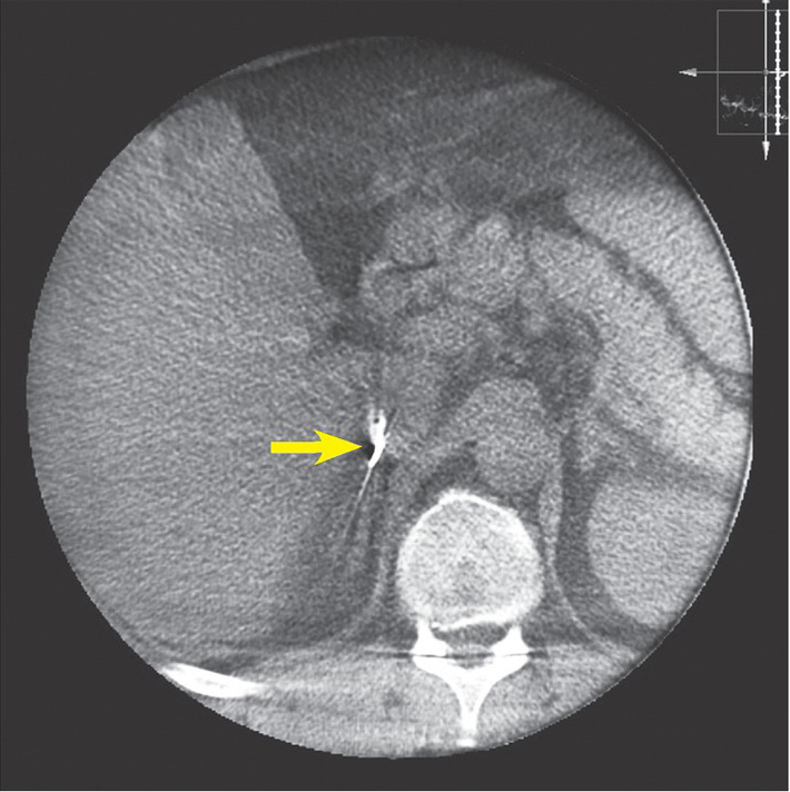
Tip of the catheter in the right adrenal vein (arrow); both limbs of the right adrenal gland are visible underneath (cone beam CT).
The average AVS procedure time decreased from 18.2 minutes in the first period to 16.8 minutes in the recent period.
When the selectivity index (SI), computed as the ratio of concentrations of cortisol from an adrenal vein and the infra-renal IVC, was at least 5, AVS was deemed successful. Lateralization index (LI), defined as the ratio of the higher over the lower cortisol–corrected aldosterone ratio, of more than 4 indicated unilateral aldosterone excess, while the values between 3 and 4 were assumed borderline.19 Suppressed plasma renin activity (PRA) values (< 0.6 ng/mL/h) were used as proof for unlikely stimulation of the contralateral adrenal cortex at a level adequate to confound interpretation of lateralization.20,21
Assays
Serum aldosterone was measured with the Active® Aldosterone RIA (Beckman Coulter, Immunotech, Czech Republic). Serum cortisol was measured with an automated chemiluminescent immunoassay (CLIA) on the Immulite® 2000 XPi (Siemens Healthcare, Gwynedd, United Kingdom). The respective within- and between-assay coefficients of variation were below 4.5% and 9.8% for aldosterone and below 6.8% and 9.4% for cortisol. PRA measurements were performed using the Angiotensin I RIA KIT (Beckman Coulter, Immunotech, Czech Republic). The respective within- and between-assay coefficients of variation were below 11.3% and 20.9%.
Statistical analysis
Descriptive statistics were calculated. Patient characteristics and outcomes were compared between periods or groups using t-test, exact Mann-Whitney test and Fisher’s exact test. Cohen’s kappa was used to assess agreement between diagnostic methods. Statistical analyses were conducted using IBM SPSS Statistics 20 (IBM Corp., Armonk, USA, 2011).
Results
Data from 235 patients with primary aldosteronism were examined. Their clinical characteristics and laboratory parameters are presented in Table 1.
Table 1.
Clinical characteristics and laboratory parameters of the patients
| Characteristic | Descriptive statistics |
|---|---|
| n | 235 |
| Male patients | 168 (71%) |
| Age (years) | 56 (32–73) |
| Body Mass Index (kg/m2) | 30.4 (18.3–48.4) |
| Systolic BP at presentation (mm Hg) | 155 (145–170) |
| Diastolic BP at presentation (mm Hg) | 90 (80–95) |
| Number of antihypertensive agents | 3 (2–4) |
| Hypokalemia | 172 (73%) |
| eGFR (ml/min/1.73 m2) | 88 (71–102) |
| Baseline aldosterone (nmol/L) | 0.7 (0.3–8.8) |
| Baseline PRA (ng/mL/h) | 0.2 (0.2–0.9) |
| Baseline ARR | 4.2 (1.1–43.8) |
| CT* normal / bilateral / unilateral | 66 (28%) / 24 (10%) / 144 (62%) |
| Tumor size on CT (mm) | 13 (8–19) |
= not performed in one patient; ARR = serum aldosterone-to-renin ratio; BP = blood pressure; eGFR = estimated glomerular filtration rate; PRA = plasma renin activity; Descriptive statistics are reported as median (interquartile range) for numeric variables and number (percentage) for categorical variables;
Most of them had a unilateral adrenal abnormality (62%) on CT scan, while bilateral adrenal thickening was present in 10% of the cases. The average adrenal nodules’ size was 13 mm and left-sided lesions were more prevalent than the right-sided ones (62% vs. 38% in total). There were 28 left-sided lesions (60%) in the first period and 91 left-sided lesions (62%) in the recent period. Finally, in 28% of the cases CT scans of both adrenals were considered normal.
The average number of AVS procedures performed per year increased statistically significantly from 7 in the 2004–2011 period to 29 in the 2012–2018 period (p < 0.001) (Figure 5). In total, AVS had to be repeated in 10% of the procedures (9% in the first period, 10% in the recent period). AVS was successful (SI ≥ 5 in both adrenal veins) in 86% of the patients and in 77% of the procedures. The overall success rate of left adrenal vein cannulation was significantly higher than that of the right adrenal vein (p = 0.001). While the success rate on the left side remained unchanged over time (94% vs. 97%; p = 0.434), there was a statistically significant improvement on the right side after the introduction of a single dedicated interventional radiologist in 2012 (66% vs. 94%; p < 0.001). Consequently, the proportion of patients with successful AVS (66% vs. 92%, p < 0.001) and of successful AVS procedures (61% vs. 82%, p < 0.001) was also significantly higher in the recent period (Figure 6). The right and left median SI values were not statistically significantly different (22.3 [interquartile range 18.2] vs. 22.4 [13.1]; p = 0.285). Decreasing the SI to ≥ 3 instead of ≥ 5 would not have improved the AVS performance on either side. Among previously tested clinical determinants of bilateral AVS success22,23, only younger age proved to be statistically significant (p = 0.004) in our cohort, whereas higher BMI and male gender did not. Adrenal hemorrhage due to vein rupture occurred during two procedures (0.8% overall), one in the initial period (1 out of 57 procedures, 1.8%) and another in the recent period (1 out of 203 procedures; 0.5%), both resolved conservatively. Primary aldosteronism persisted in both cases and was treated medically. There were no other serious adverse events associated with AVS during the study.
Figure 5.
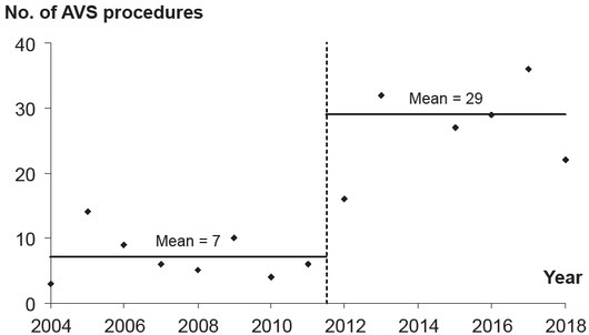
Number of adrenal vein sampling procedures per year during the study period.
Figure 6.
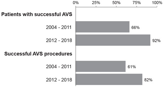
Patients with successful adrenal vein sampling (AVS) and successful AVS procedures during the study period.
CT and AVS results were compared in 181 patients with bilaterally successful AVS, excluding cases with borderline LI values between 3 and 4. The agreement amongst the two diagnostic methods was present in only 59% of cases (kappa = 0.36) (Figure 7).
Figure 7.
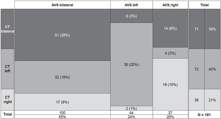
Agreement between adrenal vein sampling (AVS) and CT findings depicted with a variable-width stacked column chart. Patients with normal CT scans are included in the CT bilateral category
Among the patients with successful AVS, 44% overall (n = 79) had LI > 4 and hence proven unilateral disease. The percentage of lateralized cases did not statistically significantly differ between the two study periods (p = 0.248) or between younger (< 40 years) and older patients (p = 0.470). Adrenalectomy was recommended to all the patients with lateralized aldosterone secretion, but only 86% of them underwent surgery. All patients below 40 years of age with proven unilateral disease were operated on, but the same was true for only 84% of older subjects. The main reason for not having surgery was patient’s reluctance (n = 9). One patient was diagnosed with liver cirrhosis and was rejected by the surgeon, two patients were lost to follow-up. The proportion of patients with unilateral disease undergoing surgery did not differ statistically significantly between the periods (89% vs. 85%, p = 1.000). Finally, additional four out of 21 patients with successful AVS and borderline LI values between 3 and 4 also opted for surgery. Three of them had clear unilateral adrenal nodule on CT, whereas the remaining patient had normal glands on imaging. All other patients were treated medically.
Discussion
The present study provides an important insight in the implementation process and continued development of AVS at the Slovenian national endocrine referral center over 15 years. The overall success rate for the AVS procedures during this period
was 77%, which is similar to the recently published large multicenter AVS registry study on 1625 patients, where 80% of cases were bilaterally selective.24 Interestingly, the data from German Conn’s Registry revealed that only 31% of their initial AVS studies were successful with later increase of the success rate to 61%.25 On the other hand, the proportion of successful AVS procedures at our institution increased from 61% in 2004–2011 to 82% in 2012–2018. With 10% of procedures being repeated overall, the proportion of our patients with successful AVS rose concurrently from 66% to 92%, which is close to the success rate at the centers of excellence.19,26,27 The observed increment could be partially explained by our decision in 2012 to follow the recommendation for low-volume centers and focus the expertise on a single, dedicated interventional radiologist.12,28 This decision not only improved, but also expanded the AVS performance at our center (Figure 5).
The overall success rate improved due to superior cannulation of the right adrenal vein in the recent period (94% vs. 66%), whereas the success rate on the left side remained around 95% and unchanged over time. This was most probably not only due to the learning curve of the radiologist29,30, but mainly due to more regular pre-procedural review of CT images and intra-procedural use of high-resolution CBCT since 2012 to better map the adrenal venous anatomy, especially on the right side. The same approach has been recently used in other centers and allowed not only a better evaluation of the selectivity of right-sided adrenal vein cannulation, but also a significant decrease in the fluoroscopy time and quantity of iodine contrast injected in combination with unchanged or even lower radiation exposure.13, 14, 15, 16
Recently, another possibility to improve the catheterization success has been offered by using the newly developed ultra-rapid technique for semi-quantitative measurement of the cortisol level in adrenal veins in approximately 5 minutes, thus enabling the radiologist to reposition the catheter if the first result indicates an incorrect position.31 The rapid on-site measurement of the cortisol might be associated with a shorter procedure time and lower radiation dose than CT assisted AVS.32 However, this approach was not available at our center during the analyzed period.
Throughout the study period we strictly followed the Mayo Clinic protocol and used continuous Synacthen infusion starting 30 min before sampling and continuing throughout the procedure during sequential AVS.19 The main rationale for ACTH-stimulated AVS is to maximize the cortisol gradient between the adrenal veins and VCI. Consequently, SI is increased and so is the proportion of diagnostic AVS procedures, which is why such a practice is particularly suitable for less experienced and low-volume centers.20,21 On the other hand, some authors consider the use of ACTH-stimulation as controversial because it might have the undesirable effect of masking the lateralization of aldosterone production, thus rendering some patients with unilateral primary aldosteronism apparently unsuitable for surgery.10 Fortunately, accumulated data overall suggest that surgical outcomes are similar irrespective of whether AVS is done by ACTH stimulation or not.5,33,34
ACTH stimulation also minimizes stress-induced variations in aldosterone secretion during sequential sampling19, 20, 21, which might otherwise generate artificial between-sides gradients and lower its diagnostic accuracy.35 Additionally, according to our protocol the right adrenal vein was always being cannulated first to lessen the time lag amid the sides.28 Thereafter, a microcatheter was used to quickly cannulate the left adrenal vein36 and to keep the delay between sequential sampling under 5 minutes in most of our AVS procedures.37
Clearly, AVS studies that are not bilaterally successful should not be used to establish lateralization.20 The choice of the correct SI is pivotal for the reported catheterization success rate, diagnostic reliability of the method and clinical outcome.34,38,39 According to the expert consensus the cutoff value for the SI should be ≥ 3.0 during ACTH stimulation20, but we consistently applied an even more robust criterion (SI ≥ 5) in order to minimize the chance of misdiagnosing either unilateral or bilateral primary aldosteronism.19,21 It is conceivable that there is a progressive decrease in success rate with increasing SI cut-offs, although the recent multicenter study showed this to be less dramatic with ACTH-stimulation.34 Concordantly, decreasing the SI to ≥ 3 instead of ≥ 5 in our cohort would not improve the cannulation success rate on either side. Furthermore, the data from the same study showed post-ACTH SI cut-off of 5 to be able to clearly segregate biochemically successful and non-successful studies.34 Actually, our median SI values were much higher than the advocated threshold. There was no usual distinction between higher median right-sided and lower median left-sided SI values7,19, pointing to selective cannulation of the left adrenal vein in most cases with the microcatheter. Notably, blood sampling from the common trunk of the inferior phrenic vein and the left adrenal vein might be the preferable method of AVS due to better potential diagnostic accuracy, technical ease, lower cost and lower risk of vein rupture.40
The overall complication rate during the study was low (0.8%). Despite the almost fourfold increase of AVS procedures in the recent period, the between periods complication rates were comparable, with one adrenal hemorrhage due to vein rupture in each period (1.8% vs. 0.5%). The observation confirmed that the major determinant of the incidence of such events is the number of AVS performed by each radiologist.12
When AVS results were used as the gold standard for lateralization in a subgroup with unequivocal diagnosis of unilateral (LI > 4) or bilateral (LI < 3) disease, CT misdiagnosed the primary aldosteronism subtype in 41% of our patients despite reassessment of all normal scans at our interdisciplinary meetings. If we had relied only on imaging, 20/71 (28%) patients would have been incorrectly denied adrenalectomy and treated medically. In addition, 49/100 (49%) patients with bilateral primary aldosteronism would have been sent to unilateral adrenalectomy, and 6/77 (8%) patients with unilateral primary aldosteronism would have had removed the normal-functioning adrenal (Figure 7). By contrast, Mulatero et al. demonstrated much higher agreement of AVS and CT (77%) when imaging was performed by the same highly motivated radiologist.41 Nevertheless, the proportion of discordant AVS and CT results in our cohort closely resembles the findings of a systematic review of 38 diagnostic studies on 950 patients, where CT (or MRI) might have missed the type of primary aldosteronism in 37.8% of cases.8
Ultimately, 44% of our patients lateralized on AVS, which represents a slightly higher prevalence of unilateral disease than traditionally reported.4,7 Yet this finding was not unexpected, because several clinical characteristics of our cohort, e.g. high median number of antihypertensives, prevalent spontaneous hypokalemia and higher median aldosterone values, pointed towards more severe disease, which is consistent with unilateral primary aldosteronism. We used the most stringent LI cut-off (> 4), which is favored by the expert consensus for ACTH-stimulated AVS, in order to avoid false-positives and ensure highest possible cure rates.10,20,21 Only four out of 21 patients with borderline LI values (3–4) were referred to surgery. Use of contralateral gland suppression (e.g. lower aldosterone to cortisol ratio than the same ratio in IVC) might be potentially helpful to determine lateralization in intermediate cases8,10,38 but was not employed during the study period. Using our conservative approach to make surgical decision, close to 100% of operated patients at our center achieved complete biochemical remission of primary aldosteronism according to the international PASO outcome consensus.5
Adrenalectomy was recommended to all patients who lateralized on AVS, however a substantial proportion (14%) was ultimately treated medically. Only patients older than 40 years changed their mind and decided against the operation. These outcomes stress the importance of careful selection of patients for AVS and operation.28 Most appropriate candidates desire surgery and have a high probability of unilateral primary aldosteronism. On the other hand, AVS is not needed in individuals who prefer medical therapy and in those who are not suitable for surgery due to comorbidities or age.21,42 A simple clinical prediction criterion could probably identify some patients with bilateral primary aldosteronism who should avoid unnecessary AVS and be treated medically.18 Last but not least, the primary aldosteronism surgical outcome predictor might help finding patients who are expected to attain long-term blood pressure control after adrenalectomy to guide preoperative patient counseling and final decision for or against AVS and surgery.43
There are some limitations of the present study. Primarily, the outcomes were deducted from a retrospective analysis. However, all relevant clinical and laboratory data were logged into our AVS database virtually without missing values. Furthermore, discontinuation and/or adjustment of the antihypertensive agents before and during AVS could probably have been more rigorous, especially during the early years. Still, hypokalemia was always corrected, mineralocorticoid antagonists and potassium-wasting diuretics were discontinued on time. Most patients had resistant hypertension, so we mostly followed the expert recommendation that less interfering antihypertensive medications may be used if PRA, which was routinely measured before AVS, remained suppressed.20,21 ACTH stimulation might have the potential to mask lateralization of aldosterone production in patients with adenomas simultaneously producing cortisol, which appears more frequently than we thought earlier.10,44 During the study dexamethasone suppression testing to detect this entity was recommended only in rare patients with relatively large adrenal tumors of ≥ 3 cm and not routinely.4,17 Consequently, another possible source of error might have been unrecognized autonomous cortisol cosecretion in some patients. Finally, the technical advances in AVS techniques over the 15-year study period and their impact on the AVS success rate might not have been emphasized enough.
The main strength of our study is that our results were derived from a relatively large and a well-defined national cohort. Management of the patients was standardized and followed the Endocrine Society clinical guidelines whenever feasible4,17, which can significantly decrease the selection bias.
Conclusions
Based on the present study, we conclude that the introduction of a dedicated radiologist with higher workload and regular use of intra-procedural CBCT since 2012 have significantly enhanced the AVS performance at our center. In the future, we aim to improve the concordance of AVS results with CT findings by revising our interdisciplinary strategy with radiologists. We will also address the protocols for the selection of appropriate candidates for AVS, since we demonstrated that a substantial number of patients with proven unilateral primary aldosteronism did not proceed to surgery.
Acknowledgements
The authors would like to thank Marjana Turk Jerovsek, M.D. and Barbara Robnik, M.D. for their work with the AVS database. We appreciate the assistance of Vlasta Hocevar and Mateja Adamlje, RNs.
Disclosure
No potential conflicts of interest were disclosed.
References
- 1.Monticone S, Burrello J, Tizzani D, Bertello C, Viola A, Buffolo F. Prevalence and clinical manifestations of primary aldosteronism encountered in primary care practice. J Am Coll Cardiol. 2017;69:1811–20. doi: 10.1016/j.jacc.2017.01.052. et al. [DOI] [PubMed] [Google Scholar]
- 2.Savard S, Amar L, Plouin PF, Steichen O. Cardiovascular complications associated with primary aldosteronism: a controlled cross-sectional study. Hypertension. 2013;62:331–6. doi: 10.1161/HYPERTENSIONAHA.113.01060. [DOI] [PubMed] [Google Scholar]
- 3.Monticone S, Sconfienza E, D’Ascenzo F, Buffolo F, Satoh F, Sechi LA. Renal damage in primary aldosteronism: a systematic review and meta-analysis. J Hypertens. 2020;38:3–12. doi: 10.1097/HJH.0000000000002216. et al. [DOI] [PubMed] [Google Scholar]
- 4.Funder JW, Carey RM, Mantero F, Murad MH, Reincke M, Shibata H. The management of primary aldosteronism: case detection, diagnosis, and treatment: an endocrine society clinical practice guideline. J Clin Endocrinol Metab. 2016;101:1889–916. doi: 10.1210/jc.2015-4061. et al. [DOI] [PubMed] [Google Scholar]
- 5.Williams TA, Lenders JWM, Mulatero P, Burrello J, Rottenkolber M, Adolf C. Outcomes after adrenalectomy for unilateral primary aldosteronism: an international consensus on outcome measures and analysis of remission rates in an international cohort. Lancet Diabetes Endocrinol. 2017;5:689–99. doi: 10.1016/S2213-8587(17)30135-3. et al. [DOI] [PMC free article] [PubMed] [Google Scholar]
- 6.Hundemer GL, Curhan GC, Yozamp N, Wang M, Vaidya A. Cardiometabolic outcomes and mortality in medically treated primary aldosteronism: a retrospective cohort study. Lancet Diabetes Endocrinol. 2018;6:51–9. doi: 10.1016/S2213-8587(17)30367-4. [DOI] [PMC free article] [PubMed] [Google Scholar]
- 7.Young WF. Diagnosis and treatment of primary aldosteronism: practical clinical perspectives. J Intern Med. 2019;285:126–48. doi: 10.1111/joim.12831. [DOI] [PubMed] [Google Scholar]
- 8.Kempers MJ, Lenders JW, van Outheusden L, van der Wilt GJ, Kool LJS, Hermus AR. Systematic review: diagnostic procedures to differentiate unilateral from bilateral adrenal abnormality in primary aldosteronism. Ann Intern Med. 2009;151:329–37. doi: 10.7326/0003-4819-151-5-20090901000007. et al. [DOI] [PubMed] [Google Scholar]
- 9.Lim V, Guo Q, Grant CS, Thompson GB, Richards ML, Farley DR. Accuracy of adrenal imaging and adrenal venous sampling in predicting surgical cure of primary aldosteronism. J Clin Endocrinol Metab. 2014;99:2712–19. doi: 10.1210/jc.2013-4146. et al. [DOI] [PubMed] [Google Scholar]
- 10.Wolley M, Thuzar M, Stowasser M. Controversies and advances in adrenal venous sampling in the diagnostic workup of primary aldosteronism. Best Pract Res Clin Endocrinol Metab. 2020. p. 101400. [DOI] [PubMed]
- 11.Monticone S, Satoh F, Dietz AS, Goupil R, Lang K, Pizzolo F. Clinical management and outcomes of adrenal hemorrhage following adrenal vein sampling in primary aldosteronism. Hypertension. 2016;67:146–52. doi: 10.1161/HYPERTENSIONAHA.115.06305. et al. [DOI] [PubMed] [Google Scholar]
- 12.Rossi GP, Barisa M, Allolio B, Auchus RJ, Amar L, Cohen D. The Adrenal Vein Sampling International Study (AVIS) for identifying the major subtypes of primary aldosteronism. J Clin Endocrinol Metab. 2012;97:1606–14. doi: 10.1210/jc.2011-2830. et al. [DOI] [PubMed] [Google Scholar]
- 13.Busser WMH, Arntz MJ, Jenniskens SFM, Deinum J, Hoogeveen YL, de Lange F. Image registration of cone-beam computer tomography and pre-procedural computer tomography aids in localization of adrenal veins and decreasing radiation dose in adrenal vein sampling. Cardiovasc Intervent Radiol. 2015;38:993–7. doi: 10.1007/s00270-014-0969-z. et al. [DOI] [PubMed] [Google Scholar]
- 14.Ringe K, Wacker F, Terkamp C, Meyer B. Value of additional cone-beam CT acquisitions for adrenal vein sampling. [Abstract] J Vasc Interv Radiol. 2017;28(Suppl):S139–40. doi: 10.1016/j.jvir.2016.12.937. Abstract No. 321. [DOI] [Google Scholar]
- 15.Maruyama K, Sofue K, Okada T, Koide Y, Ueshima E, Iguchi G. Advantages of intraprocedural unenhanced ct during adrenal venous sampling to confirm accurate catheterization of the right adrenal vein. Cardiovasc Intervent Radiol. 2019;42:542–51. doi: 10.1007/s00270-018-2135-5. et al. [DOI] [PubMed] [Google Scholar]
- 16.Meyrignac O, Arcis É, Delchier M-C, Mokrane F-Z, Darcourt J, Rousseau H. Impact of cone beam - CT on adrenal vein sampling in primary aldosteronism. Eur J Radiol. 2020;124:108792. doi: 10.1016/j.ejrad.2019.108792. et al. [DOI] [PubMed] [Google Scholar]
- 17.Funder JW, Carey RM, Fardella C, Gomez-Sanchez CE, Mantero F, Stowasser M. Case detection, diagnosis, and treatment of patients with primary aldosteronism: an endocrine society clinical practice guideline. J Clin Endocrinol Metab. 2008;93:3266–81. doi: 10.1210/jc.2008-0104. et al. [DOI] [PubMed] [Google Scholar]
- 18.Kocjan T, Janez A, Stankovic M, Vidmar G, Jensterle M. A new clinical prediction criterion accurately determines a subset of patients with bilateral primary aldosteronism before adrenal venous sampling. Endocr Pract. 2016;22:587–94. doi: 10.4158/EP15982.OR. [DOI] [PubMed] [Google Scholar]
- 19.Young WF, Stanson AW, Thompson GB, Grant CS, Farley DR, van Heerden JA.. Role for adrenal venous sampling in primary aldosteronism. Surgery. 2004;136:1227–35. doi: 10.1016/j.surg.2004.06.051. [DOI] [PubMed] [Google Scholar]
- 20.Rossi GP, Auchus RJ, Brown M, Lenders JWM, Naruse M, Plouin PF. An expert consensus statement on use of adrenal vein sampling for the subtyping of primary aldosteronism. Hypertension. 2014;63:151–60. doi: 10.1161/HYPERTENSIONAHA.113.02097. et al. [DOI] [PubMed] [Google Scholar]
- 21.Monticone S, Viola A, Rossato D, Veglio F, Reincke M, Gomez-Sanchez C. Adrenal vein sampling in primary aldosteronism: towards a standardised protocol. Lancet Diabetes Endocrinol. 2015;3:296–303. doi: 10.1016/S2213-8587(14)70069-5. et al. [DOI] [PubMed] [Google Scholar]
- 22.Berney M, Maillard M, Doenz F, Matter M, Pechère-Bertschi A, Burnier M. Clinical determinants of adrenal vein sampling success. Cardiovasc Med. 2015;18:246–51. doi: 10.4414/cvm.2015.00352. et al. [DOI] [Google Scholar]
- 23.Chayovan T, Limumpornpetch P, Hongsakul K. Success rate of adrenal venous sampling and predictors for success: a retrospective study. Pol J Radiol. 2019;84:e136–e141. doi: 10.5114/pjr.2019.84178. [DOI] [PMC free article] [PubMed] [Google Scholar]
- 24.Rossi GP, Rossitto G, Amar L, Azizi M, Riester A, Reincke M. Clinical outcomes of 1625 patients with primary aldosteronism subtyped with adrenal vein sampling. Hypertension. 2019;74:800–8. doi: 10.1161/HYPERTENSIONAHA.119.13463. et al. [DOI] [PubMed] [Google Scholar]
- 25.Vonend O, Ockenfels N, Gao X, Allolio B, Lang K, Mai K. Adrenal venous sampling: evaluation of the German Conn’s registry. Hypertension. 2011;57:990–5. doi: 10.1161/HYPERTENSIONAHA.110.168484. et al. [DOI] [PubMed] [Google Scholar]
- 26.Doppman JL, Gill JR. Hyperaldosteronism: sampling the adrenal veins. Radiology. 1996;198:309–12. doi: 10.1148/radiology.198.2.8596821. [DOI] [PubMed] [Google Scholar]
- 27.Daunt N. Adrenal vein sampling: how to make it quick, easy, and successful. Radiogr Rev Publ Radiol Soc N Am Inc. 2005;25(Suppl 1):S143–58. doi: 10.1148/rg.25si055514. [DOI] [PubMed] [Google Scholar]
- 28.Young WF, Stanson AW. What are the keys to successful adrenal venous sampling (AVS) in patients with primary aldosteronism? Clin Endocrinol. 2009;70:14–7. doi: 10.1111/j.1365-2265.2008.03450.x. [DOI] [PubMed] [Google Scholar]
- 29.Siracuse JJ, Gill HL, Epelboym I, Clarke NC, Kabutey N-K, Kim I-K. The vascular surgeon’s experience with adrenal venous sampling for the diagnosis of primary hyperaldosteronism. Ann Vasc Surg. 2014;28:1266–70. doi: 10.1016/j.avsg.2013.10.009. et al. [DOI] [PubMed] [Google Scholar]
- 30.Jakobsson H, Farmaki K, Sakinis A, Ehn O, Johannsson G, Ragnarsson O. Adrenal venous sampling: the learning curve of a single interventionalist with 282 consecutive procedures. Diagn Interv Radiol. 2018;24:89–93. doi: 10.5152/dir.2018.17397. [DOI] [PMC free article] [PubMed] [Google Scholar]
- 31.Yoneda T, Karashima S, Kometani M, Usukura M, Demura M, Sanada J. Impact of new quick gold nanoparticle-based cortisol assay during adrenal vein sampling for primary aldosteronism. J Clin Endocrinol Metab. 2016;101:2554–61. doi: 10.1210/jc.2016-1011. et al. [DOI] [PubMed] [Google Scholar]
- 32.Chang C-C, Lee B-C, Chang Y-C, Wu V-C, Huang K-H, Liu K-L. Comparison of C-arm computed tomography and on-site quick cortisol assay for adrenal venous sampling: a retrospective study of 178 patients. Eur Radiol. 2017;27:5006–14. doi: 10.1007/s00330-017-4930-9. et al. [DOI] [PubMed] [Google Scholar]
- 33.Laurent I, Astère M, Zheng F, Chen X, Yang J, Cheng Q. Adrenal venous sampling with or without adrenocorticotropic hormone stimulation: meta-analysis. J Clin Endocrinol Metab. 2018. et al. [DOI] [PubMed]
- 34.Rossitto G, Amar L, Azizi M, Riester A, Reincke M, Degenhart C. Subtyping of primary aldosteronism in the avis-2 study: assessment of selectivity and lateralization. J Clin Endocrinol Metab. 2019. et al. [DOI] [PubMed]
- 35.Rossitto G, Battistel M, Barbiero G, Bisogni V, Maiolino G, Diego M. The subtyping of primary aldosteronism by adrenal vein sampling: sequential blood sampling causes factitious lateralization. J Hypertens. 2018;36:33543. doi: 10.1097/HJH.0000000000001564. et al. [DOI] [PubMed] [Google Scholar]
- 36.Noda Y, Goshima S, Nagata S, Kawada H, Tanahashi Y, Kato T. Utility of microcatheter in adrenal venous sampling for primary aldosteronism. Br J Radiol. 2020. p. 20190636. et al. [DOI] [PMC free article] [PubMed]
- 37.Almarzooqi M-K, Chagnon M, Soulez G, Giroux M-F, Gilbert P, Oliva VL. Adrenal vein sampling in primary aldosteronism: concordance of simultaneous vs sequential sampling. Eur J Endocrinol. 2017;176:159–67. doi: 10.1530/EJE-16-0701. et al. [DOI] [PubMed] [Google Scholar]
- 38.Mulatero P, Bertello C, Sukor N, Gordon R, Rossato D, Daunt N. Impact of different diagnostic criteria during adrenal vein sampling on reproducibility of subtype diagnosis in patients with primary aldosteronism. Hypertension. 2010;55:667–73. doi: 10.1161/HYPERTENSIONAHA.109.146613. et al. [DOI] [PubMed] [Google Scholar]
- 39.Lethielleux G, Amar L, Raynaud A, Plouin P-F, Steichen O. Influence of diagnostic criteria on the interpretation of adrenal vein sampling. Hypertension. 2015;65:849–54. doi: 10.1161/HYPERTENSIONAHA.114.04812. [DOI] [PubMed] [Google Scholar]
- 40.Umakoshi H, Wada N, Ichijo T, Kamemura K, Matsuda Y, Fuji Y. Optimum position of left adrenal vein sampling for subtype diagnosis in primary aldosteronism. Clin Endocrinol (Oxf) 2015;83:768–73. doi: 10.1111/cen.12847. et al. [DOI] [PubMed] [Google Scholar]
- 41.Mulatero P, Bertello C, Rossato D, Mengozzi G, Milan A, Garrone C. Roles of clinical criteria, computed tomography scan, and adrenal vein sampling in differential diagnosis of primary aldosteronism subtypes. J Clin Endocrinol Metab. 2008;93:1366–71. doi: 10.1210/jc.2007-2055. et al. [DOI] [PubMed] [Google Scholar]
- 42.Kocjan T. Rational approach to a patient with suspected primary aldosteronism. In: Lew JI, editor Clinical management of adrenal tumors InTech; 2017. [DOI]
- 43.Burrello J, Burrello A, Stowasser M, Nishikawa T, Quinkler M, Prejbisz A. The primary aldosteronism surgical outcome score for the prediction of clinical outcomes after adrenalectomy for unilateral primary aldosteronism. Ann Surg. 2019. et al. [DOI] [PubMed]
- 44.Arlt W, Lang K, Sitch AJ, Dietz AS, Rhayem Y, Bancos I. Steroid metabolome analysis reveals prevalent glucocorticoid excess in primary aldosteronism. JCI Insight. 2017;2:e93136. doi: 10.1172/jci.insight.9313610.1172/jci.insight.93136. et al. [DOI] [PMC free article] [PubMed] [Google Scholar]


